Key Points
A BCL2 IHC score is a strong prognostic factor independent of the IPI and MYC protein/rearrangement status in DLBCL treated with R-CHOP.
The BCL2 scoring system we propose is a simple, at-a-glance, and highly reliable system, which was confirmed by an image analysis.
Abstract
Overexpression of the BCL2 is associated with a poor prognosis in diffuse large B-cell lymphoma (DLBCL). The assessment of MYC immunohistochemistry (IHC) is becoming optimized, whereas the criteria for BCL2 positivity are highly variable. Furthermore, data on the frequency and prognostic value of BCL2 positivity are conflicting. We aimed to evaluate BCL2 expression by IHC and assess the prognostic significance of the histopathologically scored BCL2 expression in 456 patients with DLBCL uniformly treated with standard immunochemotherapy (rituximab plus cyclophosphamide, doxorubicin, vincristine, and prednisone, R-CHOP). We initially designed 4-grade BCL2 scoring criteria, from 0 to 3+, and found that ∼40% of DLBCL showed strong BCL2 expression (score 3+). The scores from the pathologist’s visual estimation were confirmed to be reliable using a digital image analysis. A retrospective survival analysis revealed that BCL2 score 3+ was a significant prognostic factor independent of the international prognostic index (IPI), the IHC-determined cell of origin, and the MYC protein/rearrangement status in a training set (n = 218). The adverse prognostic impact of BCL2 score 3+ was confirmed in a validation set (n = 238). We also developed a prognostic model consisting of 3 groups with a combined BCL2 score and MYC protein/rearrangement status. Patients with BCL2 score 3+ showed a higher treatment failure rate; therefore, alternative therapeutic strategies should be considered for these patients. A highly selective BCL2 inhibitor, venetoclax, was recently introduced as breakthrough therapy. Our BCL2 scoring system could readily be used by pathologists to evaluate patients with DLBCL who might benefit from BCL2-targeted therapies.
Introduction
Diffuse large B-cell lymphoma (DLBCL) represents the largest entity of non-Hodgkin lymphoma and is recognized as heterogeneous at both the clinical and the molecular levels.1 Standard therapy such as rituximab, cyclophosphamide, doxorubicin, vincristine, and prednisone (R-CHOP) cures two-thirds of the patients, even those in advanced stages. Predictive biomarkers have been explored for the remaining 30% of the patients at higher risk of treatment failure. The International Prognostic Index (IPI) is a well-established clinically defined score. However, a robust prognostic factor based on the cell biology of DLBCL has not yet been determined. Gene expression profiling studies provide the prognostic value based on the cell of origin (COO) subgroup, revealing that activated B-cell–type DLBCL has a poor outcome compared with germinal center (GC) B-cell–type DLBCL.2,3 Nevertheless, independent of these factors, patients can relapse after R-CHOP therapy, suggesting the existence of additional oncogenic events that are responsible for chemoresistance.
The BCL2 proteins are central regulators of the mitochondrial apoptotic pathway.4 BCL2 was initially discovered as the protooncogene involved in the t(14;18)(q32;q21) chromosomal translocation in follicular lymphoma.5 The antiapoptotic protein BCL2 is overexpressed in many cancers, and BCL2 overexpression is associated with drug resistance.6 Recently, venetoclax, a highly selective BCL2 inhibitor, was developed and reported to be efficacious in several hematological cancers7,8 ; thus, BCL2 expression in tumor cells has attracted tremendous attention. An increased BCL2 protein expression is led by not only chromosomal translocations/amplifications but also other oncogenic pathways through transcriptional or posttranscriptional mechanisms.6 Therefore, the BCL2 protein is the most appropriate biomarker for inhibitor efficacy because the protein is the ultimate product and the real target of the inhibitor. In DLBCL, the ratio of BCL2-positive cases is highly variable, ranging from 24% to 80% among the study population in the previous studies that used immunohistochemistry (IHC); therefore, its prognostic relevance is controversial.9-25 Although this discrepancy largely derives from the use of different cutoff values ranging from 1% to 75% (supplemental Table 1, available on the Blood Web site), few studies have sought to establish definite criteria for the evaluation of BCL2 by IHC.
Currently, considerable attention has been devoted to studies on DLBCL with MYC and BCL2 abnormalities. The oncoprotein MYC causes uncontrolled cell proliferation when it is deregulated. However, in the absence of survival factors, MYC-sensitized cells undergo a default pathway of cellular death due to a growth-inhibitory mechanism.26 In other words, tumors with a high proliferation ability require a second hit to suppress cell death.27 Thus, the combined deregulation of MYC and BCL2 collaborate successfully in tumor progression. In DLBCL, MYC translocations occur in 5% to 14% of patients.28 When MYC alterations are associated with BCL2 (less frequently with BCL6) rearrangement, they are called “double-hit” or “dual-hit” DLBCLs (DHL).29,30 Recent studies show that ∼30% of DLBCLs coexpress high levels of MYC and BCL2 proteins, which are hereafter referred to as “dual-expresser” DLBCL (DEL).22,31 These groups show a more aggressive behavior and more frequent treatment failure after conventional therapy than non-DHL/non-DEL groups; therefore, they are regarded as a new biomarker-defined subset. These facts strongly show that exact determination of BCL2 and MYC status will be of central importance in the treatment of DLBCL.
The aim of this study is to evaluate BCL2 protein expression by IHC and assess the prognostic significance of the histopathologically scored BCL2 expression in a total of 456 patients with newly diagnosed DLBCL treated with R-CHOP therapy. The BCL2 staining level was validated quantitatively by image analysis assessing the pixel values on the digital slide images. Similarly, we measured MYC protein expression by IHC with a visual estimate and an image analysis, and the genetic status was analyzed by fluorescent in situ hybridization (FISH). The prognostic value of the combined BCL2 protein expression score and the MYC IHC/FISH status was also evaluated.
Materials and methods
Patient selection
We studied 456 patients who were newly diagnosed as having DLBCL between March 2003 and November 2015 and were followed up at The Cancer Institute Hospital, Japanese Foundation for Cancer Research (Tokyo, Japan). The patients were selected based on the availability of both the clinical information and the histologic material for a definite diagnosis. All patients were treated with R-CHOP for 3 to 8 cycles with or without radiotherapy. Cases were excluded if the patient had a history of low-grade lymphoma. All patients were negative for HIV antibody. This study was approved by the institutional review boards, and all patients provided written informed consent.
Histopathological analysis and digital image acquisition
Diagnostic formalin-fixed, paraffin-embedded tissue specimens were obtained, and 2-µm-thick sections were used for the morphological, IHC, and FISH analyses. IHC was performed using a Dako Autostainer with EnVision chain polymer-conjugated method or Leica Bond-III with Bond polymer Refine Detection kit, with the following antibodies (clone): CD5 (4C7), CD10 (56C6), CD20 (L26), BCL2 (124), BCL2 (3.1), BCL6 (PG-B6p), MUM1/IRF4 (MUM1p), Ki-67 (MIB1), and MYC (Y69). COO was assigned based on the Hans algorithms.16 The slides stained for BCL2 or MYC were scanned with the ScanScope AT (Aperio Technologies) at ×20 magnification and were analyzed using the Aperio ImageScope software. The FISH analysis was performed using 3 break-apart probes, BCL2 (Dako), BCL6 (Dako), and MYC (Vysis). In the present study, cases whose cytomorphology were consistent with DLBCL were included, and those resembling lymphoblastic lymphoma or lymphomas intermediate between DLBCL and Burkitt lymphoma were excluded. In situ hybridization assay for Epstein-Barr virus (EBV)-encoded small RNAs and FISH for BCL2 and BCL6 were not performed on all cases of the validation set because of the scarcity of specimens. This limitation resulted in the inclusion of DLBCL, not otherwise specified (∼90%), high-grade B-cell lymphoma with MYC and BCL2 and/or BCL6 rearrangements (∼5%, only cases with a DLBCL morphology), and EBV-positive DLBCL, not otherwise specified (∼0% to 5%, estimated) for overall cases according to the 2016 revision of the World Health Organization classification of lymphoid neoplasms.32
BCL2 IHC evaluation on the slides and image analysis
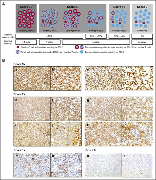
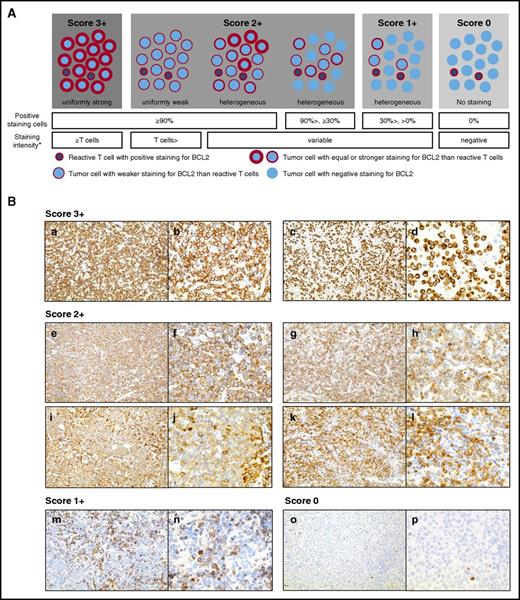
We assessed BCL2 IHC with a microscope on the diagnostic whole sections in all 456 cases, comparing it with the hematoxylin-eosin, CD5, CD20, and Ki-67 stained slides. An estimation of the percentage of BCL2-positive cells was performed by viewing the tumor area at ×10 and ×20 objectives, and the staining extent was divided largely into 3 categories as follows: (1) virtually all tumor cells were positive; (2) the majority of the tumor cells was either negative or was only focally weakly positive; or (3) a situation in between these 2. Then, at ×40 magnification, the cytoplasmic staining intensity of the tumor cells was compared with that of the admixed reactive T cells on the same slide. Based on the staining degree and the intensity, the results were classified into the 4-grade scores represented in Figure 1. For the quantitative evaluation, the digital slide images were obtained from the same IHC-stained slides of the training set and were analyzed with the Aperio Positive Pixel Count Algorithm v.9 (Figure 2).
Scoring of BCL2 staining by pathologists. (A) The schema of the BCL2 IHC scoring based on the pattern, positive cell ratio, and intensity. *The staining intensity of all tumor cells. (B) Representative images of the BCL2 expression in DLBCLs. BCL2 protein expression by IHC in representative cases is shown at ×20 objective (left) and at ×40 objective (right). A BCL2 score of 3+ without a BCL2 rearrangement (a-b) and with a BCL2 rearrangement (c-d) by FISH analysis. Variable staining patterns of the cases with a BCL2 score 2+ (e-l). Note that compared with the score of 3+, the cytoplasmic staining intensity of the tumor cells was not uniformly strong. A BCL2 score of 1+ corresponded to negative staining in the majority of the tumor cells, and only weak positive staining in a small number of the tumor cells (m-n). A BCL2 score of 0 corresponded to only T-cell staining (o-p).
Scoring of BCL2 staining by pathologists. (A) The schema of the BCL2 IHC scoring based on the pattern, positive cell ratio, and intensity. *The staining intensity of all tumor cells. (B) Representative images of the BCL2 expression in DLBCLs. BCL2 protein expression by IHC in representative cases is shown at ×20 objective (left) and at ×40 objective (right). A BCL2 score of 3+ without a BCL2 rearrangement (a-b) and with a BCL2 rearrangement (c-d) by FISH analysis. Variable staining patterns of the cases with a BCL2 score 2+ (e-l). Note that compared with the score of 3+, the cytoplasmic staining intensity of the tumor cells was not uniformly strong. A BCL2 score of 1+ corresponded to negative staining in the majority of the tumor cells, and only weak positive staining in a small number of the tumor cells (m-n). A BCL2 score of 0 corresponded to only T-cell staining (o-p).
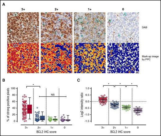
Correlation analysis between the pathologist’s IHC scores for BCL2 staining and the quantified values obtained by image analysis. (A) A digital slide image is shown for the BCL2 IHC each for score 3+ to 0 (top). The bottom figure is the corresponding artificial image produced with the positive pixel count algorithm (PPC). The positive brown DAB (3,3′-diaminobenzidine) staining is colored according to the level of pixel intensity with red (strong), orange (medium), yellow (weak), and blue (negative, nuclear counter stain). The thresholds for the weak, medium, and strong intensities of staining were set as defaults. Because BCL2-positive reactive T cells are always admixed in the tumor areas, these small T cells are manually selected and separated from the large tumor cells in each ROI, and the total sum of these individual areas are evaluated, respectively. Clusters of large tumor cells and individual small T cells are manually delineated by the gray lines and light blue circles, respectively. (B) In each ROI, the number of strong-positive pixels was automatically counted, and the percentage of total number of positive-staining pixels was plotted. The box and plots show the distribution of the percentage. The median values of the score of 3+ group showed a significantly higher than the score of 2+ group (P < .0001, Mann-Whitney U test for pairwise comparison with Bonferroni correction), whereas no significant difference was observed among score 0 to 2+ groups. The horizontal line within the box indicates the median value, and the lower and upper portions indicate the 25% and 75% interquartile range, respectively. The error bars represent the 5% and 95% quantiles. (C) To evaluate the staining intensity of the tumor cells, the average intensity values of the tumor cells are compared after normalization to the T cells. The intensity ratio (tumor/normal T cell) was plotted; a significant positive trend was observed for the tumor staining intensity levels with the IHC score groups (P < .0001, respectively, Mann-Whitney U test for pairwise comparison with Bonferroni correction). *Statistically significant; NS, not significant.
Correlation analysis between the pathologist’s IHC scores for BCL2 staining and the quantified values obtained by image analysis. (A) A digital slide image is shown for the BCL2 IHC each for score 3+ to 0 (top). The bottom figure is the corresponding artificial image produced with the positive pixel count algorithm (PPC). The positive brown DAB (3,3′-diaminobenzidine) staining is colored according to the level of pixel intensity with red (strong), orange (medium), yellow (weak), and blue (negative, nuclear counter stain). The thresholds for the weak, medium, and strong intensities of staining were set as defaults. Because BCL2-positive reactive T cells are always admixed in the tumor areas, these small T cells are manually selected and separated from the large tumor cells in each ROI, and the total sum of these individual areas are evaluated, respectively. Clusters of large tumor cells and individual small T cells are manually delineated by the gray lines and light blue circles, respectively. (B) In each ROI, the number of strong-positive pixels was automatically counted, and the percentage of total number of positive-staining pixels was plotted. The box and plots show the distribution of the percentage. The median values of the score of 3+ group showed a significantly higher than the score of 2+ group (P < .0001, Mann-Whitney U test for pairwise comparison with Bonferroni correction), whereas no significant difference was observed among score 0 to 2+ groups. The horizontal line within the box indicates the median value, and the lower and upper portions indicate the 25% and 75% interquartile range, respectively. The error bars represent the 5% and 95% quantiles. (C) To evaluate the staining intensity of the tumor cells, the average intensity values of the tumor cells are compared after normalization to the T cells. The intensity ratio (tumor/normal T cell) was plotted; a significant positive trend was observed for the tumor staining intensity levels with the IHC score groups (P < .0001, respectively, Mann-Whitney U test for pairwise comparison with Bonferroni correction). *Statistically significant; NS, not significant.
MYC IHC evaluation on the slides and image analysis
MYC IHC was evaluated using digital images. The region of interest (ROI) was manually selected within the scanned image from the whole sections or from the tissue microarray and extracted as a TIFF image in each case. For the training set, MYC IHC was assessed by the visual estimation of 3 hematopathologists (N. Tsuyama, S.S., and K.T.) with the images displayed on monitors. The same images were used for the quantitative evaluation by manual counting and the image analysis. The image analysis was performed using Aperio Nuclear Algorithm v.9. Details of the cell-counting procedure and tissue microarray construction were described in the supplemental Methods.
Statistical analysis
The characteristics at the diagnosis between the groups were compared using a χ2 test. Progression-free survival (PFS) was defined as the time from diagnosis to disease progression/relapse, last follow-up, or death from any cause. Overall survival (OS) was defined as the time from diagnosis to last follow-up or death from any cause. The PFS and OS were estimated by the Kaplan-Meier method, and the differences were evaluated by the log-rank test. A multivariate analysis was performed using the Cox proportional hazards regression model. The Bonferroni correction was used in multiple testing for variables or endpoints. The analyses were performed using GraphPad Prism version 7 and Microsoft Excel 2013. P values < .05 were considered significant.
Results
Clinicopathological characteristics of the 456 patients with DLBCL
The summary of the patients’ characteristics is shown in Table 1 and supplemental Table 2. Information on all FISH and IHC results for MYC, BCL2, and BCL6 was available for 218 of the 456 patients. These 218 patients were selected as a training set to assess the prognostic value of MYC and BCL2 status, whereas the remaining 238 patients were used as a validation set. The patient characteristics were generally similar between the 2 cohorts except that the validation set had more male patients in addition to having fewer patients with bulky tumors.
BCL2 IHC score and correlation with image analysis, FISH, and clinicopathological features
We first assessed BCL2 IHC on 218 cases in the training set. According to our criteria, 89 (41%), 55 (25%), 37 (17%), and 37 (17%) cases scored 3+, 2+, 1+, and 0, respectively. For cases difficult to score either 3+ or 2+, in which more than half of the tumor cells showed strong positivity, we restricted the score 3+ only to those that displayed a uniformly strong-stained pattern. If the samples were admixed with weakly stained tumor cells, the cases were scored as 2+. In addition, cases with a heterogeneous staining pattern at a low magnification were also scored as 2+ even if a strong-stained area was observed in part of the section. The pathologists’ IHC score significantly correlated with the quantitative values (the number of strong-positive pixels and the cytoplasmic staining pixel intensity) by the image analysis (Figure 2).
Among 218 cases, 20 (9%) were positive for BCL2 rearrangement by FISH. Seventeen out of the 20 (85%) cases showed higher BCL2 IHC scores (score 3+, n = 15; score 2+, n = 2; Table 1), suggesting an upregulated BCL2 due to translocation. One case showed a score of 1+. Interestingly, 2 cases scored 0 despite the presence of a BCL2 rearrangement; these cases were weakly positive when stained with another BCL2 antibody (clone 3.1), which was produced by a different immunogen (supplemental Figure 1). This finding implies that these cases had a BCL2 mutation.33 Among the 198 cases negative for BCL2 rearrangement, 74 (37%) exhibited a score of 3+. In these 74 cases, 51 (69%) were of non-GC type. The remaining 23 GC-type cases (31%) might have other mechanisms for BCL2 deregulation than translocation or the activation of the transcription factor nuclear factor-κB pathway.
The baseline clinicopathological features stratified by the BCL2 IHC scores are shown in Table 1. Patients with a score of 3+ presented with a significantly lower rate to achieve complete remission (CR) (P < .001), higher relapse rate after CR (P < .001), and a poorer 3-year PFS (P < .001) than patients with a score of 0 to 2+. Cases with a MYC rearrangement were significantly more frequent in the group with the score of 3+.
MYC IHC percentage and correlation with FISH and clinicopathological features
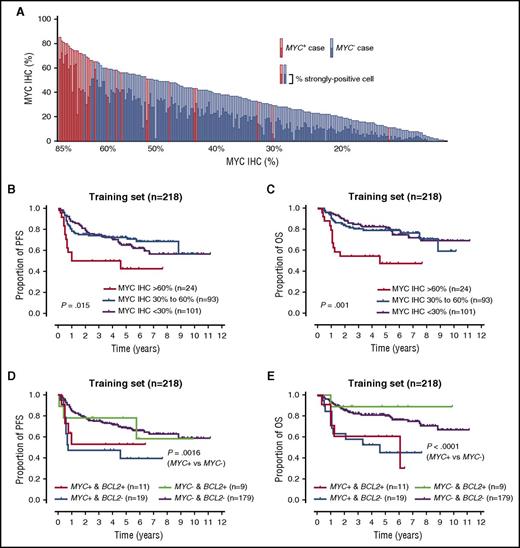
The results on MYC IHC percentage obtained by the 2 types of quantitative evaluation, manual counting and the image analysis, were in extremely good agreement (R2 = .9221, P < .0001; supplemental Figure 2A-B). Thus, we adopted the results of the image analysis for the following clinicopathological analyses. The number of cases scored >60% accounted for only 24/218 (11%), with 20/24 (83%) harboring a MYC alteration (rearrangement, n = 19; amplification, n = 1) (supplemental Figure 2C). This finding suggested a substantial correlation between protein expression and gene rearrangement, although 2 (2%) out of the 101 cases with <30% showed a MYC rearrangement; MYC-IG fusion FISH showed that 1 case had MYC-IG κ fusion signals, but the other had no evidence of either MYC-IGH or MYC-IG light chains. Significant differences were observed in the MYC IHC percentages between MYC-rearrangement-positive and -negative groups (Figure 3A).
Survival analysis according to the MYC protein expression and MYC/BCL2 rearrangement status. (A) Distribution and association of MYC staining intensity and rearrangement status. Each lymphoma cell was judged for staining intensity as strong, medium, weak, or none by image analysis. The percentage of cells with either positive intensity and those with strong intensity in each case was automatically calculated. The y-axis represents the proportion of MYC-positive cells. The x-axis represents the individual case of the training set (n = 218), arrayed according to their overall percentage of MYC-positive cells from left (highest) to right (lowest). Cases with MYC rearrangement are shown as red bars, and cases without rearrangement are shown as blue bars. Light blue/red bars display the overall percentage, and dark-colored bars represent the percentage of cells with strong staining intensity in each case. There are significant differences of MYC IHC percentages between patients with and without MYC rearrangement (percentages of both positive-stained and strongly stained cells; P < .0001, respectively, Mann-Whitney U test). (B-C) PFS and OS of patients with DLBCL in the training set based on percentages of MYC protein expression obtained by the image analysis. An optimal cutoff for MYC protein expression is determined using X-Tile software (version 3.6.1; Yale University School of Medicine, New Haven, CT) with highest χ2 value. (D-E) PFS and OS of the patients with DLBCL in the training set based on MYC/BCL2 rearrangements by FISH analysis. BCL2−, BCL2 rearrangement-negative; BCL2+, BCL2 rearrangement-positive; MYC−, MYC rearrangement-negative; MYC+, MYC rearrangement-positive.
Survival analysis according to the MYC protein expression and MYC/BCL2 rearrangement status. (A) Distribution and association of MYC staining intensity and rearrangement status. Each lymphoma cell was judged for staining intensity as strong, medium, weak, or none by image analysis. The percentage of cells with either positive intensity and those with strong intensity in each case was automatically calculated. The y-axis represents the proportion of MYC-positive cells. The x-axis represents the individual case of the training set (n = 218), arrayed according to their overall percentage of MYC-positive cells from left (highest) to right (lowest). Cases with MYC rearrangement are shown as red bars, and cases without rearrangement are shown as blue bars. Light blue/red bars display the overall percentage, and dark-colored bars represent the percentage of cells with strong staining intensity in each case. There are significant differences of MYC IHC percentages between patients with and without MYC rearrangement (percentages of both positive-stained and strongly stained cells; P < .0001, respectively, Mann-Whitney U test). (B-C) PFS and OS of patients with DLBCL in the training set based on percentages of MYC protein expression obtained by the image analysis. An optimal cutoff for MYC protein expression is determined using X-Tile software (version 3.6.1; Yale University School of Medicine, New Haven, CT) with highest χ2 value. (D-E) PFS and OS of the patients with DLBCL in the training set based on MYC/BCL2 rearrangements by FISH analysis. BCL2−, BCL2 rearrangement-negative; BCL2+, BCL2 rearrangement-positive; MYC−, MYC rearrangement-negative; MYC+, MYC rearrangement-positive.
The overall interobserver agreement for the visual estimates among any 2 of the 3 hematopathologists was good to excellent, and the high-percentage agreement (88%) was achieved in the cases that scored >60% (supplemental Table 3; supplemental Figure 2D). In terms of MYC deregulation status (MYC IHC > 60% and/or MYC rearrangement/amplification positive), which we finally adopted for our prognostic model, the concordance rate between TMA and WS was satisfactory (97%, 100/103) (supplemental Figure 3).
Prognostic impact of the BCL2 and MYC statuses
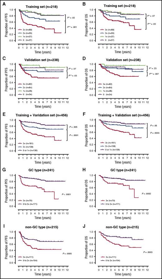
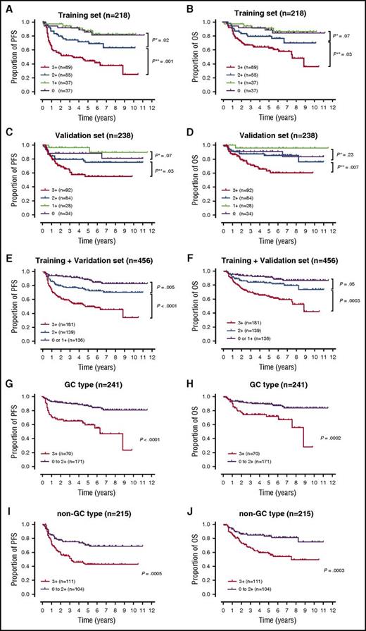
In the training set, the BCL2 score of 3+ group showed significantly inferior outcomes compared with patients with a score of 2+ (PFS, P = .001; OS, P = .03; Figure 4A-B). The adverse prognostic effects of a score of 3+ against the score of 2+ group were confirmed in the validation set (PFS, P = .03; OS, P = .007; Figure 4C-D). Although the survival difference between the patients with a score of 2+ and a score of 0 to 1+ was not confirmed in the validation set, 3 grades of BCL2 scores (score 3+, 2+, and 0/1+) were in good correlation with both PFS and OS in the entire cohort (Figure 4E-F). Furthermore, the adverse impact of a BCL2 score of 3+ was observed within both GC and non-GC types (Figure 4G-J). The univariate and multivariate analyses revealed that a BCL2 score of 3+ was a significant prognostic factor for both PFS and OS independent of the IPI and MYC status in the training set (Table 2; supplemental Table 4).
Survival analysis according to the BCL2 IHC scores. (A-B) PFS and OS of patients with DLBCL in the training set stratified by 4-grade BCL2 IHC scores, from 0 to 3+. Between score 3+ and 2+: HR for PFS = 2.41; 95% CI, 1.49-3.90; P* = .001; HR for OS = 1.98; 95% CI, 1.12-3.49; P* = .030. (C-D) PFS and OS of patients with DLBCL in validation set. Between score 3+ and 2+: HR for PFS = 1.86; 95% CI, 1.09–3.17; P* = .03. HR for OS = 2.44; 95% CI, 1.31–4.54; P* = .007. (E-F) PFS and OS of patients with DLBCL in the entire cohort stratified by 3-grade BCL2 scores; 0 to 1+, 2+, and 3+. (G-H) PFS and OS of patients with GC-type and (I-J) with non-GC–type DLBCL based on BCL2 scores, 3+ or 0 to 2+. P**, BCL2 score 2+ vs score 0 to 1+; P**, BCL2 score 3+ vs score 2+.
Survival analysis according to the BCL2 IHC scores. (A-B) PFS and OS of patients with DLBCL in the training set stratified by 4-grade BCL2 IHC scores, from 0 to 3+. Between score 3+ and 2+: HR for PFS = 2.41; 95% CI, 1.49-3.90; P* = .001; HR for OS = 1.98; 95% CI, 1.12-3.49; P* = .030. (C-D) PFS and OS of patients with DLBCL in validation set. Between score 3+ and 2+: HR for PFS = 1.86; 95% CI, 1.09–3.17; P* = .03. HR for OS = 2.44; 95% CI, 1.31–4.54; P* = .007. (E-F) PFS and OS of patients with DLBCL in the entire cohort stratified by 3-grade BCL2 scores; 0 to 1+, 2+, and 3+. (G-H) PFS and OS of patients with GC-type and (I-J) with non-GC–type DLBCL based on BCL2 scores, 3+ or 0 to 2+. P**, BCL2 score 2+ vs score 0 to 1+; P**, BCL2 score 3+ vs score 2+.
MYC IHC > 60% and MYC rearrangement-positive were both significantly associated with a poor PFS and OS (Table 2; Figure 3B-E; supplemental Figure 3). The 60% cutoff was determined optimal according to the highest χ2 value based on log-rank test in PFS analyses using the X-Tile software.34 In our cohort, no outcome difference was observed between the patients with and without a BCL2 rearrangement in both the MYC rearrangement-positive and -negative groups (Figure 3D-E). Also, different cutoff points for MYC IHC did not affect the outcome in both patients with MYC ≤ 60% and with MYC rearrangement-positive (supplemental Figure S5).
Taken together, our analyses revealed that (1) MYC rearrangements, MYC expression (IHC, >60%), and BCL2 expression (IHC, score 3+) affected the prognosis of DLBCL patients, but not BCL2 rearrangements; and (2) gene rearrangement and protein expression were correlated in MYC but not in BCL2.
Prognostic impact of combined BCL2 IHC score and MYC deregulation
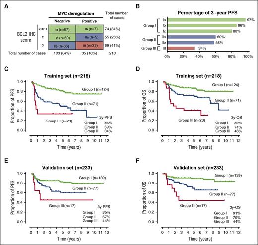
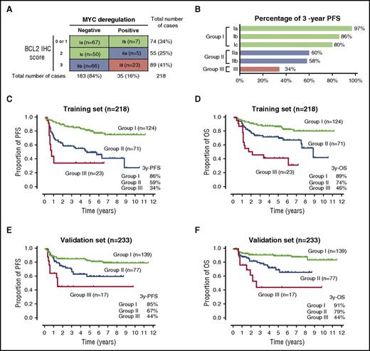
According to the above-mentioned results, we decided to use the BCL2 IHC score and MYC deregulation (MYC IHC > 60% and/or MYC rearrangement/amplification positive) to develop a prognostic model. Based on the combined parameters and their 3-year PFS rates, we divided the patients in the training set into 3 groups (Figure 5A-B). The 3 groups represented 3 separate prognostic groups for both the PFS and the OS (3-year PFS and OS rates ranging from 34% to 89% and 46% to 92%, respectively; Figure 5C-D). Similar outcomes were observed in the validation set and in the groups determined using an alternative clone 3.1 for BCL2 scoring (Figure 5E-F; supplemental Figure 6).
Prognostic groups according to the combined BCL2 IHC score and MYC deregulation status. (A) Prognostic group classification according to the BCL2 IHC score and MYC deregulation status. Group I (n = 124) had a BCL2 score of 0 to 2+ and non-MYC–deregulated DLBCL or a BCL2 score of 0 to 1+ and MYC-deregulated DLBCL; group II (n = 71) had a BCL2 score of 3+ and non-MYC–deregulated DLBCL or a BCL2 score of 2+ and MYC-deregulated DLBCL; and group III (n = 23) had a BCL2 score of 3+ and MYC-deregulated DLBCL. (B) Percentage of 3-year PFS rate of the various groups. (C-D) PFS and OS curves of patients with DLBCL treated with R-CHOP according to the prognostic groups. (E-F) PFS and OS curves of patients with DLBCL treated with R-CHOP according to the prognostic groups in the validation set.
Prognostic groups according to the combined BCL2 IHC score and MYC deregulation status. (A) Prognostic group classification according to the BCL2 IHC score and MYC deregulation status. Group I (n = 124) had a BCL2 score of 0 to 2+ and non-MYC–deregulated DLBCL or a BCL2 score of 0 to 1+ and MYC-deregulated DLBCL; group II (n = 71) had a BCL2 score of 3+ and non-MYC–deregulated DLBCL or a BCL2 score of 2+ and MYC-deregulated DLBCL; and group III (n = 23) had a BCL2 score of 3+ and MYC-deregulated DLBCL. (B) Percentage of 3-year PFS rate of the various groups. (C-D) PFS and OS curves of patients with DLBCL treated with R-CHOP according to the prognostic groups. (E-F) PFS and OS curves of patients with DLBCL treated with R-CHOP according to the prognostic groups in the validation set.
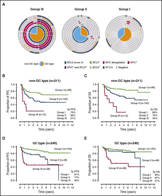
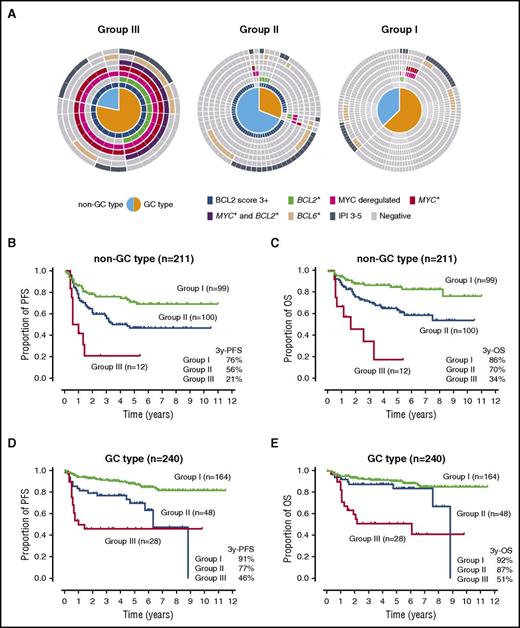
Next, we examined the relationship between the prognostic group, the COO type, and survival outcomes (Figure 6). GC-type DLBCL was more prevalent in group III than in group II (78% and 31%, P < .0001), and all DHL belonged to the GC type of group III. Sixty-six percent of patients in group II were of non-GC type without MYC/BCL2 rearrangement but with strong BCL2 expression. Group III consistently showed dismal outcomes regardless of COO types, whereas group II and I were indiscernible in OS in GC-type DLBCL. Because the proportion of IPI 3-5 in group II was different between the COO types, we tentatively divided patients in group II according to IPI, resulting in that the patients with a low IPI showed a better prognosis in GC type (supplemental Figure 7).
Prognostic groups, COO types, and survival outcomes. (A) Distributions of COO types by the Hans criteria, gene rearrangements, and protein expressions in 3 prognostic groups. (B) PFS and (C) OS curves of non-GC-type DLBCLs according to the prognostic groups. (D-E) PFS and OS curves of GC-type DLBCLs according to the prognostic groups. BCL2+, BCL2 rearrangement-positive; BCL6+, BCL6 rearrangement-positive; MYC+, MYC rearrangement-positive.
Prognostic groups, COO types, and survival outcomes. (A) Distributions of COO types by the Hans criteria, gene rearrangements, and protein expressions in 3 prognostic groups. (B) PFS and (C) OS curves of non-GC-type DLBCLs according to the prognostic groups. (D-E) PFS and OS curves of GC-type DLBCLs according to the prognostic groups. BCL2+, BCL2 rearrangement-positive; BCL6+, BCL6 rearrangement-positive; MYC+, MYC rearrangement-positive.
Discussion
In the clinical settings, an accurate and prompt decision of the BCL2 expression status is becoming more crucial for the treatment of patients with DLBCL. A highly selective BCL2 inhibitor, venetoclax, was recently designated as a breakthrough therapy for refractory or relapsed chronic lymphocytic leukemias.7,35,36 With regard to DLBCL, in vitro studies using cell lines showed that the level of BCL2 expression by IHC and the sensitivity to venetoclax were correlated, and a lack of BCL2 was associated with resistance to venetoclax.7,37
By applying the combination of the staining intensity and the ratio, we achieved a good correlation between the pathologist’s score and the quantified values for BCL2 scoring obtained by the image analysis, suggesting a highly reproducible method. Previous reports evaluating BCL2 protein expression in DLBCL used a different cutoff and lacked specific information on the staining intensity, impeding how best to use those data to determine the prognosis for the subsequent studies; whereas by taking into consideration both the intensity and the percentage, the observers’ agreement for BCL2 IHC improved.38 Another issue in assessing BCL2 expression is that IHC measures the protein expression in the cytoplasm of both the tumor and the admixed reactive T cells at various intensity levels. By careful selection of tumor area in the image analysis, we obtained quantitative pixel values for both the staining area and the intensity of the tumor cells. The pathologist’s score that was defined as positive in the present study (score 3+) was confirmed to have significantly higher pixel values. From a practical point of view, however, we adopted the pathologist’s score for the survival analysis because the traditional use of a microscope is faster and more accessible than using image analysis. Notably, a BCL2 score of 3+ was feasible to predict outcome. Taken together, the scoring strategy presented herein can be generally accepted by pathologists and is another potential method that is better than setting a threshold percentage.
BCL2 mutations are frequently detected in GC-type DLBCL, are associated with a BCL2 translocation,39,40 and can cause pseudo-negative BCL2 protein expression by IHC.40 The most common mutation region in DLBCL is the flexible loop domain (amino acid 31-92) that contains the epitope recognized by the standard BCL2 antibody clone 124, which was raised against amino acid 41-54. Mutations resulting in an amino acid substitution within the residues lead to failure in detecting protein expression by IHC. This event is almost equivalent to the discrepancy between the BCL2 rearrangement and the BCL2 protein expression. In our analyses, the frequency of the pseudo-negative BCL2 protein expression was low (2 of 20 BCL2 rearrangement-positive DLBCL), and the BCL2 rearrangement was not correlated with the prognosis or the BCL2 protein expression; however, we still recommend using BCL2 FISH for the BCL2-negative GC-type DLBCL, because FISH may be useful for selecting the patients who could benefit from the BCL2 inhibitors. Venetoclax is a BCL2-selective BCL2 homology 3 (BH3) mimetic. Therefore, mutations in flexible loop domain causing pseudo-negative BCL2 protein expression do not impede venetoclax binding to the BH3 domain (amino acid 97-107). Mutations in the BH3 domain in DLBCL have been reported in only ∼1.5% of the total nonsynonymous mutations.39,40 Although BCL2 mutations were not examined in the present study, a comprehensive assessment of BCL2 abnormalities will be needed for patients who are potentially eligible to receive BCL2-targeted therapies.
With regard to determining MYC-positive DLBCL by IHC, previous studies used a ≥40% cutoff.22-24,31,32 However, it can cause discrepancies in the pathologist’s interpretation because a considerable number of cases are scored from 30% to 60%, and as demonstrated here and elsewhere,41-43 the observers’ agreement is lower in this range. Therefore, Kluk et al proposed a concurrent analyses of protein expression and gene rearrangement; MYC IHC ≤ 60% and/or MYC rearrangement-negative cases require further confirmation by IHC or FISH for MYC deregulation,42 because a higher cutoff value of MYC IHC was related to both a higher sensitivity for detecting MYC rearrangement and a better agreement of the MYC IHC evaluation between pathologists.42,44 This finding was also confirmed in the present study. Considering the above, as a prognostic marker, we defined cases with an MYC IHC > 60% and/or MYC rearrangement/amplification-positive as MYC-deregulated DLBCL.
Another significant finding here is that 3 distinct prognostic groups were recognized using a combined BCL2 IHC score and MYC-deregulated status. Group III, consisting of patients with a BCL2 score of 3+ and MYC-deregulated DLBCL, showed the poorest prognosis with 3-year PFS and OS rates at 34% and 46%, respectively, under R-CHOP therapy. An alternative therapeutic strategy should be considered for this group. Group II mainly consisted of patients with a BCL2 score of 3+ and non-MYC-deregulated DLBCL. Although this group II showed a better survival than that of group III, we revealed that R-CHOP-only therapy did not contribute to the long-term survival because of the impact of the BCL2 score of 3+. All DHLs involving MYC and BCL2 were included in group III. Therefore, this group is virtually regarded as an expansion of the DHL with the subset of DLBCL with MYC rearrangements and strong BCL2 expression (score 3+). Meanwhile, cases with MYC rearrangement without strong BCL2 expression or DHLs involving MYC and BCL6 were included in groups II and I. Although the number of these cases was relatively small, their distribution may explain conflicting results in the literature in which MYC rearrangements were associated with very poor prognosis in some studies but not in others. Since the introduction of the DEL subgroup and the feasible use of MYC IHC, many researchers have focused on dividing DLBCL into 2 groups, double-positive or not.22,24,31,45 However, the challenge remains how to interpret patients with intermediate levels of MYC/BCL2 protein expression. Group III is virtually regarded also as a DEL subgroup with stricter cutoff. An advantage of our approach is its ability to predict the outcome of such IHC-borderline cases, although these findings should be validated in other study populations.
COO classification and IPI have both been powerful prognostic factors in DLBCL in the R-CHOP era, but in patients with DEL, recent studies have provided different views regarding the value of COO. One study24 demonstrated that a prognostic significance of COO was confounded by the strong negative impact of DEL, which was predominantly seen in non-GC type, whereas others46 claimed COO persisted as a predictor independent of MYC/BCL2 coexpression. Although in the present study we showed that DLBCL with BCL2 score 3+ had poor prognoses in both the COO types, different outcomes according to COO types were exhibited when BCL2 score 3+ was combined with MYC-deregulated status. The majority of group II patients had BCL2 score 3+, but those of GC type showed a better prognosis when they had a low IPI. However, different assays for COO assignment were used in the above-mentioned studies. We applied only the IHC-based criteria by Hans, and therefore, the present findings regarding COO are tentative and need to be reinvestigated using other more reliable assays like the GEP-based assay.46
In summary, we analyzed 456 patients with de novo DLBCL uniformly treated with R-CHOP and provided additional evidence for the prognostic value of BCL2 protein expression. The BCL2 scoring system we propose is a simple, at-a-glance, and highly reliable system, which was confirmed by an image analysis. Therefore, it will be readily used by pathologists in the forthcoming era of BCL2 inhibitors. With the system, we revealed that patients with a BCL2 score of 3+ showed a higher treatment failure rate, for whom alternative therapeutic strategies should be considered, including BCL2 inhibitors. In addition, our 3-risk group system defined with the combined BCL2 score and MYC-deregulated status helps in solving the difficulty to manage “IHC-borderline cases” by the conventional criteria. This prognostic model is not in agreement with current World Health Organization definitions that do not group MYC IHC and FISH with the same significance: the former being a prognostic marker for DLBCL and the latter being used to diagnostically move cases into the entity, high-grade B-cell lymphomas with MYC and BCL2 and/or BCL6 translocations. Our findings have implications for the evaluation of BCL2 and MYC statuses, and thus for the stratification and the treatment of patients with DLBCL.
The online version of this article contains a data supplement.
The publication costs of this article were defrayed in part by page charge payment. Therefore, and solely to indicate this fact, this article is hereby marked “advertisement” in accordance with 18 USC section 1734.
Acknowledgments
The authors thank the members of the Ganken Ariake Lymphoma Study Group and the Tokyo Lymphoma Study Group for their advice. They also thank Sayuri Sengoku for her administrative assistance and Keiko Shiozawa and Tomoyo Kakita for their technical assistance.
This study was supported in part by grants from the Japan Society for the Promotion of Science, Ministry of Health, Labor, Welfare of Japan, and Japan Agency for Medical Research and Development, and the Naito Foundation.
N. Tsuyama is a PhD candidate at Tokyo Medical and Dental University, and this work is submitted in partial fulfillment of the requirement for a PhD.
Authorship
Contribution: N. Tsuyama and K.T. conceived and designed the study and wrote the manuscript; N. Tomita supervised the study and interpreted the clinical and statistical data; N. Tsuyama, S.S., and K.T. made the histopathological diagnoses and analyzed the immunohistochemical data; N. Tsuyama collected and analyzed the FISH and digital image data; S.B. constructed the tissue microarray and produced the FISH and IHC data; Y.M., N.N., K.U., M.Y., Y.T., and K.H. contributed to the patient care and collected the clinical data; N.I. helped with the statistical analyses; N. Tomita and M.K. revised the manuscript. All authors gave final approval of the submitted and published versions.
Conflict-of-interest disclosure: The authors declare no competing financial interests.
Correspondence: Kengo Takeuchi, Pathology Project for Molecular Targets, The Cancer Institute, Japanese Foundation for Cancer Research. 3-8-31 Ariake, Koto, Tokyo 135-8550, Japan; e-mail: kentakeuchi-tky@umin.net.

