Abstract
Background. Recent studies have highlighted the interest of non-invasive procedures to assess extramedullary hematopoiesis in patients with overt myelofibrosis, primary (PMF) or secondary (sMF) to essential thrombocytemia (ET) or to polycythemia vera (PV) (1-4). Several nuclear medicine imaging techniques have been employed without clear consensus about the predominant value of one over other in these mostly preliminary works (2-4).
Population and objectives. Because the pathophysiology of PMF or sMF involves some inflammatory pathways we studied the interest of 18F-FDG (18F-fluorodeoxyglucose) PET/CT in patients with PMF (5). The main objective was to know whether the inflammatory component of PMF could be well detected by this technique. Secondly, we looked for some kind of correlation between the FDG uptake on medullary and extramedullary sites of hematopoiesis and the severity of the disease.
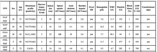
Five patients with diagnosis of MF (3 PMF and 2 sMF) were assessed (see Table 1 for baseline characteristics and results)
Methods.18F-FDG PET/CT imaging was performed following a 60 min. uptake after intravenous injection of a weight-adapted FDG activity (2,5 MBq/kg) on a Siemens Biograph mCT. Images were reviewed by board certified nuclear medicine physicians. Spleen size was measured on axial slices. SUVmax were assessed in a 5 mm diameter spheric volume of interest in homogenous parts of liver and spleen, and in the L2 vertebral body for axial skeleton assessment. Spleen uptake was considered as abnormal in case of a diffuse uptake exceeding hepatic baseline uptake. Skeleton uptake was considered as abnormal in case of diffuse increased uptake.
Results. UPN01LP: slight higher metabolic activity in the axial skeleton and diffuse spleen uptake ; UPN02AP: no hypermetabolic activity in the spleen and slightly higher uptake in the axial skeleton ; UPN03AA: diffuse bone hypermetabolic activity and high metabolic activity in the spleen ; UPN05HL: diffuse bone hypermetabolic activity and higher metabolic activity in the spleen ; UPN06DF: no diffuse hypermetabolic activity in the spleen, slight higher uptake in the axial skeleton.
In summary, those patients with overt myelofibrosis and constitutional signs tend to show a diffuse higher activity both in the axial skeleton and the spleen whereas those without overt myelofibrosis and without constitutional signs tend to present a normal metabolic activity in the axial skeleton and in the spleen. These patterns are in accordance with the progression and severity of the disease. The higher uptake of 18FDG in the axial skeleton in patients with overt MF contrasts with what is observed with some hybrid imaging techniques that show a lower radionuclide (111In/SPECT/CT) uptake at this level translating a lower hematopoietic activity (3).
Conclusions. Based on these preliminary results we consider 18FDG-PET/CT procedures are able to detect the inflammatory component of PMF or sMF and the intensity of FDG uptake appears to be correlated to the severity of the disease. Those patients with a high metabolic activity in the axial skeleton and the spleen might be candidates for specific therapies, although these statements need to be confirmed in a larger series of patients.
Bibliography. 1 Derlin T et al. Assessment of bone marrow inflammation in patients with myelofibrosis: an 18F-fluorodeoxyglucose PET/CT study. Eur J. of Nucl Med Mol Imaging 2015 2 Agool A et al. Radionuclide imaging of bone marrow disorders. Eur J Nucl Med Mol Imaging. 2011. 3 Ojeda-Uribe M et al. Assessment of sites of marrow and extramedullary hematopoiesis by hybrid imaging in primary myelofibrosis patients. Cancer Medicine 2016. 4 Vercellino L et al. Assessing bone marrow activity in patients with myelofibrosis: results of a pilot study of 18F-FLT PET. J. of Nucl Med 2017. 5 Desterke C et al. Inflammation as a Keystone of Bone Marrow Stroma Alterations in Primary Myelofibrosis. Mediators Inflamm. 2015
No relevant conflicts of interest to declare.
Author notes
Asterisk with author names denotes non-ASH members.


