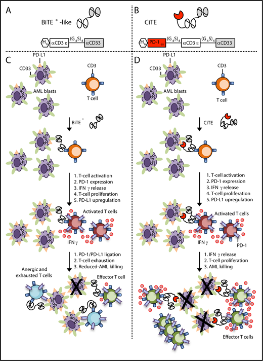
In this issue of Blood, Herrmann et al1 have developed a novel checkpoint inhibitory T-cell–engaging (CiTE) antibody for acute myeloid leukemia (AML) by fusing the extracellular domain of programmed cell death protein 1 (PD-1ex) to a CD3 × CD33 bispecific T-cell engager (BiTE; see figure panels A-B). Incorporation of the PD-1ex domain increased human T-cell activation and led to efficient cytotoxicity against CD33+/PD-L1+ cells in vitro and in a murine xenograft model. Importantly, the PD-1ex domain is not sufficient to redirect T cells to PD-L1+ cells lacking the CD33 target. This preferential targeting of CD33+/PD-L1+ cells should reduce the immune-related adverse events associated with on-target/off-tumor effects of global checkpoint blockade with anti-PD1/PD-L1 monoclonal antibodies (mAbs).
Design and properties of BiTE and CiTE molecules. (A) Schematic representation of the BiTE-like molecule. scFv domains, consisting of 1 variable heavy chain (VH) joined to 1 variable light (VL) domain, are connected into a single polypeptide chain. One of the 2 distal scFv domains (white) is specific for CD3ε, and the other (gray) is specific for CD33. Black connecting lines represent flexible Gly4Ser linker between the domains. (B) Schematic representation of the CiTE molecule. The CiTE molecule was constructed by fusing the PD-1ex to the CD3 × CD33 BiTE. (C) Mechanism of BiTE action. To induce activation of and cytotoxic activity by a T cell, a BiTE protein must engage both a T cell and tumor cell (AML blast) simultaneously. Single-cell binding to a T cell or a target cell causes no activation. Simultaneous binding of multiple BiTE molecules to both T cell and tumor cell promotes the formation of an immunological synapse leading to T-cell activation, release of cytokines (IFN-γ), and cytotoxicity of the tumor cell. This T-cell activation leads to upregulation of checkpoint molecules like PD-1 on T cells and PD-L1 on AML blasts. PD-L1 interacts with PD-1 on T cells to suppress the T-cell–mediated tumor cytotoxicity. (D) Mechanism of CiTE action. Blockade of the interaction between PD-1 on T cells and PD-L1 of the tumor by the PD-1ex domain of the CiTE prevents T-cell anergy and exhaustion, leading to enhanced T-cell proliferation and tumor cell killing.
Design and properties of BiTE and CiTE molecules. (A) Schematic representation of the BiTE-like molecule. scFv domains, consisting of 1 variable heavy chain (VH) joined to 1 variable light (VL) domain, are connected into a single polypeptide chain. One of the 2 distal scFv domains (white) is specific for CD3ε, and the other (gray) is specific for CD33. Black connecting lines represent flexible Gly4Ser linker between the domains. (B) Schematic representation of the CiTE molecule. The CiTE molecule was constructed by fusing the PD-1ex to the CD3 × CD33 BiTE. (C) Mechanism of BiTE action. To induce activation of and cytotoxic activity by a T cell, a BiTE protein must engage both a T cell and tumor cell (AML blast) simultaneously. Single-cell binding to a T cell or a target cell causes no activation. Simultaneous binding of multiple BiTE molecules to both T cell and tumor cell promotes the formation of an immunological synapse leading to T-cell activation, release of cytokines (IFN-γ), and cytotoxicity of the tumor cell. This T-cell activation leads to upregulation of checkpoint molecules like PD-1 on T cells and PD-L1 on AML blasts. PD-L1 interacts with PD-1 on T cells to suppress the T-cell–mediated tumor cytotoxicity. (D) Mechanism of CiTE action. Blockade of the interaction between PD-1 on T cells and PD-L1 of the tumor by the PD-1ex domain of the CiTE prevents T-cell anergy and exhaustion, leading to enhanced T-cell proliferation and tumor cell killing.
T-cell recruiting antibody constructs like BiTEs are composed of the single-chain variable fragments (scFvs) of at least 2 antibodies of different specificities, one for a tumor-associated surface antigen, and the other for a surface antigen on an effector cell, such as CD3ε on T cells (see figure panel A). Through their dual specificities, BiTEs bring tumor cells into close proximity to T-cell effectors in an HLA-independent fashion to form artificial synapses. These synapses induce activation of T cells followed by cytotoxic responses mediated through release of perforin, granzyme, cytokines, and expression of Fas ligand (see figure panel C). Various combinations of whole antibodies and their fragments have yielded >60 different formats of bispecific antibodies targeting AML.2 Currently, 7 different T-cell redirecting bispecific antibodies targeting CD33 (NCT02520427, NCT03224819, NCT03144245, and NCT03516760), CD123 (NCT02152956 and NCT02730312), or CLL1 (MCLA 117; NCT03038230) are in early phase clinical trials for AML.
One mechanism limiting the activity of bispecific antibodies appears to be T-cell anergy and exhaustion driven by, among others, the PD-1/PD-L1 axis (see figure panel C). In 2014, blinatumomab, a BiTE targeting CD3 and CD19, was approved for the treatment of B-cell acute lymphoblastic leukemia (B-ALL). Correlative studies showed that PD-L1 expression was significantly higher on B-ALL cells of the nonresponders to blinatumomab as compared with responders and to controls.3 Subsequent in vitro studies demonstrated that the blockade of the PD1/PD-L1 axis with mAb restores blinatumomab activity. Similar data have been described with the sister molecule of blinatumomab, AMG330, a T-cell recruiting BiTE molecule targeting CD33 in AML. AMG330 upregulated PD1 on T cells and PD-L1 on AML blasts in vitro.4,5 Cytotoxic potential, T-cell activation, and proliferation were all strongly enhanced upon blockade of the PD-1/PD-L1 axis with mAbs.4,5 Clinically, a pediatric nonresponder to blinatumomab exhibited a remarkable reduction in bone marrow blasts from 45% to 1% following combination therapy with blinatumomab and pembrolizumab (anti-PD1 mAb).3 In AML, we observe upregulation of PD-1 on T cells and PD-L1 on blasts in patients treated with Flotetuzumab (NCT02152956).6
The novel CiTE molecule reported by Herrmann et al is unique due to its incorporation of a PD-1ex domain (low micromolar binding affinity for PD-L1) rather than an anti-PD-L1 scFv (KD = 9 nM) into the CD33 × CD3 BiTE-like molecule (see figure panel B). This new CiTE was engineered to bind with greater affinity toward CD33 (KD = 29 nM) than CD3 (KD = 121 nM) or PD-L1 (low micromolar range) in order to provide for preferential binding to CD33+ target cells. Checkpoint inhibition mediated by the novel CiTE was dependent upon the engagement of CD33 on target cells. Furthermore, the CiTE selectively targeted CD33+/PD-L1+ dual positive cells over CD33+/PD-L1− and CD33−/PD-L1+ single positive cells due to the avidity-dependent binding of both the CD33 scFv and the PD-1ex domains. This increased specificity for binding PD-1 ligands on CD33+ target cells in the CiTE molecule may provide significant clinical benefits compared with administration of nontargeted PD-L1 blocking antibodies. The lack of tumor selectivity associated with current PD-L1–blocking antibodies can induce an indiscriminate reactivation of all antigen-experienced T cells, including functionally silenced yet potentially harmful autoreactive T cells, leading to immune-related adverse events during and after treatment.7
Herrmann et al compared the PD-1ex domain-containing CiTE to a conventional CD33 × CD3 BiTE as well as to a single-chain triplebody (CD33 × CD3 × PD-L1) in which the PD-1ex module was replaced by a high-affinity anti-PD-L1 scFv. All 3 molecules induced T-cell activation in a CD33-restricted manner, provided efficient in vitro killing of CD33+ AML cells at very low concentrations (picomolar range), and cleared a human CD33+ cell line in a murine xenograft model. The CiTE significantly increased (1) T-cell secretion of interferon-γ (IFN-γ) and granzyme B, (2) killing of CD33+PD-L1+ dual expressing targets, and (3) in vitro T-cell proliferation compared with the traditional CD33 × CD3 BiTE (see figure panel D). Importantly, the CiTE exhibited less binding to CD33−/PD-L1+ targets compared with the single-chain triplebody. This decreased binding of the CiTE to CD33-negative targets may have reduced the body weight loss (potentially immune-related adverse event) associated with the single-chain triplebody in a murine xenograft model.
The CD33 target antigen appears during commitment of hematopoietic stem cells to the myelomonocytic lineage and is expressed on ∼90% of AML myeloblasts. It is also expressed on monocytes, myeloid dendritic cells, and less so, on macrophages and granulocytes.2 The ligands for PD-1 and the PD-1ex domain included in the new CiTE are PD-L1 and PD-L2. Both ligands are constitutively expressed on B cells, dendritic cells, and macrophages, and proinflammatory signals are known to induce much higher levels of both PD-L1 and PD-L2.8 Taken together, these expression patterns suggest that the new CiTE may target normal myeloid cell populations in addition to AML blasts. This targeting may exacerbate cytokine release syndrome, the major dose-limiting toxicity observed to date with bispecific antibody therapy. Future preclinical studies examining the new CiTE molecule in humanized NSG mice reconstituted with human T cells and carrying established primary human AML tumors are warranted.
Early phase clinical trials combining T-cell recruiting antibody constructs with PD-1/PD-L1 antibodies are ongoing. The increased specificity afforded by the new PD-1ex domain-containing CiTE described by Herrmann et al might be an important step forward if immune-related adverse events are limiting in these initial trials.
Conflict-of-interest disclosure: J.F.D. receives research funding from Amphivena Therapeutics, MacroGenics, and Novimmune; serves as a consultant for Amphivena Therapeutics, Celgene, Incyte, Karyopharm Therapeutics, RiverVest Venture Partners, and Tioma Therapeutics; is an Advisory Board Member for Cellworks Group and RiverVest Venture Partners; and has equity ownership in Magenta Therapeutics and WUGEN. M.P.R. receives research funding from Amphivena Therapeutics and serves as a consultant for RiverVest Venture Partners.


