Abstract
The clue that surface Ig (sIg) is implicated in the pathogenesis of follicular lymphoma (FL) is that in FL the vast majority of the sIg variable regions are structurally modified by insertion of mannose residues into the antigen-binding sites. This is tumor-specific and reflects positive selection of sequence motifs for glycan addition introduced during somatic hypermutation. The termination at high mannoses is very unusual in cell surface molecules and it confers an ability to interact with local lectins expressed by macrophages. In this respect, the mannose cloaking of the sIg receptor resembles that of human immunodeficiency virus (HIV), which acquires a similar mannose coat to facilitate binding to macrophages.
The lectin DC-SIGN is upregulated in FL, likely via local IL-4, and is the strong lectin candidate. To address the question of the functional implications of interaction between DC-SIGN and sIgM we used a FL-derived cell line (WSU-FSCCL) which expresses sIg-mannoses. We found that two recombinant derivatives of DC-SIGN (DC-SIGN-Fc or DC-SIGN-HA) bind to the sIgM but not to a cell line which express sIgM without mannose insertion. Although the mannoses are an integral part of the sIgM, and anti-IgM induces a Ca2+ flux, DC-SIGN binds but does not mimic anti-IgM. In the WSU-FSCCL cell line it remains on the surface IgM without inducing a Ca2+ response and without mediating endocytosis of the sIgM.
However, DC-SIGN does act on the sIgM as revealed by the effects of pre-exposure of the cells to either DC-SIGN derivative, which, although there is no blocking of access of anti-IgM, alters the ability of the cell to respond to stimulation. Pre-exposure appears to partially paralyze the subsequent sIgM-induced Ca2+ flux indicating a lectin-mediated modification of sIgM function.
On engagement by anti-IgM, sIg undergoes conventional endocytosis and this was confirmed in WSU-FSCCL. Inhibitors which block signaling did not affect endocytosis, indicating bifurcation of signaling and endocytic pathways. Knowing that DC-SIGN derivatives, again in contrast to anti-IgM, did not induce endocytosis of sIgM, we then asked if the paralysis of the sIgM-mediated Ca2+ flux by pre-exposure to lectin affected anti-IgM-induced endocytosis, and found that it did not. This confirms the separation of signaling and endocytosis and indicates that whatever change is occurring in sIgM due to lectin exposure does not remove the sIgM from the endocytic machinery.
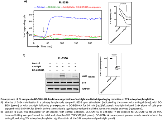
We then investigated primary FL cases, using a splenic FL and 2 lymph node FLs, all carrying N-glycosylation motifs and able to bind DC-SIGN. We focused on the DC-SIGN-HA derivative, which gives no detectable Ca2+ flux signal by itself. Binding of the lectin again paralyzed subsequent anti-IgM-induced Ca2+ flux in all cases (Fig.1A), confirming the results with the cell line. Further investigation revealed that the lectin prevents early events after antigen binding to the sIg receptor reducing SYK phosphorylation and leading to a reduced Ca2+ mobilization (Fig.1B).
In conclusion, modulation of the B-cell receptor by mannose addition to the antigen-binding site allows lectin access from innate microenvironmental cells. The effect of this is to provide a low level/null signal without loss by endocytosis. The novel finding is that this interaction then lowers the function of the B-cell receptor and perhaps blocks potential interference by antigen. There may be a parallelism with reports of modification of T-cell receptor function by galectins, with both pointing to the role of post-translational modification adding another layer of control on the operation of the major immune receptors. In the case of FL, this has been exploited to maintain tumor cells in the hostile environment of the germinal center. The apparently lymphoma-specific adaptation offers opportunities for targeted inhibition of the interaction.
Forconi:Janssen-Cilag: Consultancy; Abbvie: Consultancy. Packham:Aquinox: Research Funding.
Author notes
Asterisk with author names denotes non-ASH members.


