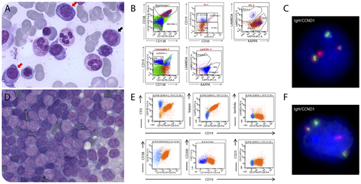
During the diagnostic workup of a 72-year-old man with anemia (83 g/L) with increased free κ light chain (140 mg/L), a bone marrow smear revealed infiltration by 25% of plasma cells (panel A, red arrows; original magnification ×60, May-Grünwald-Giemsa stain) together with rare atypical lymphoid cells (panel A, black arrow). Flow cytometry revealed, by fluorescent in situ hybridization, monotypic κ light chain–restricted plasma cells (CD38+, CD56+, CD19−) (panel B) that harbor the t(11;14)(q13;q32) translocation on sorted CD138 cells (panel C); atypical lymphoid cells also had κ monotype (panel B). Given the spontaneous improvement of the anemia, a watch-and-wait strategy was adopted. During the first year of follow-up, a splenomegaly developed, with moderate uptake (standardized uptake value, 5.63) on positron emission tomography, and splenectomy was performed. The spleen was infiltrated by small B cells (panel D; original magnification ×40, May-Grünwald-Giemsa stain) with the immunological profile of mantle cell lymphoma (CD5+, CD23−, CD200−, κ monotype) (panel E), which was confirmed by histology and the detection of the t(11;14)(q13;q32) translocation (panel F). No treatment was initiated.
This case illustrates the synchronous association of a mantle cell lymphoma and a myeloma expressing the same light chain and exhibiting the same t(11;14) translocation, suggesting a common origin of these 2 components from the same B-cell clone. Molecular analysis for the immunoglobulin heavy chain rearrangement could confirm the common origin, as previously described in literature.
For additional images, visit the ASH Image Bank, a reference and teaching tool that is continually updated with new atlas and case study images. For more information, visit http://imagebank.hematology.org.


