Background:
Venoarterial extracorporeal membrane oxygenation (VA-ECMO) is often required to support infants with congenital defects, congenital heart disease, and other causes of reversible cardiopulmonary insufficiency. Although ECMO can be a life-saving modality, bleeding, inflammation, and thrombosis are well-described complications of ECMO. Adult porcine models of ECMO have been used to recapitulate the physiology and hematologic consequences of ECMO cannulation. However, these models lack the unique physiology and persistence of fetal forms and quantities of coagulation proteins and fibrinogen found in human infants. Furthermore, anticoagulation and hemostatic strategies developed for adults or using adult porcine models are often extrapolated to infants without specific considerations for developmental hemostasis. Therefore, development of an animal model of ECMO that faithfully reproduces both the physiology and coagulation profile of human infants is important to improving anticoagulation strategies and gaining a better mechanistic understanding of hemostatic challenges in infants. We hypothesized that an infant porcine mode (piglets) supported with VA-ECMO would closely recapitulate the physiology and hematology of human infants on ECMO.
Methods:
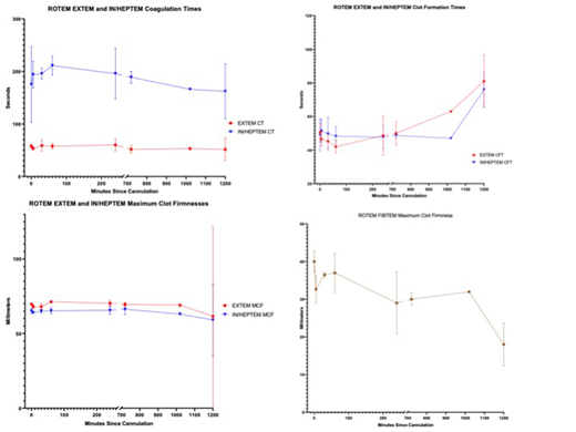
Four healthy piglets (5.7-6.4 kg) were cannulated via the jugular vein and carotid artery and supported for 20 hours or until adult whole blood for transfusion was exhausted. Heparin was used with a goal activated clotting time (ACT) of 180-220 seconds. Blood gas (ABG) was performed hourly, and blood was transfused from an adult donor to maintain hematocrit ≥24%. Rotational thromboelastometry (ROTEM) assays were performed at each of seven time points. Specifically, EXTEM, INTEM, and FIBTEM were used to determine the coagulation time (CT) and maximum clot firmness (MCF) of the extrinsically-mediated, intrinsically-mediated, and acellular fibrin clots, respectively.
Results:
All animals (n=4) had slow but significant hemorrhage at cannulation, arterial line, and bladder catheter sites. All animals required the maximum blood transfusion volume available. All animals became anemic after exhaustion of blood for transfusion, and two required sacrifice before the 20-hour endpoint for critical anemia. ABG showed progressively declining hematocrit and adequate oxygenation. ROTEM demonstrated decreasing FIBTEM clot firmness with preservation of EXTEM and INTEM coagulation times. Histology was overall unremarkable.
Conclusion:
Our infant porcine model faithfully reproduces the physiology and hematostatic profile of human infants during VA-ECMO, including transfusion dependence and heparin-induced coagulopathy. Weak whole-blood clot firmness by ROTEM suggests defects in fibrinogen. This model serves as an important means to study the complex derangements in hemostasis during VA ECMO in infants, including subtle derangements due to adult blood product transfusions, as well as to investigate novel approaches to anticoagulation and hemostasis during extracorporeal life support.
Arepally:Veralox Therapeutics: Membership on an entity's Board of Directors or advisory committees; Apotex Pharmaceuticals: Consultancy; Biokit: Patents & Royalties.
Author notes
Asterisk with author names denotes non-ASH members.


