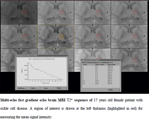
Background:Children with sickle cell disease (SCD) are at a high risk for neurocognitive impairment which may be due to iron overload in brain tissue or hemoglobin polymerization and endothelial dysfunction.Primary objectivewas measuring brain iron content (using R2* values) in the caudate and thalamic regions through quantitative brain MRI in Egyptian adolescents and young adults with multi-transfused SCD in comparison to beta thalassemia major (BTM) and age- and sex-matched healthy controls.Secondary objectiveswere evaluating the impact of brain iron content on neurocognitive functions of SCD patients and its association with MRI assessment of liver iron concentration (LIC) and cardiac iron (myocardial T2*).Methods: 32 children and young adults with SCD (mean age: 15.3 ± 3.7, 19 males and 13 females), 15 BTM (mean age: 19.4 ± 4.3, 7 males and 8 females) and 11 healthy control age- and gender-matched were recruited. Thorough clinical assessment, hematological and serum ferritin were performed. Brain MRI study using multi-echo fast gradient echo sequence was performed only for 15 patients with SCD, 15 patients with BTM and 11 controls and brain R2* values of both caudate and thalamic regions (right and left sides) were calculated. LIC and myocardial T2 were performed for; 15 with SCD and 15 with BTM. 32 SCD patients were examined for the neurocognitive functions; Wechsler IV Intelligence scale (verbal, perceptual, memory, processing and total IQ), Benton Visual Retention Test and Brief Psychiatric Rating Scale (BPRS).Results:For SCD patients their mean transfusion index was 174.70±63.98ml/kg/year and mean iron overload/day 0.30±0.12 mg/kg. 30 (93.8%) all SCD patients were on regular chelation therapy; 16 were on deferiprone and 16 were on combined chelation over last 5 years. Of those 32 SCD patients; 20 received concomitantly hydroxyurea therapy. Mean total IQ for SCD patients was 86.9±10.7; 68.9% had under- threshold <90 IQ and 27.5% had average (90-109) IQ. 12.5% of SCD patients had moderate to severe anxiety and 60.8% had of SCD patients had depression. No significant differences were found between SCD, BTM as regards LIC (p=0.102) No significant differences were found between SCD, BTM and control group in all regions of interests in brain MRI except that left thalamus R2* higher in BTM patients than both SCD and controls (p=0.032). R2* values of different regions of brain in relation with the studied parameters of SCD patients was not significant except that mean right caudate R2* was higher in female 17.4±0.8 than male 15.6±1.7 (p=0.044). The correlation coefficients showed no significant association between brain R2* and LIC or heart R2* values of SCD patients. There were positive correlation between left caudate R2* and both age and HbS%, negative correlation between transfusion index and right thalamus R2*, negative correlation between HbA% and left caudate R2* among SCD patients.Conclusion:Brain iron content in adolescents and young adults with SCD was not significantly different from either controls or BTM; SCD had high prevalence of neurocognitive dysfunction, which could not be explained by brain iron content or distribution.
No relevant conflicts of interest to declare.
Author notes
Asterisk with author names denotes non-ASH members.


