Abstract
Introduction
Hematological malignancies can lead to bone lesions, most commonly the osteolytic lesions found in multiple myeloma (MM). Cases of osteolytic lesions have been rarely reported in acute lymphoblastic leukemia (ALL), non-Hodgkin lymphoma, Waldenström macroglobulinemia, chronic lymphocytic leukemia (CLL), acute myeloid leukemia (AML), and myeloproliferative neoplasms (MPN). This review sheds light on the association between ALL and osteolytic bone lesions.
Methods
A literature search was performed for English language articles using PubMed, Scopus, and Google Scholar, for any date up to May 2022. The following keywords were used "ALL," "acute lymphoblastic leukemia," osteolytic bone lesions," and "osteolysis." Search results were initially reviewed by title and abstract, and articles were selected for more in-depth analysis if deemed relevant. Pertinent articles were examined in-depth for this report.
Results
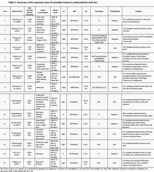
To our knowledge, we found 15 cases of patients with ALL who developed osteolytic lesions. They were reported in 14 articles published between April 1998 and May 2022. Most patients were males (12 out of 15). The median age was 29 years (ranging from 22 to 78 years), with most patients being young (in the third and fourth decades). B-ALL was the most common type of ALL associated with osteolytic lesions (14 cases). All patients presented with bone pain, and hypercalcemia was found in 80% of the reported cases. Osteolytic lesions were detected by plain radiography(x-ray) in approximately half of the patients. The osteolytic lesions were confirmed in the remaining patients by CT, MRI, or PET scans. The most affected bones were the vertebrae, skull, and pelvis. The clinical outcomes among these patients were variable; two of the reported cases died after the diagnosis of osteolytic lesions.
Discussion
Bone metastases are classified as osteolytic, osteoblastic, or mixed based on the primary interference mechanism with bone remodeling. The destruction of normal bone characterizes osteolytic bone metastasis, occurring in multiple myeloma (MM), renal cell carcinoma, melanoma, non-small cell lung cancer, non-Hodgkin lymphoma, thyroid cancer, or Langerhans-cell histiocytosis. Osteolytic metastasis in adults with ALL is an extremely rare condition. The exact pathophysiology of osteolysis in ALL patients is unknown.PTHrP secretion by lymphoblasts has been shown to be the primary cause of osteolysis in ALL patients. Additionally, serum from ALL patients with osteolysis can induce in vitro proliferation of osteoclasts, indicating the mechanism of lymphoblasts induces osteoclasts activation in vivo.
Based on our review, there was no association between osteolytic bone lesions and the Philadelphia chromosome detected in only one patient. Osteolytic bone lesions in ALL patients mainly present with bone pain. Pathological fracture of the vertebral body leading to spinal cord compression is a complication of osteolytic lesions in patients with MM. However, there is no case of spinal cord compression in ALL patients attributed to osteolytic lesions of the vertebra. Given that our review is based on case reports, we could not conclude if the presence of osteolytic bone lesions is a prognostic factor for adverse outcomes or indicates an "aggressive" form of leukemia in adult patients with ALL.
Conclusion
Osteolytic bone lesions in adult patients with ALL is an infrequent finding and mainly occurs in young adults. Plain radiography is often sufficient in diagnosing osteolytic bone lesions; however, other imaging modalities are sometimes required. Based on our review, the osteolytic lesions do not indicate a poor prognosis in adult patients with ALL.
Disclosures
No relevant conflicts of interest to declare.
Author notes
Asterisk with author names denotes non-ASH members.


