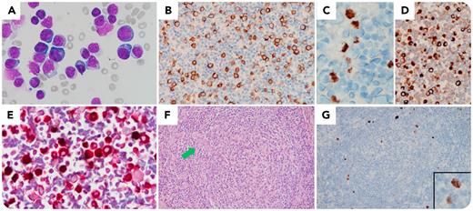
A 70-year-old man with multiple comorbidities, including coronary artery disease, chronic kidney disease, and Sjögren syndrome, presented with a huge mediastinal tumor, generalized lymphadenopathy, and right pleural effusion. The laboratory examination showed anemia, thrombocytopenia, hypoalbuminemia, polyclonal hypergammaglobulinemia, and elevated C-reactive protein. There was no evidence of HIV, M protein, or osteolytic lesions. The mediastinal tumor was surgically removed and disclosed a well differentiated liposarcoma. Thoracentesis revealed abundant plasmablasts without marked anaplasia (panel A; objective 100×). According to cell block immunohistochemistry, they were positive for CD138 (panel B; objective 40×), human herpesvirus 8 (HHV-8) latency-associated nuclear antigen (LANA) (panel C; objective 100×), and IgM (panel D; objective 40×), but negative for CD3, CD20, CD30, and Epstein-Barr virus. Interestingly, these LANA+ plasmablasts were lambda-restricted (panel E, LANA-brown; lambda-red double immunostain; objective 100×). Lymphadenectomy specimen showed the histomorphology of multicentric Castleman disease (MCD) (panel F; objective 20×; a penetrating vessel indicated by the green arrow) with scattered LANA+ plasmablasts in the mantle zones (panel G; objective 20×; insert, speckled nuclear staining pattern). The overall picture indicated an HHV-8–associated MCD presenting in pleural effusion. Unfortunately, the patient died of ischemic bowel disease shortly before any treatment.
This unique case demonstrates that HHV-8–associated MCD may present in pleural effusion and that HHV-8–positive plasmablasts may mimic primary effusion lymphoma (PEL). LANA+ lambda-restricted plasmablasts without cellular atypia and with generalized lymphadenopathy were not typical for PEL and deserve further workup.
For additional images, visit the ASH Image Bank, a reference and teaching tool that is continually updated with new atlas and case study images. For more information, visit http://imagebank.hematology.org


