TO THE EDITOR:
Inhibitors against coagulation factor IX (FIX) develop in 10% of patients with severe hemophilia B shortly after initiation of replacement therapy.1 The inhibitors neutralize the infused FIX, and this is the most challenging issue in the management of hemophilia B. The deleterious F9 gene mutations, including large deletions and nonsense and frameshift mutations, disrupt expression of FIX and constitute the major risk factor for alloantibody development against FIX administered in replacement therapy.2,3 The Centers for Disease Control and Prevention Hemophilia B Mutation Project,3 providing a large patient cohort, found that the inhibitor risk varied from 0.4% (missense, splice site, or promoter mutations) to over 5% (nonsense and frameshift mutations) and 27% (large deletions >50 base pairs [bp]). This was confirmed by the European PedNet study,1 which observed a risk of 33% for deletions >50 bp. However, neither study reported the inhibitor risk according to deleted FIX domains, although this could be a major determinant of the inhibitor risk.
In our study, we surveyed the genetic mutations and status of inhibitors in 28 children with hemophilia B from January 2015 through May 2022 and compared the incidences and titers of inhibitors among patients with mutations causing complete absence or different truncation of FIX. All patients investigated had met 2 criteria: (1) younger than 18 years and (2) exposure over 100 days with regular follow-up on status of inhibitors.
The FIX coagulant activity was measured by activated partial thromboplastin time-based clotting assay. The FIX inhibitor titers were determined using the Nijmegen modification of the Bethesda assay,4 and positive results were reexamined within 1 to 4 weeks to confirm the existence of inhibitors. The genomic DNA was extracted from peripheral blood leukocytes, and the entire F9 gene coding regions, including intron-exon boundaries, were directly sequenced to identify F9 gene mutation. CNVkit software was used to obtain copy number variation information. Interpretation of sequence variants was performed according to the American College of Medical Genetics and Genomics guidelines.5 The study was approved by the Ethics Review Committee of Beijing Children’s Hospital, and the informed consent form was acquired from the patients/guardians appropriately.
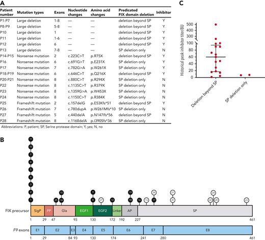
In total, 28 unrelated patients with severe hemophilia B were identified harboring deleterious F9 mutations, including 13 (46.4%) large deletions, 11 (39.3%) nonsense mutations, and 4 (14.3%) frameshift mutations (Figure 1A). Among them, 19 (67.9%) patients had developed inhibitors, of whom 13 were placed on low-dose immune tolerance induction (ITI) using prothrombin complex concentrate at 25 to 50 FIX international units (IU)/kg every other day in combination with rituximab at 375 mg/m2 weekly (maximum 600 mg) for 4 weeks.6
The detailed data for the current study cohort. (A) The spectrum of the F9 mutations in this cohort. (B) The distribution of mutations on F9 encoding regions and FIX structure domains among 28 children with hemophilia B. Black circles represent patients with inhibitor, white circles represent non-inhibitor patients, and the number in the circle represents the number of patients. (C) The peak inhibitor titers of patients with only serine protease (SP) domain deletion and FIX truncation beyond the SP domain.
The detailed data for the current study cohort. (A) The spectrum of the F9 mutations in this cohort. (B) The distribution of mutations on F9 encoding regions and FIX structure domains among 28 children with hemophilia B. Black circles represent patients with inhibitor, white circles represent non-inhibitor patients, and the number in the circle represents the number of patients. (C) The peak inhibitor titers of patients with only serine protease (SP) domain deletion and FIX truncation beyond the SP domain.
The F9 gene has 8 exons and directs synthesis of FIX precursor protein, containing signal peptide, propeptide, and the 415-residue mature protein. The signal peptide is encoded by exon 1, the propeptide sequence and the γ-carboxyglutamic acid (Gla)-rich domain are encoded by exons 2 and 3, and the 2 epidermal growth factor (EGF) domains are encoded by exons 4 and 5. Exon 6 encodes the activation peptide (AP) domain and a short N terminal motif of the SP domain, and the remaining SP domain is encoded by exons 7 and 8 (Figure 1B).7
Unlike large deletions extended to the whole F9 gene leading to complete absence of FIX in circulation, the mutations only involving a part of the encoding sequence are presumed to still direct synthesis of truncated FIX. It has been suggested that partially expressed protein may have impact on the induction of tolerance.8 Sauna et al9 showed that in patients with hemophilia A harboring intron 22 inversion mutations, even the coagulation factor VIII (FVIII) fragment expressed intracellularly may induce the tolerance toward FVIII and prevent the development of FVIII inhibitor.
In the patient cohort investigated, 7 patients had deletions involving 8 exons of the F9 gene, and 2 patients had either exon 1 or exons 1 to 6 deleted, which all led to a complete absence of FIX expression. All 3 patients with either deletions of exon 6 or exons 5 to 8 and 8 patients with either nonsense mutations or frameshift mutations were predicated to have FIX truncations involving SP domain, part or all EGF, and AP domains (Figure 1A). Almost all patients (17/18, 94.4%) with FIX structure deletions beyond the SP domain developed inhibitors after initiation of replacement therapy (Table 1; Figure 1B).
In contrast, among 10 patients with only SP domain deletion, inhibitors developed in only 2 (20.0%) patients, whose median historical peak inhibitor titers, 6.8 and 7.7 BU/mL, were also much lower than those of patients with FIX structure deletion beyond the SP domain (median, 60.0 BU/mL; range, 1.6 to 500.0 BU/mL) (Figure 1C).
The size of truncated FIX also affected the outcome of inhibitor eradication therapy. For 13 patients receiving inhibitor eradication treatment, 7 (53.8%) patients achieved success with inhibitor titer <0.6 BU/mL on 2 consecutive measurements. Six patients failed after a median treatment time of 19.3 months (range, 6.4-30.2 months), and the median titer at the last inhibitor assay was 12.0 BU/mL (range, 1.0-57.6 BU/mL). In patients with deletion beyond the SP domain (n = 11), 5 cases (45.5%) were successful and 6 cases (54.5%) failed in ITI. In patients with only SP deletion (n = 2), 100% of patients achieved ITI success (Table 1).
Christophe et al10 showed that antibodies isolated from hemophilia B patients recognized epitopes mostly on Gla and SP domains. In our study, we found that patients with mutations that predicated the truncation of FIX up to the SP domain were much less likely to develop inhibitors than patients who had mutations causing more domains missing, and when patients with only SP deleted developed inhibitors upon exposure to administered FIX, the titer of the inhibitors was much lower, and they were more prone to be eradicated by ITI and immune suppression therapy.
We have compiled data of 55 patients with available information on both inhibitor development and deletion mutations from a hemophilia B variant database (http://www.factorix.org/). Consistent with findings in our study, among patients in whom inhibitors developed, 77.3% had mutations leading to FIX truncation beyond the SP domain.
The SP domain of FIX shares high homology with other 138 members of the S1 protease family; the EGF domains also share consensus sequence among 600 extracellular proteins.11 The Gla domain, however, is only found in limited number of proteins, such as vitamin K–dependent coagulation factors, protein C/S, and transthyretin,12 which may predispose the Gla domain as the primary target for alloantibody.
In conclusion, F9 mutations causing deletions beyond the SP domain are associated with higher risk for inhibitor development and poor prognosis for inhibitor-eradication therapy.
Acknowledgments
The authors thank all the patients and their parents.
This work was supported by grants from Capital Health Development Research Project (no. 2022-2-2093), and National Natural Science Foundation of China (No. 82270133).
Authorship
Contribution: W.W. and R.W. conceived and designed the study, analyzed and interpreted the data, and wrote and revised the manuscript; G.L. and J.S. collected the data, performed the statistical analysis, analyzed and interpreted the data, and wrote the manuscript; Z.L. and Z.C. interpreted the data and contributed to the discussion of important intellectual content; and all authors approved the final version of the manuscript.
Conflict-of-interest disclosure: The authors declare no competing financial interests.
Correspondence: Runhui Wu, Hemophilia Comprehensive Care Center, Hematology Center, Beijing Children’s Hospital, Capital Medical University, National Center for Children’s Health, No 56 Nanlishi Rd, West District, Beijing 100045, China; e-mail: runhuiwu@hotmail.com; and Wenman Wu, Department of Clinical Laboratory Medicine, Ruijin Hospital affiliated to Shanghai Jiao Tong University School of Medicine, 197 Ruijin Second Rd, Shanghai 200025, China; e-mail: wenmanwu@shsmu.edu.cn.
References
Author notes
For original data, contact the corresponding author, Runhui Wu (runhuiwu@hotmail.com).
G.L. and J.S. contributed equally to this work.


