JAK1/2 inhibitor therapy reduces the degree of splenomegaly and improves systemic symptoms of myelofibrosis (MF) patients, but does not delay disease progression and modestly prolongs overall survival. HDM2 antagonists (nutlins) have been reported to selectively eliminate MF CD34 + cells by activating the p53 pathway. Nutlins alone target malignant cells with wild type (WT) p53 and in clinical trials, HDM2 antagonists have led to reductions in spleen size, improvement of systemic symptoms and reduction in MPN driver mutation allelic burdens (Lu et al, Blood.2012; Mascarenhas et al, Blood. 2019). Although most patients with MF have WT TP53, mutations are present in a limited number of such patients and are associated with poor outcomes. We have searched for combinations of drugs to serve as a more effective and tolerable MF stem cell depleting therapy.
Exportin 1 (XPO1) mediates the nuclear export of various proteins and RNAs (Azmi, et al. Nat Rev Clin Oncol. 2021) including tumour-suppressor proteins and growth regulators, such as RB1, p53, p63, p73, p21, cyclin B1 or D1 (Azmi, 2021). XPO1 is frequently overexpressed and/or mutated in human cancers and functions as an oncogenic driver. Suppression of XPO1-mediated nuclear export has been shown to be a potential therapeutic strategy to treat MF patients (Yan, et al. Clin Cancer Res. 2019). Selinexor (SEL) is an investigational, oral XPO1 inhibitor that may inhibit multiple pathways relevant in MF including JAK/STAT, ERK, and AKT (Tantravahi, ASH 2021).
In the current study, we tested the effects of SEL on apoptosis, and cell cycle arrest in JAK2V617F cells with either TP53WT or TP53mut. We also evaluated the ability of SEL alone to eliminate MF CD34 + cells and in combination with other candidate drugs such as HDM201 (HDM2 antagonist), ruxolitinib (RUX), pan-BET inhibitor (JQ1) or a Bcl-2/Bcl-xL inhibitor (navitoclax).
Treatment with SEL alone induced apoptosis of both TP53WT (UKE-1) and TP53mut (SET-2) MPN CD34 + cell lines in a dose-dependent fashion. The transcript levels of TP53 as well as its downstream genes, such as p21, PUMA, NOXA (intrinsic apoptotic pathway) and DR5 and Trail (extrinsic apoptotic pathway) were increased by 2-3 fold after SEL treatment of TP53WT and TP53mut cells. Importantly, SEL also increased TP63 and TP73 by ~2 fold but decreased the transcript levels of the anti-apoptotic Bcl-xL and c-Myc (by 20% and 50%), TP63 and TP73 are TP53 family members which promote p53 downstream events independently of WT p53. In addition, treatment with SEL for 2 days increased the proportion of cells in sub-G0/G1 phase from 16% (control) to 60% in UKE-1 cells and from 15% (control) to 53% in SET-2 cells.
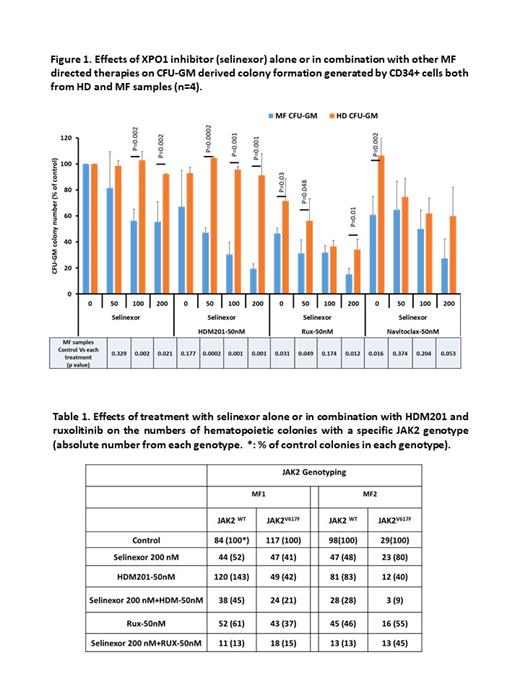
SEL and HDM201 alone induced apoptosis of primary MF but not healthy donor (HD) CD34 + cells in a dose dependent fashion. The combination of SEL with suboptimal doses of either HDM201, RUX, JQ1 or navitoclax showed additive pro-apoptotic effects on MF CD34 + cells. Colony formation assays showed that SEL, HDM201 and navitoclax alone decreased CFU-GM colony numbers generated by MF CD34 + cells in a dose-dependent fashion, yet had no effects on HD CD34 + cells, while RUX reduced both MF and ND colony formation. Combination treatment with SEL and 50nM of HDM201 or RUX led to a greater degree of reduction of MF CFU-GM colony numbers, but only SEL+HDM201 did not affect HD CFU-GM colony numbers (Figure 1).
Furthermore, JAK2 genotyping showed that treatment with SEL, Rux and HDM201 alone decreased the absolute number of JAK2V167F+ colonies. Furthermore, SEL+HDM201 and SEL+RUX decreased the number of JAK2V617F+ colonies to greater extent than either drug alone, but SEL+HDM201 treatment led to the persistence of a greater numbers of JAK2WT colonies than SEL+RUX (Table 1). SEL+navitoclax did not further decrease JAK2V167F+ colonies than SEL alone (data not showed).
These data suggested that SEL induces apoptosis and inhibits MPN CD34 + cell proliferation by affecting both p53-dependent and -independent pathways. SEL alone induces apoptosis and inhibits MF progenitor cells but not HD cells. The effects of SEL combined with either RUX or an HDM2 antagonist more effectively depleted mutated MF CD34+ cells than either drug alone. The combination of SEL+HDM201 spared the reservoir of WT progenitors, and reduced JAK2 mutated progenitor cells to a great degree suggesting that this combination might be associated with less hematological toxicity and greater efficacy.
Disclosures
Hoffman:Dexcel Pharma: Research Funding; TD2: Research Funding; Karyopharm: Research Funding; Silence Therapeutics: Consultancy; Kartos Abbvie: Research Funding; Curis: Research Funding; Summitomo: Research Funding; Dompe: Patents & Royalties; Cellinkos: Consultancy; Protagonist Therapeutics: Consultancy.


