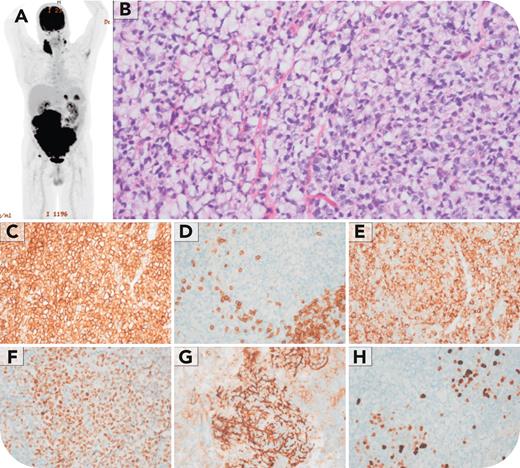
A 64-year-old man with a history of hypertension and abdominal aortic aneurysm presented for surveillance. The aneurysm was unchanged, but a 19-cm abdominal mass (positron emission tomography-computed tomography, maximum standardized uptake value 22.7) was seen (panel A). A complete blood count and peripheral smear showed mild anemia (hemoglobin 12.8 g/dL), but was otherwise unremarkable (white blood cell count 5.8 × 10⁹/L, normal differential count, no atypical cells). A core needle biopsy of the mass showed fibrovascular tissue replaced by a predominantly diffuse, focally nodular population of small cells, many of which had signet ring morphology (panel B; original magnification ×400, hematoxylin and eosin stain). Immunohistochemistry showed that the follicular lymphoma (FL) cells were positive for CD20, BCL2 and BCL6 (panels C, E, and F; original magnification ×400), and negative for CD3 (panel D; original magnification ×400). CD21 highlighted intact follicular dendritic cell meshworks within the tumor nodules (panel G; original magnification ×400). Ki-67 showed a low proliferative index (panel H; original magnification ×400). Conventional cytogenetics demonstrated a normal male karyotype. Molecular studies were not performed. The patient was diagnosed with FL and started treatment with cyclophosphamide, doxorubicin, prednisone, rituximab, and vincristine to which he is responding well.
Signet ring cell FL is an exceedingly rare variant, representing just 0.05% of FL cases. It poses significant diagnostic challenges due to its overlapping morphologic features with other conditions, including adenocarcinomas and other FL subtypes. Recognition of this variant is essential to prevent misdiagnosis.
For additional images, visit the ASH Image Bank, a reference and teaching tool that is continually updated with new atlas and case study images. For more information, visit https://imagebank.hematology.org.


