Abstract
To study the role of bone marrow endothelial cells (BMEC) in the regulation of hematopoietic cell trafficking, we have designed an in vitro model of transendothelial migration of hematopoietic progenitor cells and their progeny. For these studies, we have taken advantage of a human BMEC-derived cell line (BMEC-1), which proliferates independent of growth factors, is contact inhibited, and expresses adhesion molecules similar to BMEC in vivo. BMEC-1 monolayers were grown to confluency on 3 μm microporous membrane inserts and placed in 6-well tissue culture plates. Granulocyte-colony stimulating factor (G-CSF )–mobilized peripheral blood CD34+ cells were added to the BMEC-1 monolayer in the upper chamber of the 6-well plate. After 24 hours of coincubation, the majority of CD34+ cells remained nonadherent in the upper chamber, while 1.6 ± 0.3% of the progenitor cells had transmigrated. Transmigrated CD34 cells expressed a higher level of CD38 compared with nonmigrating CD34+ cells and may therefore represent predominantly committed progenitor cells. Accordingly, the total plating efficiency of the transmigrated CD34+ cells for lineage-committed progenitors was higher (14.0 ± 0.1 v 7.8% ± 1.5%). In particular, the plating efficiency of transmigrated cells for erythroid progenitors was 27-fold greater compared with nonmigrating cells (8.0% ± 0.8% v 0.3% ± 0.1%) and 5.5-fold compared with unprocessed CD34+ cells (2.2% ± 0.4%). While no difference in the expression of the β1-integrin very late activation antigen (VLA)-4 and β2-integrin lymphocyte function-associated antigen (LFA)-1 was found, L-selectin expression on transmigrated CD34+ cells was lost, suggesting that shedding had occurred during migration. The number of transmigrated cells was reduced by blocking antibodies to LFA-1, while L-selectin and VLA-4 antibodies had no inhibitory effect. Continuous coculture of the remaining CD34+ cells in the upper chamber of the transwell inserts resulted in proliferation and differentiation into myeloid and megakaryocytic cells. While the majority of cells in the upper chamber comprised proliferating myeloid precursors such as promyelocytes and myelocytes, only mature monocytes and granulocytes were detected in the lower chamber. In conclusion, BMEC-1 cells support transmigration of hematopoietic progenitors and mature hematopoietic cells. Therefore, this model may be used to study mechanisms involved in mobilization and homing of CD34+ cells during peripheral blood progenitor cell transplantation and trafficking of mature hematopoietic cells.
MICROVASCULAR endothelial cells isolated from human bone marrow aspirates have been shown to support long-term proliferation of myeloid and megakaryocytic progenitor cells in vitro.1-3 Subsets of CD34+ hematopoietic progenitor cells adhere to monolayers of cultured bone marrow endothelial cells (BMEC).3 In vivo, BMEC act as gatekeepers separating the bone marrow stroma from the sinusoidal lumen, and may therefore play an important role in the trafficking of hematopoietic cells.4,5 Transendothelial migration of hematopoietic progenitor cells occurs during mobilization in response to cytokines or chemotherapy and during homing of circulating progenitors. Mechanisms involved in progenitor cell migration are still poorly understood. It has been shown that resting and circulating CD34+ hematopoietic progenitor cells express a variety of adhesion molecules including selectins and integrins.6 Potential ligands for L-selectin, β1 and β2 integrins, which are expressed on CD34+ progenitors, are found on endothelial cells, including BMEC, particularly after cytokine stimulation.1,7,8 Extracellular matrix proteins and stromal cells also provide ligands, such as fibronectin for the β1-integrins very late activation antigen (VLA)-4 and VLA-5.9-13
By analogy to studies in mature leukocytes, L-selectin expressed on progenitor cells and putative ligands such as CD34 on BMEC are likely to be important for the initial, reversible binding of the progenitors to the bone marrow endothelium, which has been described as “rolling.”14,15 E- and P-selectin expressed on the endothelial cells could also contribute, since ligands have been found on hematopoietic progenitor cells.16 Stable adhesion and transendothelial migration of mature leukocytes depends on the interaction of integrins with their endothelial ligands. The concept that integrins may also be important for progenitor cell trafficking is supported by the finding that circulating progenitors express lower levels of VLA-4 and the β2-integrin lymphocyte function-associated antigen (LFA)-1 compared with progenitors of the bone marrow.17,18 Furthermore, in vivo administration of antibodies to VLA-4 was found to mobilize hematopoietic progenitors into the circulation.19
In the bone marrow microenvironment, progenitors proliferate and give rise to precursor cells that are usually not found in the circulation. Changes in the expression of adhesion molecules during differentiation might account for the low migratory capacity of maturating precursor cells, due to stronger binding to the bone marrow stroma. Furthermore, more mature precursor cells may not be able to migrate through an endothelial cell layer. Mature leukocytes regain the ability to migrate and are released into the circulation after transendothelial migration.
In this study, an in vitro model of hematopoietic cell trafficking was developed using the BMEC-1 cell line, which retains morphology, phenotype, and function of primary BMEC, and grows contact inhibited independent of exogenous growth factors.20 The cells express a pattern of adhesion molecules similar to BMEC in vivo. In vitro, cultured primary BMEC change the expression of surface molecules important for progenitor cell trafficking during serial passaging before they undergo senescence. For instance, the glycoprotein CD34 which is expressed on BMEC in vivo21 and may act as a selectin-binding adhesion molecule,22 is downregulated during culturing of primary BMEC.1 2 However, the BMEC-1 cell line constantly expresses CD34. Because migration of mature and immature hematopoietic cells including progenitor cell homing takes place during steady-state hematopoiesis, BMEC-1 monolayers used for the experiments were not prestimulated with cytokines. In this report, we demonstrate that confluent layers of the BMEC-1 cell line in vitro mimick the function of the bone marrow endothelium in vivo allowing selective transendothelial migration of hematopoietic progenitor cells and mature monocytes and granulocytes, while proliferating myeloid precursors were unable to transmigrate.
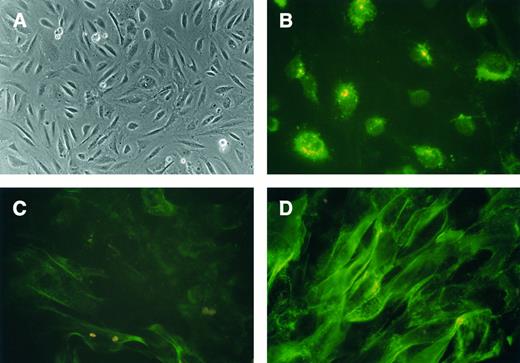
Characterization of BMEC-1 cells. (A) Phase-contrast microscopy of a confluent monolayer of spindle-shaped BMEC-1 cells (original magnification × 100). (B) Characteristic granular immunofluorescence was observed after staining with MoAb to factor VIII/vWF (original magnification × 400). (C) ICAM-1 was weakly positive on resting BMEC-1 cells (original magnification × 400). (D) Expression of ICAM-1 was upregulated after stimulation with IL-1β (original magnification × 400).
Characterization of BMEC-1 cells. (A) Phase-contrast microscopy of a confluent monolayer of spindle-shaped BMEC-1 cells (original magnification × 100). (B) Characteristic granular immunofluorescence was observed after staining with MoAb to factor VIII/vWF (original magnification × 400). (C) ICAM-1 was weakly positive on resting BMEC-1 cells (original magnification × 400). (D) Expression of ICAM-1 was upregulated after stimulation with IL-1β (original magnification × 400).
MATERIALS AND METHODS
Immortalized bone marrow endothelial cells.The BMEC-1 cell line was generated by introducing the SV40-large T antigen into an early passage of primary BMEC.1 20 The resulting cell line BMEC-1 was derived from a single transfected clone and cultured in medium 199 (GIBCO-BRL Life Technologies, Grand Island, NY) with 10% to 20% fetal bovine serum (FBS; HyClone, Logan, UT). The cells were passaged weekly by trypsinization. No growth supplements were required for propagation of the cell line. By direct and indirect immunofluorescence, the phenotype of BMEC-1 cells was characterized using monoclonal antibodies to factor VIII/vWF (Dako, Carpinteria, CA), CD34 (HPCA-2, Becton Dickinson, San Jose, CA), intercellular adhesion molecule (ICAM)-1 (84H10, Immunotech, Marseille, France), vascular cell adhesion molecule (VCAM)-1 (1G11, Immunotech), CD62P (AC1.2, Becton Dickinson), CD62E (1.2B6, Immunotech), CD31 (HEC7, a gift from W.A. Muller, The Rockefeller University, New York, NY). Expression of adhesion molecules was also analyzed after stimulation with interleukin-1β (IL-1β) or tumor necrosis factor-α (TNF-α) (100 U/mL) for 12 to 24 hours.
For the transmigration experiments, 5 × 10⁵ BMEC-1 cells were seeded on 3 μm transwell microporous membranes (Transwell, Costar, Cambridge, MA). After 3 days, the monolayers achieved full confluency and were suitable for transmigration studies. The transwell inserts with the monolayers were placed in a 6-well tissue culture plate, thus separating an upper from a lower chamber in each well. To assess the integrity of BMEC-1 monolayers grown on transwell inserts, 1 mL medium 199/20% FBS containing 14C-albumin (ARC, St Louis, MO) was added to the upper chamber. After 6 hours, the medium in the lower chamber was removed. Both the medium of the lower chamber and an aliquot of the medium before incubation were measured in a β-counter (Rackbeta 1214, Wallac, Gaithersburg, MD) allowing calculation of the percentage of albumin diffused. In repeated experiments, diffusion of radioactive labeled albumin was always less than 10% within 6 hours (6.8% ± 1.3%, mean ± standard deviation [SD], n = 7). Once confluent, contact-inhibited BMEC-1 monolayers maintained their integrity for several weeks, as measured by albumin diffusion.
To assess the migratory capacity of peripheral blood monocytes and lymphocytes in this system, 1 × 10⁶ mononuclear cells were isolated from peripheral blood samples by Ficoll-Hypaque (Pharmacia, Uppsala, Sweden) density gradient centrifugation and added to the upper chamber. After 6 hours, the transmigrated cells were recovered from the lower chamber. The proportion of monocytes (CD14+), T cells (CD2+), and B cells (CD19+) was measured by flow cytometry and the percentage of transmigrated cells was calculated for each cell population.
Transmigration of hematopoietic progenitor cells.After informed consent, peripheral blood mononuclear cells (PBMNC) were obtained from patients with ovarian cancer who had received no previous cytotoxic therapy. Progenitor cells were mobilized with cyclophosphamide plus granulocyte colony-stimulating factor (G-CSF ). MNC were separated by Ficoll density gradient centrifugation. PB CD34+ cells were isolated with immunomagnetic beads (Dynal A.S., Oslo, Norway) using the CD34 monoclonal antibody (MoAb) 11.1.6. The cells detached from the beads during incubation in Iscove's modified Dulbecco's medium (IMDM) (GIBCO) containg 20% FBS at 37°C/5% CO2 for 18 hours. A total of 1 × 10⁶ CD34+ cells were added to the upper chamber of each transwell insert placed in the 6-well plate. After 24 hours, hematopoietic cells from the upper and lower chambers were recovered and further characterized by flow cytometry and agarose progenitor cell assay.
Transmigration of proliferating hematopoietic cells.Continuous coculture of BMEC-1 and CD34+ cells in the upper chamber of the transwell inserts resulted in proliferation and differentiation of CD34+ cells into myeloid and megakaryocytic precursors without addition of exogenous cytokines.20 To analyze migration of proliferating hematopoietic cells, 1 × 10⁶ CD34+ cells were added to the upper chamber of the transmigration system. The cells in the upper chamber were demidepopulated after 24 hours, after 5 days, and then every 5 days up to day 30. The transmigrated cells in the lower chamber were completely removed after 24 hours, 5 days, and then alternating after 3 or 2 days up to day 30. During the study, the endothelial monolayer retained its cellular integrity and no passaging of the cells was required. At day 15 and day 30, the hematopoietic cells from the upper and lower chambers were characterized by Wright-Giemsa stain, flow cytometry, and agarose progenitor cell assay. To remove the cells from the chambers by pipetting, neither vigorous shaking nor scraping was required. Thus, endothelial cells were not detected in the cytospin preparations or clonogenic assays.
Flow cytometry.A total of 1 × 10⁴ to 1 × 10⁵ cells were incubated for 30 minutes at 4°C with the fluorescein isothiocyanate (FITC) or phycoerythrin (PE)-conjugated MoAb CD34-PE, CD45-FITC (clone HPCA-2, HLe-1; Becton Dickinson), CD34-FITC, CD38-FITC, L-selectin-FITC, VLA-4-FITC, LFA-1-PE (clone QBEnd-10, T16, DREG56, HP2.1, 25.3; Immunotech). Isotype-identical antibodies served as controls (IgG1 and IgG2a, FITC/PE-conjugated, Immunotech). The cells were analyzed using a Coulter Elite flow cytometer (Coulter, Hialeah, FL). For coexpression analysis, a FL-1/FL-2 contour plot was used. To calculate the percentage of positive cells, a proportion of 1% false positive events was accepted in the negative control sample. The mean fluorescence intensity was calculated from the fluorescence histogram and expressed in arbitrary units. For the characterization of the proliferating hematopoietic cells, CD14-FITC, CD15-FITC, CD33-FITC, Glycophorin A-PE (clone RMO52, 80H5, D3HL60.251, D2.10; Immunotech) and CD41a-PE (clone HIP8; PharMingen, San Diego, CA) were used. The distinct populations of CD14++ (due to mature monocytes) and CD15++ (due to granulocytic precursors and granulocytes) were enumerated in a FL-1/FL-2 dot plot. To measure the proportion of lymphocytes in MNC samples, CD2-FITC/CD19-PE (Immunotech) was used.
Cell counts and cytospin preparation.Cell numbers and concentrations were assessed using a hemocytometer or automated cell counter (Coulter). Standard cytospin-preparations were stained with Wright-Giemsa. A differential count of at least 100 cells was performed for each cytospin-preparation.
Progenitor cell assay (agarose assay).A total of 1 × 10³ (24 hours) or 1 × 10⁴ to 1 × 10⁵ (day 15 and day 30) cells were plated in triplicate in 35-mm tissue culture dishes (Corning, Corning, NY) containing 1 mL IMDM, 20% FBS, 0.36% agarose (FMC Bioproducts, Rockland, ME), and a combination of five cytokines: human Kit-ligand (20 ng/mL; kindly provided by Immunex, Seattle, WA), human IL-3 (50 ng/mL; Immunex), mutein IL-6 (20 ng/mL; kindly provided by Imclone Systems Inc, New York, NY), human granulocyte colony-stimulating factor (100 ng/mL; Amgen, Thousand Oaks, CA), and human erythropoietin (6 U/mL; Amgen). The plates were cultured for 14 days at 37°C, 100% humidity, and 5% CO2 . Colonies (>40 cells) were scored using an inverted microscope. By this technique, erythroid (mainly burst-forming units-erythroid [BFU-E]) and myeloid colonies (colony-forming units granulocyte, macrophage, and granulocyte-macrophage [CFU-G, CFU-M, CFU-GM]) could be detected.
Blocking of progenitor cell migration.A total of 5 × 10⁵ CD34+ progenitor cells in 100 μL phosphate-buffered saline (PBS) containing 1% bovine serum albumin (BSA; Sigma, St Louis, MO) were incubated for 30 minutes at 4°C with 5 μg of the MoAB IgG1 isotype control (clone 107.3; PharMingen), L-selectin (clone DREG56; Endogen, Cambridge, MA), VLA-4 (CD49d, clone HP2.1; Immunotech), LFA-1 (CD11a, clone TS 1/11; Endogen), and LFA-1 (CD18, clone TS 1/18; Endogen). These antibodies have been shown to functionally block the respective adhesion molecule.23-25 The suspension was added to the upper chamber of the transmigration system containing 1 mL of medium 199/10% FBS. After 24 hours, the number of transmigrated cells was assessed and divided by the number of migrated cells without antibody pretreatment (relative number of transmigrated cells).
Statistical analysis.Data are expressed as mean ± standard error of the mean (SEM) of at least three independent experiments. To detect differences between migrating and nonmigrating cells, the t-test for paired samples was applied. A P value < .05 was considered statistically significant.
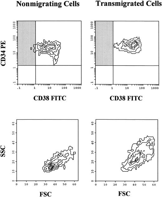
Coexpression of CD38 and CD34 on nonmigrating and transmigrated progenitor cells. Contour plots of one representative experiment are shown. The fluorescence intensity of CD38 was greater on the transmigrated cells, while no difference was observed regarding the expression level of CD34. More primitive, CD34+/CD38− progenitor cells are indicated by the shaded area. This particular population was virtually absent (<1%) in the transmigrated cells, compared with 1% to 5% in the nonmigrating cells. The difference was not due to selective migration of smaller cells, because the forward scatter (FSC) of the transmigrated cells tended to be even greater.
Coexpression of CD38 and CD34 on nonmigrating and transmigrated progenitor cells. Contour plots of one representative experiment are shown. The fluorescence intensity of CD38 was greater on the transmigrated cells, while no difference was observed regarding the expression level of CD34. More primitive, CD34+/CD38− progenitor cells are indicated by the shaded area. This particular population was virtually absent (<1%) in the transmigrated cells, compared with 1% to 5% in the nonmigrating cells. The difference was not due to selective migration of smaller cells, because the forward scatter (FSC) of the transmigrated cells tended to be even greater.
RESULTS
Immunophenotype of BMEC-1 cells and transmigration of peripheral blood monocytes and lymphocytes.To confirm that the cell line used in this study retains the characteristics of endothelial cells and expresses adhesion molecules similar to primary bone marrow endothelial cells, the phenotype of BMEC-1 cells was analyzed by direct and indirect immunofluorescence. Factor VIII/vWF showed a characteristic granular pattern of reactivity (Fig 1). Furthermore, the cells were positive for CD34 (Table 1). A basal expression of ICAM-1 was found, which was upregulated after stimulation with IL-1β (Fig 1). VCAM-1, E-selectin, and P-selectin were positive after cytokine stimulation (Table 1). Similar to previous studies using resting endothelial cells,26 peripheral blood monocytes efficiently transmigrated the BMEC-1 layer (78.9% ± 5.7% of the monocytes were recovered from the lower chamber after 6 hours), while the proportion of transmigrated lymphocytes was substantially lower (3.7% ± 0.3% of the T cells). Particularly, B cells were not detected in the lower chamber (<0.1%).
Number and phenotype of transmigrated progenitor cells.A total of 1 × 10⁶ CD34+ hematopoietic progenitor cells were added to the upper chamber of the transmigration system. After 24 hours, 1.6% ± 0.3% of the CD34+ cells had transmigrated into the lower chamber, while 71.0% ± 2.5% stayed nonadherent in the upper chamber. As assessed by flow cytometry, transmigrated CD34+ cells expressed a higher level of CD38 compared with nonmigrating cells (Table 2). This indicates that transmigrated progenitors are more mature, since expression of CD38 on progenitor cells is related to differentiation and lineage commitment.27 More primitive, CD34+/CD38− progenitor cells were virtually not detected after transmigration as indicated in Fig 2 by the shaded areas. The difference was not due to selective transmigration of smaller cells, since their forward scatter tended to be even greater. As shown in Table 2, the greater fluorescence intensity of CD38 was statistically significant in a series of eight experiments, while no difference was found in the expression level of CD34 glycoprotein.
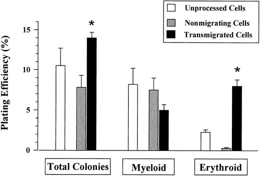
Plating efficiency of transmigrated and nonmigrating progenitors.The difference in the phenotype of migrating and nonmigrating cells is supported by data from agarose colony assays demonstrating a significantly higher plating efficiency of transmigrated versus nonmigrating CD34+ cells for lineage-committed colony-forming units (Fig 3). The plating efficiency was also greater compared with nonprocessed CD34+ cells that were not subjected to the transmigration assay. However, the increased plating efficiency was related to a higher number of erythroid progenitors, while no significant difference was found for the number of myeloid progenitors. Because the number of erythroid progenitors was lower in the nonmigrating population compared with the nonprocessed CD34+ cells, this could be due to a more effective migration of erythroid progenitors.
Plating efficiency of progenitor cells. Equal numbers of nonmigrating, transmigrated, and unprocessed CD34+ cells were plated. The plating efficiency of the transmigrated progenitors for total colonies was greater compared with both nonmigrating and nonprocessed CD34+ cells. The difference was related to the greater number of erythroid progenitors (BFU-E).
Plating efficiency of progenitor cells. Equal numbers of nonmigrating, transmigrated, and unprocessed CD34+ cells were plated. The plating efficiency of the transmigrated progenitors for total colonies was greater compared with both nonmigrating and nonprocessed CD34+ cells. The difference was related to the greater number of erythroid progenitors (BFU-E).
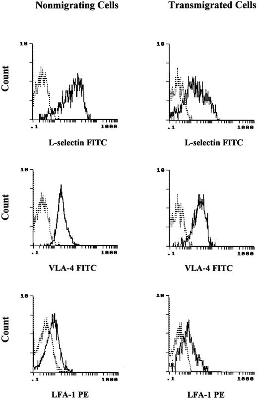
Expression of adhesion molecules.In Fig 4, the fluorescence histogram of one representative experiment is shown. While L-selectin was expressed on the majority of the nonmigrating CD34+ cells, only low levels were found on the transmigrated progenitors. No difference of VLA-4 expression was observed. VLA-4 was positive on most of the nonmigrating, as well as transmigrated progenitor cells. LFA-1 was expressed at a low fluorescence level on the progenitor cells independent of whether they had transmigrated or not. The mean fluorescence intensities obtained from repeated experiments are shown in Table 2. The difference in L-selectin expression between nonmigrating and transmigrated progenitors was statistically significant.
Expression of adhesion molecules on nonmigrating and transmigrated progenitor cells. While L-selectin was positive on most of the nonmigrating CD34+ cells, the expression was markedly reduced after transmigration. No difference in the expression of VLA-4 and LFA-1 was observed when nonmigrating and transmigrated progenitors were compared. Progenitor cells were positive for both VLA-4 and LFA-1. While VLA-4 was positive at a high level, the fluorescence intensity of LFA-1 was only low.
Expression of adhesion molecules on nonmigrating and transmigrated progenitor cells. While L-selectin was positive on most of the nonmigrating CD34+ cells, the expression was markedly reduced after transmigration. No difference in the expression of VLA-4 and LFA-1 was observed when nonmigrating and transmigrated progenitors were compared. Progenitor cells were positive for both VLA-4 and LFA-1. While VLA-4 was positive at a high level, the fluorescence intensity of LFA-1 was only low.
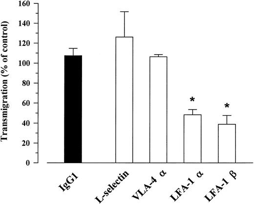
Blocking of transendothelial migration. CD34+ cells were either preincubated with the respective blocking MoAb, an isotype matched IgG1, or PBS/BSA 1% alone (control without antibody), and added to the upper chamber of the transmigration system. The number of transmigrated cells was measured after 24 hours. Transmigration was expressed as percent of the control without antibody pretreatment, *P < .05.
Blocking of transendothelial migration. CD34+ cells were either preincubated with the respective blocking MoAb, an isotype matched IgG1, or PBS/BSA 1% alone (control without antibody), and added to the upper chamber of the transmigration system. The number of transmigrated cells was measured after 24 hours. Transmigration was expressed as percent of the control without antibody pretreatment, *P < .05.
Blocking of progenitor cell migration.Preincubation with an irrelevant, isotype matched IgG1 antibody had no effect on the number of transmigrated CD34+ cells, as indicated by a relative transmigration of nearly 100% compared with cells that had not been preincubated with antibody (Fig 5). Similarly, antibodies to L-selectin and VLA-4 did not block the migration of hematopoietic progenitor cells. In the presence of blocking MoAbs to the LFA-1 alpha and beta chain, the number of transmigrated cells was reduced to 40% and 30% of the IgG1 isotype control, respectively. In a series of four experiments, the difference was statistically significant.
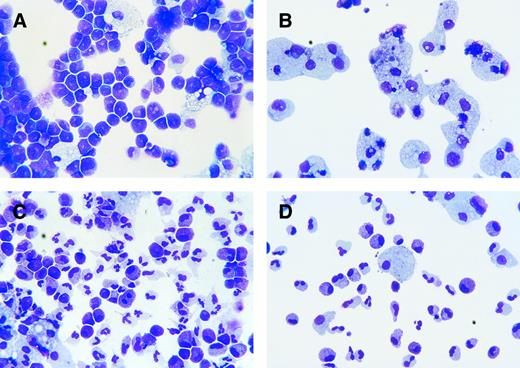
Transmigration of proliferating hematopoietic cells.On day 15, 2.1 ± 0.4 × 10⁶ cells were found in the upper chamber before demidepopulation, mainly comprising granulocytic precursors such as promyelocytes, mature monocytes, rarely granulocytes, and occasionally megakaryocytes (Fig 6 [see page 74], Table 3). However, the vast majority of the transmigrated cells were mature monocytes and macrophages with foamy cytoplasm. A total number of 4.1 ± 0.7 × 10⁵ cells had transmigrated during 48 hours. Flow cytometry confirmed the morphologic data (Table 3). Nonmigrating cells were brightly positive for CD15 characteristic for granulocytic precursors, while the monocytic marker CD14 was highly expressed on the transmigrated cells. Only few cells with megakaryocytic differentiation (CD41a+) were detected. At this time point, early progenitor cells were still efficiently transmigrating as demonstrated by a comparable plating efficiency of nonmigrating and transmigrated cells. On day 30 finally, the upper chamber contained 3.2 ± 0.5 × 10⁵ cells, whereas 1.3 ± 0.2 × 10⁵ cells had transmigrated into the lower chamber within 48 hours. The majority of nonmigrating, but also of transmigrated cells, comprised mature granulocytes and monocytes. Accordingly, CD15++ cells were also detected in the lower chamber. Only very few colony-forming units were observed at that time point.
Transmigration of proliferating hematopoietic cells. (A) Wright-Giemsa stained proliferating cells in the upper chamber at day 15. The majority of the cells consisted of granulocytic precursors such as promyelocytes and some mature monocytes/macrophages (original magnification × 400). (B) Transmigrated cells in the lower chamber at day 15. The transmigrated cells recovered comprised almost exclusively mature monocytes/macrophages (original magnification × 400). (C) Proliferating cells in the upper chamber at day 30. In addition to myeloid precursors and monocytes, also mature granulocytes and bands were found (original magnification × 400). (D) Transmigrated cells from the lower chamber at day 30. Both mature granulocytes and monocytes transmigrated into the lower chamber. Precursors earlier than metamyelocytes were virtually not found among the transmigrated cells (original magnification × 400).
Transmigration of proliferating hematopoietic cells. (A) Wright-Giemsa stained proliferating cells in the upper chamber at day 15. The majority of the cells consisted of granulocytic precursors such as promyelocytes and some mature monocytes/macrophages (original magnification × 400). (B) Transmigrated cells in the lower chamber at day 15. The transmigrated cells recovered comprised almost exclusively mature monocytes/macrophages (original magnification × 400). (C) Proliferating cells in the upper chamber at day 30. In addition to myeloid precursors and monocytes, also mature granulocytes and bands were found (original magnification × 400). (D) Transmigrated cells from the lower chamber at day 30. Both mature granulocytes and monocytes transmigrated into the lower chamber. Precursors earlier than metamyelocytes were virtually not found among the transmigrated cells (original magnification × 400).
DISCUSSION
The in vitro transmigration model used in this study allows analysis of transendothelial migration of hematopoietic progenitors and subsets of hematopoietic cells during differentiation. Phenotype and plating efficiency of peripheral blood progenitor cells that had transmigrated through immortalized bone marrow endothelial cells in vitro were different from that of nonmigrating cells. The relatively low number of migrating progenitor cells compared with monocytes or granulocytes26 indicates that progenitors migrate less avidly than mature leukocytes. This model might therefore also be useful to evaluate conditions that promote migration and could play a role in progenitor cell mobilization. Granulocytic precursors such as promyelocytes showed virtually no migratory capacity. Adhesion molecules such as β1 and β2 integrins are expressed in lower levels on circulating progenitor cells compared with noncirculating CD34+ cells or mature leukocytes, which could account for their moderate migration in vitro.18 However, expression of integrins on the cell surface, even in small amounts, may be a prerequisite for transendothelial migration and homing to the bone marrow as demonstrated by the ability of LFA-1 antibodies to reduce in vitro transmigration. Furthermore, it was shown that administration of VLA-4 antibodies in vivo was associated with both mobilization of progenitors and inhibition of homing to the bone marrow.19 28 Indeed, blocking of VLA-4 could also increase the number of circulating progenitor cells by inhibiting homing and thus increasing the time progenitors stay in circulation, rather than recruiting progenitor cells from the bone marrow.
In our in vitro model, blocking VLA-4 antibodies were not able to inhibit transendothelial migration. VLA-4 may particularly be important for the final homing of progenitor cells in the bone marrow stroma, which provides the ligands fibronectin and VCAM. The latter is constitutively expressed on bone marrow stromal cells.9,10 In a recent study however, expression of VCAM-1 at low levels was also found on resting bone marrow endothelium of mice in vivo.10 Also ICAM-1, the ligand for LFA-1, might be constitutively expressed on BMEC in vivo, similar to microvascular endothelium of other tissues.29 Bone marrow endothelial cells analyzed directly after isolation by cell sorting were shown to be positive for ICAM-1, while epression of VCAM-1 and E-selectin was only weak.2 The immortalized bone marrow endothelial cell line BMEC-1 retains a stable phenotype of first-passage BMEC, which could explain that basal levels of ICAM-1 are detectable. Interaction of β2-integrins with constitutively expressed ICAM on endothelial cells could be important for adhesion and transendothelial migration of circulating progenitors in the bone marrow. This is in accordance with our finding that antibodies to LFA-1 significantly reduced the number of migrated cells. Blocking of β2-integrin with CD18 antibodies also reduces migration of mature monocytes through resting endothelium in vitro26 suggesting that similarities exist in adhesion and migration of mature and immature hematopoietic cells. In other studies, however, progenitor cells did not adhere to ICAM-1–transfected cells in a significant amount suggesting that LFA-1 expressed on CD34+ cells is in a low affinity state.17 In our transmigration assay, the affinity state of integrins could be modulated by signaling through other adhesion molecules during sequential steps of adhesion and transmigration.
The initial, reversible steps of adhesion termed as “rolling,” which involves selectins and their ligands, might be required to activate integrins. This idea is supported by the finding that ligation of L-selectin can upregulate the affinity of β2-integrins in mature leukocytes.30 Assays that use endothelial cells can detect sequential steps of adhesion and transendothelial migration in vitro. Furthermore, ligation of CD34, which is known to act as a selectin ligand, upregulated the affinity state of LFA-1 expressed on hematopoietic progenitor cells.31 In our study, the loss of L-selectin on transmigrated cells suggests that shedding may have occurred during transmigration after initial binding to a ligand on the endothelial cells. This finding is in accordance with observations in transmigration assays for mature leukocytes such as lymphocytes.32 Similarly, in mature leukocytes no difference was observed in the expression of LFA-1 and VLA-4 after transmigration. In contrast to selectins, which are regulated by rapid proteolytic cleavage (shedding), the function of integrins is mainly not modulated by expression level, but rather by changes in the receptor affinity. Ligation of β2-integrins during migration also causes shedding of L-selectin,33 which might similarly occur in our transmigration assay, as involvment of β2-integrins was confirmed by the blocking experiments. Signaling through L-selectin by receptor binding does not only increase affinity state and even expression level of integrins, but also enhances proliferation of hematopoietic progenitor cells.34 The more mature phenotype of the transmigrated cells as assessed by CD38 expression and the higher plating efficiency for committed progenitors, which are contained in the CD38bright population further supports this notion. However, a higher number of BFU-E accounts for the higher plating efficiency of transmigrated cells, while no significant difference was found in the plating efficiency for myeloid CFU.
During continuous coculture, immature myeloid precursor cells such as promyelocytes and myelocytes did not migrate, while mature hematopoietic cells and colony-forming progenitor cells were found in the transmigrated compartment. This indicates that the capacity of bone marrow endothelial cells to allow selective migration of both mature and very early hematopoietic cells is still retained by the BMEC-1 cell line in vitro. This suggests that transit of hematopoietic cells is regulated, at least partially, at the level of the sinusoidal bone marrow endothelium. The gain or loss of adhesion molecules expressed on hematopoietic cells may finally dictate whether cells migrate into the peripheral circulation. In addition, differentiation-dependent expression of surface molecules with adhesive function could also be important for modulating the affinity of hematopoieitc cells to bind to stromal cells or matrix proteins.35 In our transmigration assay, erythroid progenitors seem to migrate more avidly than myeloid. One may speculate that lineage-related expression of adhesion molecules9 could result in a different migratory capacity.
In conclusion, the human bone marrow endothelial cell line BMEC-1 allows migration of progenitors and mature hematopoietic cells. Differences in the phenotype and plating efficiency between migrating and nonmigrating cells suggest that more primitive and lineage-committed progenitors differ in their ability to migrate. On the other hand, signaling through adhesion molecules during transendothelial migration could also initiate differentiation and proliferation, which takes place in vivo after homing to the bone marrow microenvironment. The in vitro transmigration model provides a suitable technique to further analyze mechanisms of progenitor cell trafficking.
ACKNOWLEDGMENT
We thank Barbara Ferris and Michael Querijero for outstanding technical assistance.
Supported by Gar Reichman fund of the Cancer Research Institute (M.A.S.M.), National Institutes of Health Grant No. K08-HL-02926, Dorothy Rodbell Cohen Foundation for Sarcoma Research, and The Rich Foundation (S.R.). R.M. is the recipient of a fellowship from the Dr Mildred Scheel Stiftung für Krebsforschung (Bonn, Germany).
Address reprint requests to Robert Möhle, MD, Laboratory of Developmental Hematopoiesis, Memorial Sloan-Kettering Cancer Center, 1275 York Ave, Mailbox 101, New York, NY 10021.

