Abstract
Two subtypes of G-protein–coupled cannabinoid receptors have been identified to date: the CB1 central receptor subtype, which is mainly expressed in the brain, and the CB2 peripheral receptor subtype, which appears particularly abundant in the immune system. We investigated the expression of CB2 receptors in leukocytes using anti-CB2 receptor immunopurified polyclonal antibodies. We showed that peripheral blood and tonsillar B cells were the leukocyte subsets expressing the highest amount of CB2 receptor proteins. Dual-color confocal microscopy performed on tonsillar tissues showed a marked expression of CB2 receptors in mantle zones of secondary follicles, whereas germinal centers (GC) were weakly stained, suggesting a modulation of this receptor during the differentiation stages from virgin B lymphocytes to memory B cells. Indeed, we showed a clear downregulation of CB2 receptor expression during B-cell differentiation both at transcript and protein levels. The lowest expression was observed in GC proliferating centroblasts. Furthermore, we investigated the effect of the cannabinoid agonist CP55,940 on the CD40-mediated proliferation of both virgin and GC B-cell subsets. We found that CP55,940 enhanced the proliferation of both subsets and that this enhancement was blocked by the CB2 receptor antagonist SR 144528 but not by the CB1 receptor antagonist SR 141716. Finally, we observed that CB2 receptors were dramatically upregulated in both B-cell subsets during the first 24 hours of CD40-mediated activation. These data strongly support an involvement of CB2 receptors during B-cell differentiation.
Δ9-TETRAHYDROCANNABINOL (Δ9-THC) is the principal psychoactive component in preparations of Cannabis sativa (marijuana, hashish).1 Its effects are mainly mediated via the CB1 central cannabinoid receptor,2 which belongs to the family of G-protein–coupled seven-transmembrane domain proteins.3,4 Activation of CB1 receptor, which is coupled to a Gi protein, leads to inhibition of adenylyl cyclase5and N-type voltage-dependent calcium channels.6 Moreover, the CB1 receptor is functionaly coupled to mitogen-activated protein kinase (MAPK) cascade7 and regulates the krox-24 (egr-1) gene.8 In addition to a wide range of physiological effects on the central nervous system, cannabinoid ligands have been reported to affect the immune system.9,10 At high concentrations, they modulate proliferative responses of T lymphocytes,11,12 cytotoxic T-cell activity,13humoral response,14 microbiocidal activity, cytokine production, and antigen processing by macrophages.15-17Cannabinoid ligands also act at physiological concentrations by inhibiting the synthesis of tumor necrosis factor-α (TNF-α) by human large granular lymphocytes18 and the activation of mast cells.19 All of these effects strongly suggested that cannabinoid-induced immune modulation may be mediated at least in part through a cannabinoid receptor-associated mechanism.
Recently, a second cannabinoid receptor has been cloned.20This receptor, CB2, shows 44% identity with the CB1 receptor. It has been defined as peripheral cannabinoid receptor, because it is mainly localized in cells of the immune system. Among these cells, low levels of CB2 receptors have been localized in B-cell areas of different rat lymphoid tissues such as spleen, lymph nodes, and Peyer’s patches.21 The CB2 receptor, which is also linked to a Gi protein, displays pharmacological and biochemical properties similar to those of the CB1 receptor. It inhibits adenylyl cyclase activities,22 activates the MAPK pathway, and induces krox-24 expression,23 but does not modulate the activity of calcium channels.24 Recently, it has been shown that, via CB2 receptor, cannabinoid ligands cause a dose-dependent increase of B-cell proliferation induced through cross-linking of surface Igs or ligation of the CD40 antigen.25
With CB2 receptors being considerably more abundant in the immune system than CB1 receptors,26 27 we used antibodies (Abs) raised against the C-terminal tail of the CB2 receptor to investigate its expression in leukocytes. With the highest level being observed in B cells, we studied the expression of CB2 receptors in tonsillar B cells and found that it was modulated during B-cell differentiation. Finally, we assessed the function of the CB2 receptor by demonstrating that (1) cross-linking of CD40 by monoclonal antibodies (MoAbs) increased CB2 receptor expression on both virgin and GC B cells and (2) CB2 receptors act as coreceptors of CD40-induced proliferation of these two B-cell subsets.
MATERIALS AND METHODS
Production of anti-CB2 receptor Abs.
Synthetic peptide derived from the predicted aminoacid-sequence of the carboxylic tail of the human CB2 receptor (Y-P-D-S-R-D-L-D-L-S-D-C) and bovine serum albumin (BSA)-conjugated peptide used as immunogen were from Neosystem (Strasbourg, France). Rabbits were injected subcutaneously with 2 mg BSA-peptide in 250 μL water and 250 μL complete Freund’s adjuvant. Animals were boosted monthly under the same conditions and blood was taken 10 days after the fifth injection. Abs directed against the C-terminal part of the human CB2 receptor were immunopurified on a Bio-Rad Affi-Gel 10 modified with the peptide (Bio-Rad, Hercules, CA) as already described.28 Briefly, 10 mL of immune serum was incubated overnight at 4°C with 1 mL modified gel. After extensive washings, anti-CB2 receptor Abs were eluted with 100 mmol/L glycine-HCl, pH 1.8, and neutralized with 1 mol/L Tris-NaOH. The pooled fractions were supplemented with 10 mg/mL BSA, concentrated, and dialyzed on a Filtron microsep 30 kD (Filtron, Nortborough, MA). Concentrated Abs were stored in 50% glycerol at −20°C.
Immunoblotting experiments.
Membranes of wild-type hamster ovary cells (CHO-WT) and of CHO cells stably transfected with the CB2 receptor (CHO-CB2)27 were prepared by homogeneizing cells with polytron in 5 mmol/L Tris, pH 7.4, containing 1 mmol/L EDTA, 20 μg/mL aprotinin, and 1 mmol/L 4-(2-aminoethyl)-benzenesulfonyl fluoride hydrochloride (AEBSF). The homogenate was centrifuged for 15 minutes at 2,000g. The nuclear free supernatant was centrifuged for 1 hour at 100,000g. Immunoblotting experiments were performed on the membrane pellets after electrophoresis on a 4% to 20% sodium dodecyl sulfate (SDS)-polyacrylamide gel (Novex, San Diego, CA) and transfer onto nitrocellulose filters. Proteins were electroblotted on nitrocellulose filters (Novex). Nonspecific binding was blocked with 10% casein in TBS buffer (20 mmol/L Tris-HCl, 150 mmol/L NaCl, pH 7.5) for 1 hour at room temperature. The blotted filters were washed with 0.1% Tween 20 in TBS buffer and then incubated for 3 hours at room temperature with anti-CB2 receptor Abs (1:2,000 dilution). After another washing, peroxidase-conjugated antirabbit Ig (1:8,000 dilution; Sigma, St Louis, MO) was added for 45 minutes at room temperature. After 5 extensive washes, immune complexes were detected using the ECL kit on Hyperfilm-MP (Amersham, Buckinghamshire, UK) following the supplier’s instructions.
Antibodies.
The following Abs were used for flow cytometry. Phycoerythrin-conjugated human CD4, CD8, CD20, and CD38 MoAbs were purchased from Becton Dickinson (San Jose, CA). Tricolor (phycoerythrin-Cy5)-conjugated human CD3 MoAbs were from Caltag (South San Francisco, CA). Biotinylated human CD44 MoAbs were from Leinco Technologies (Ballwin, MO). Biotinylated antihuman IgD Abs were from Tagoimmunologicals (Burlingame, CA). The human purified CD77 MoAbs were from Immunotech and were stained with the biotinylated antirat IgG (mark-1) MoAbs (Immunotech). All of the biotinylated Abs were labeled with Tricolor-conjugated streptavidin (Caltag). Rabbit anti-CB2 Abs were labeled with fluorescein isothiocyanate (FITC)-conjugated donkey antirabbit IgG Abs from Jackson Immunoresearch (West Grove, PA).
The following Abs were used for confocal laser scanning microscopy: purified human CD3, CD38 MoAbs, anti-Ki67 MoAbs, antihuman IgD MoAbs, and antifollicular dendritic cell MoAbs (HJ2) were from Dako. These MoAbs were all shown with FITC-conjugated donkey antimouse IgG Abs. Rabbit anti-CB2 receptor Abs were stained with Cy3-conjugated donkey antirabbit IgG Abs from Jackson Immunoresearch.
Cells and tissues.
Cells of the human myelocytic HL60 cell line (ATCC, Rockville, MD) were grown in RPMI 1640 (GIBCO, Grand Island, NY) medium supplemented with 10% heat-inactivated fetal calf serum, 0.26 mg/mL glutamine, 180 IU/mL penicillin, and 0.18 mg/mL streptamycin.
Mononuclear cells were isolated from Ficoll Hypaque density centrifugation of peripheral blood obtained from 3 consenting healthy donors (Caucasian men 26, 38, and 44 years of age). Tonsils were obtained after obtaining approvals from children (Caucasian females 8, 8, and 10 years of age) undergoing tonsillectomy (Clinique du Parc, Montpellier, France). For confocal microscopic studies, tonsils were frozen in liquid nitrogen and maintained at −80°C until staining and analysis. For flow cytometric studies, tonsils were immediately minced, labeled, and analyzed. B cells were purified from tonsils with magnetic beads using the Variomacs system (Tebu, Le Perray en Yvelines, France). In the first step, tonsil T cells and monocytes were depleted, respectively, with CD3- and CD14-coated beads. In the second step, B cells were incubated with biotinylated anti-IgD Abs and streptavidin-coated beads to isolate IgD+ and IgD− B cells. The purity of both B-cell populations was greater than 95% as assessed by fluorescence-activated cell sorting (FACS). Isolation of GC (CD38+CD44−) B cells were thus performed by negative selection. IgD− B cells were submitted to two rounds of depletion with different anti-CD44 MoAbs (clone NKI-P2 and clone J173 purchased from Immunotech), followed by incubation with magnetic beads coated with antimouse IgG Abs.
Leukocyte staining for flow cytometry.
Cell surface phenotyping was performed by incubating cells with appropriate phycoerythrin and tricolor MoAbs in phosphate-buffered saline (PBS) for 30 minutes at 4°C, following the supplier’s instructions. Cells were fixed in 1% formaldehyde overnight at 4°C, washed once, and permeabilized for 10 minutes with a solution of 0.1% saponin in PBS containing 1% BSA. Purified anti-CB2 receptor Abs (1:1,000 dilution) were added to 106 cells in 100 μL of the 0.1% saponin/1% BSA solution for 30 minutes. After two washes with 0.03% saponin in PBS containing 0.3% BSA, cells were incubated with FITC-conjugated donkey antirabbit IgG Abs (1:100 dilution) for 30 minutes, washed once with the 0.03% saponin/0.3% BSA solution, and washed once with PBS alone. Negative controls were performed by 1 hour of preincubation of anti-CB2 receptor Abs with the C-terminal synthetic peptide at 20 μg/mL. The fluorescence intensity mean of each subset was calculated by substracting the fluorescence of the irrelevant controls (anti-CB2 receptor Abs + peptide) from that of the relevant labeling (anti-CB2 receptor Abs).
Tissue staining for confocal microscopy.
Serial cryostat sections (9-μm thick) of tonsils were fixed in acetone for 5 minutes at room temperature. Sections were simultaneously incubated with mouse antihuman leukocyte antigen MoAbs and rabbit anti-CB2 receptor Abs under 100 μL of PBS containing 0.5% BSA. After three washes in the same buffer, sections were simultaneously stained with FITC-conjugated donkey antimouse IgG Abs and Cy3-conjugated donkey antirabbit IgG Abs, both at 1:200 dilution in PBS containing 0.5% BSA. After two washes in the same buffer and one wash in PBS without BSA, sections were mounted in a solution of glycerol/PBS containing the antibleaching reagent DABCO at 50 mg/mL (Sigma). Specificity controls were performed by 1 hour of preincubation of anti-CB2 receptor Abs with the C-terminal synthetic peptide at 20 μg/mL.
Dual fluorescence analysis was performed using a laser confocal microscope (LSM410; Zeiss, Oberkochen, Germany) equipped with a Plan NEOFLUAR water immersion lens (16×; numerical aperture [NA] = 0.50). Signals were collected separately after excitations of FITC and Cy3 at 488 and 543 nm, respectively. FITC emission was collected using a transmission filter centered at 530 nm and Cy3 emission using a 590-nm long-pass filter.
RNA preparation and reverse transcription-polymerase chain reaction (RT-PCR) analysis.
Subpopulations of B cells were obtained, purified, and labeled for cell surface phenotyping as described above. Cell sorting of 2 × 10⁵ cells of each B-cell subsets was performed using the Normal-C mode of a FacstarPlus cytometer (Becton Dickinson, Erembodegen, Belgium). Purification of each B-cell subset was checked by reanalyzing another sorting run. This procedure led to B-cell subpopulation purities ranging from 95% to 99.5%. The mRNA purification and conversion to single-strand cDNA were performed using a PolyATtract series 9600J mRNA Isolation System with cDNA Synthesis Reagents (Promega, Charbonnières, France) according to the manufacturer’s instructions.
Briefly, 105 sorted cells were centrifuged and suspended in 20 μL of extraction buffer. Each sample was then transfered to the well of a V-bottom 96-well plate. After hybridization with a synthetic biotinylated oligo (dT) probe and incubation with streptavidin-coated magnetic beads, 3′-polyadenylated RNA was captured using a 96 pins Multi-Magnet (Promega, Charbonnièrès, France). After successive washes, purified mRNA was eluted into 20 μL of water. mRNA was converted to single-strand cDNA by adding 10 μL of reverse transcriptase master mix (containing AMV Reverse Transcriptase) in each well of the 96-well microplate.29
Quantitation of CB2 receptor mRNA levels was performed in the exponential phase of amplification as previously described.30 Independent PCR amplifications of CB2 receptor and β2-microglobulin (used as an external control) were run in parallel as already described.26
Reaction products were analyzed using a nonisotopic microplate assay supplied by Sanofi Diagnostics Pasteur (Marnes-la-Coquette, France). PCR products and amplicons were captured by specific immobilized nucleotide probes complementary to β2-microglobulin and CB2-receptor sequences located between primers. Quantitation was performed by hybridization with a biotinylated labeled probe and incubation with an avidin-peroxidase conjugate. The probes used were as follows: CB2 receptor capture probe, 5′-gccaacctcacatccagcctcattcgggc-3′; CB2 receptor detection probe, 5′-biotin-tgggaaccaacagatgagga-3′; β2-microglobulin capture probe, 5′-caattctctctccattcttcagtaagtcaac-3′; and β2-microglobulin detection probe, 5′-biotin-agaaagaccagtccttgctg-3′. Sandwich hybridization assays were performed as recently described,31 with slight modifications. Briefly, 96-well microplates (Maxisorb Nunc, Rosk, Denmark) were coated with 200 μL of the capture oligonucleotide solutions (0.5 μg/mL in PBS buffer). After overnight incubation at room temperature, plates were washed twice, dried for 20 minutes at 55°C, and then sealed for long-term storage. Wells were prehybridized for 30 minutes at 37°C with 200 μL of hybridization buffer; after supernatants were discarded, 200 μL of biotin-oligonucleotide probes (50 ng/mL in hybridization buffer) containing 7 μL of heat-denaturated (10 minutes at 95°C) PCR products were distributed in each well and incubated for 60 minutes at 37°C with gentle shaking. Microplates were washed six times with 200 μL of washing buffer and a second incubation with 200 μL of extravidin-peroxidase conjugate (Sigma; 1:5,000 dilution in PBS containing 0.3% BSA) was performed for 30 minutes at 37°C under shaking. Plates were washed six times and the immobilized hybrid complex was detected by addition of 200 μL of ortho-phenylene diamine chromogenic substrate solution (OPD) for 15 minutes at 37°C. Fifty microliters of 4N H2SO4 was added to block the enzymatic reaction. Buffer solutions and reagents were distributed in microplates using a Biomek 1000 automated laboratory workstation (Beckman, Paris, France) equipped with a spectrophotometer that measures optical densities at 492 nm.
Plasmid construction and generation of a stable cell line.
A 1.08-kb HindIII BamHI fragment encompassing the CB2 receptor coding sequence was ligated into the mammalian expression vector pcDNA3 (In Vitrogen, San Diego, CA), placing transcription of cDNA under the strong immediate early promoter of human cytomegalovirus. Transfection of HL60 cell line with recombinant plasmid was performed by electroporation. Briefly, 2 × 10⁷ cells were washed in PBS, resuspended in 1 mL PBS containing 20 μg vector, and incubated on ice for 10 minutes. Electroporation was performed in a 0.4-cm in diameter cuvette by using a Bio-Rad Gene Pulser at 320 mV and 250 μF. After 10 minutes of incubation at room temperature, electroporated cells were grown in the medium described above. The day after, cells were seeded at 5 × 10⁵/mL in the presence of geneticin (600 μg/mL medium) in 24-well microplates. Cells were screened 3 to 4 weeks later for expression of CB2 receptors by flow cytometry. Positive cells were cloned in 96-well microplates with a FacstarPlus cytometer to get a stable transfected HL60 cell line.
Staining of wild-type (HL60-WT) and CB2 receptor-transfected (HL60-CB2) cells was performed by incubating cells with anti-CB2 receptor Abs (1:1,000 dilution) with or without C-terminal synthetic peptide at 20 μg/mL, as described above for leukocytes. FITC-conjugated antirabbit IgG Abs (1:100 dilution) was used for flow cytometry, while the same reagent linked to Cy3 fluorochrome was used for confocal microscopy.
CD40-mediated proliferation of B-cell subsets.
CD32+-L cells suspended at 2 × 10⁶/mL were treated for 1 hour with 75 μg/mL mitomycin C in RPMI 1640 supplemented with 0.5% heat-inactivated calf serum, 2 mmol/L L-glutamine, 100 U/mL penicillin, 100 μg/mL streptomycin, and 5 mmol/L HEPES buffer. These cells were washed four times and plated at 5 × 10³ cells per well in round-bottomed 96-well microtiter trays. CD40 MoAbs (MAB89) were added at 100 ng/mL and incubated for 2 hours at 37°C. Purified B-cell subsets were seeded at 105 cells per well simultaneously with CP55,940 ligand (generously provided by Pfizer, Groton, CT) at the indicated concentrations. When the cannabinoid compounds SR 141716α (CB1 receptor antagonist) and SR 144528α (CB2 receptor antagonist) from Sanofi Recherche were used, they were preincubated for 30 minutes with B cells before the addition of CP 55,940. DNA synthesis was determined 72 hours later by pulsing the cells with 1 μCi/well [3H] thymidine for the last 16 hours of the culture period. Each point was the mean of four replicates. The data shown are representative of two separate experiments performed with two different donors.
Modulation of CB2 receptor expression in B-cell subsets.
B-cell subsets were activated by CD40 MoAbs as described above, except that CD32+-L cells and B cells were seeded at 5 × 10⁴ and 106 cells per well, respectively, in 24-well microtiter trays. CB2 receptor expression was checked at 24 and 48 hours using the flow cytometric and RT-PCR analyses.
Statistical analysis.
Data were analyzed using the Dunnett’s analysis of variance test. A cut-off value of P ≤ .05 was used to indicate statistical significance. Each experiment was repeated at least twice.
RESULTS
Production and characterization of anti-CB2 receptor polyclonal Abs.
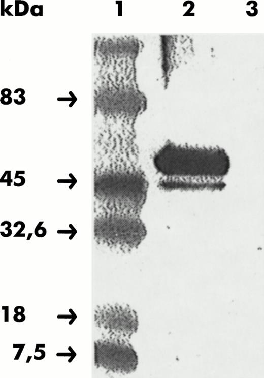
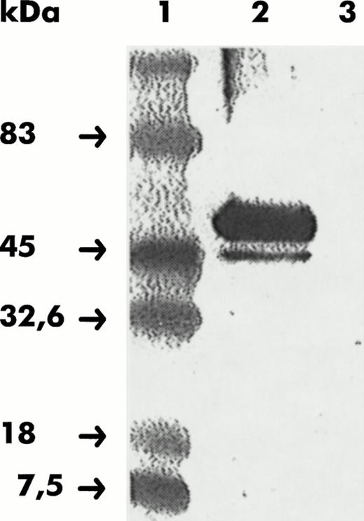
Several rabbits were immunized with 1 of the 18 BSA-conjugated peptides corresponding to different intracellular and extracellular parts of the CB2 receptor. Among these, the peptide corresponding to the intracellular 11-aminoacid sequence of the C-terminal part was the only one that led to specific anti-CB2 receptor Abs. After five injections, Abs from immune serum were immunopurified on a gel modified with the synthetic peptide. The specificity of purified Abs was evaluated in immunoblotting experiments performed with membrane of CHO cells stably transfected with the CB2 receptor (CHO-CB2). Two bands were shown when 75 μg of protein from CHO-CB2 membranes were electrophoresed, whereas no band was observed when the same amount of protein from the wild-type cell line (CHO-WT) was analyzed (Fig 1). The major band corresponded to a molecular weight of approximately 46 kD, consistent with the deduced amino acid sequence of the human CB2 receptor cDNA. The minor band corresponded to a molecular weight of approximately 45 kD, which could represent a degraded receptor or another form of the receptor differently glycosylated.
Reactivity of CHO membranes to anti-CB2 receptor Abs assayed in Western blot. CHO-WT and CHO-CB2 membranes were resolved by SDS-PAGE, transferred to nitrocellulose, and shown with anti-CB2 receptor Abs raised against the C-terminal CB2 receptor peptide. Lane 1, molecular weight markers; lane 2, CHO-CB2 membranes; lane 3, CHO-WT membranes.
Reactivity of CHO membranes to anti-CB2 receptor Abs assayed in Western blot. CHO-WT and CHO-CB2 membranes were resolved by SDS-PAGE, transferred to nitrocellulose, and shown with anti-CB2 receptor Abs raised against the C-terminal CB2 receptor peptide. Lane 1, molecular weight markers; lane 2, CHO-CB2 membranes; lane 3, CHO-WT membranes.
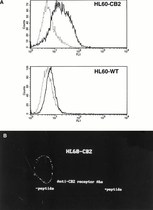
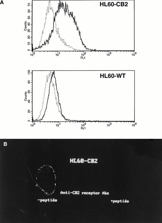
The specificity of immunopurified anti-CB2 receptor Abs to recognize CB2 receptors in their native forms was achieved by studying their binding on HL60 cells transfected with the human CB2 receptor cDNA (HL60-CB2). Flow cytometric analysis showed that a positive staining was obtained in HL60-CB2 cell line but not in the wild-type cell line (HL60-WT), as shown in Fig 2A. Moreover, inhibition of the labeling was observed when anti-CB2 receptor Abs were preincubated with the C-terminal synthetic peptide confirming the specificity of anti-CB2 receptor Abs. We next examined the subcellular distribution of CB2 receptors by confocal microscopy. Figure 2B shows a localization of CB2 receptors mainly associated with the plasma membrane of HL60-CB2 cells.
(A) Flow cytometric analysis of the labeling of HL60 cells transfected with CB2 receptor cDNA (HL60-CB2) by anti-CB2 receptor Abs. After formaldehyde fixation and saponin permeabilization, HL60-CB2 (top histogram) and wild-type HL60 cells (bottom histogram) were labeled with anti-CB2 receptor Abs preincubated (dotted line) or not (solid line) with the C-terminal peptide of the CB2 receptor. (B) Confocal microscopic analysis of the localization of CB2 receptors in HL60-CB2 cells. The left side corresponds to HL60-CB2 stained with anti-CB2 receptor Abs and the right side corresponds to HL60-CB2 stained with anti-CB2 receptor Abs preincubated with the C-terminal peptide as negative control.
(A) Flow cytometric analysis of the labeling of HL60 cells transfected with CB2 receptor cDNA (HL60-CB2) by anti-CB2 receptor Abs. After formaldehyde fixation and saponin permeabilization, HL60-CB2 (top histogram) and wild-type HL60 cells (bottom histogram) were labeled with anti-CB2 receptor Abs preincubated (dotted line) or not (solid line) with the C-terminal peptide of the CB2 receptor. (B) Confocal microscopic analysis of the localization of CB2 receptors in HL60-CB2 cells. The left side corresponds to HL60-CB2 stained with anti-CB2 receptor Abs and the right side corresponds to HL60-CB2 stained with anti-CB2 receptor Abs preincubated with the C-terminal peptide as negative control.
Expression of CB2 receptors in mononuclear cells isolated from peripheral blood and tonsils.
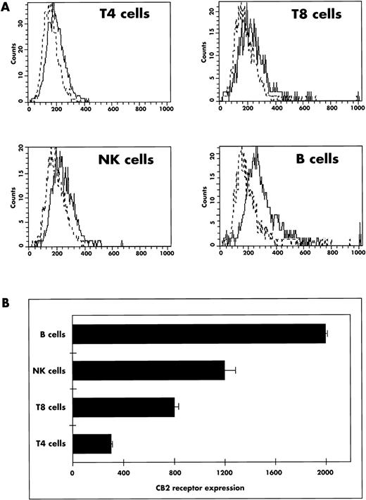
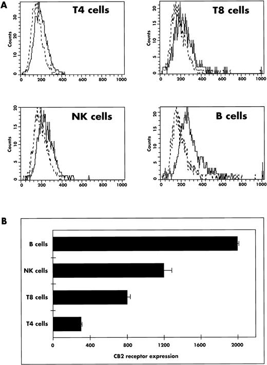
The expression of CB2 receptors was first assayed by flow cytometry in peripheral blood mononuclear cells isolated from three different human donors. As shown in Fig 3A, the levels of CB2 receptor expression in these cells was relatively low as compared with HL60-CB2 cells. The quantitation of CB2 receptors in leukocytes showed that B lymphocytes (CD20+) expressed the highest level of CB2 receptors, followed by NK cells (CD56+; Fig3B). Among T-cell subsets, T8 (CD3+CD8+) lymphocytes displayed a higher level of CB2 receptors than did T4 cells (CD3+CD4+).
Expression of CB2 receptors in peripheral blood mononuclear cells. (A) Mononuclear leukocytes were isolated and labeled for flow cytometric analysis as reported in Materials and Methods. Each staining profile for CB2 receptor expression (solid line) was overlayed with the negative control (dotted line) performed by preincubating anti-CB2 receptor Abs with the C-terminal synthetic peptide. The four staining profiles were obtained after positionning a region of interest on cells expressing CD3+ CD4+ (T4 cells), CD8+ CD3+ (T8 cells), CD56+ (NK cells), or CD20+ (B cells). The histograms shown are all from one donor and are representative of three different donors. (B) Mean ± SD of CB2 receptor fluorescence intensities in peripheral blood leukocyte subsets from three different donors analyzed by flow cytometry as described in (A). For each leukocyte subset, the fluorescence intensity reported on the abcissa was calculated in arbitrary units by subtracting the irrelevant control (anti-CB2 receptor Abs + specific peptide) from the anti-CB2 receptor Ab labeling.
Expression of CB2 receptors in peripheral blood mononuclear cells. (A) Mononuclear leukocytes were isolated and labeled for flow cytometric analysis as reported in Materials and Methods. Each staining profile for CB2 receptor expression (solid line) was overlayed with the negative control (dotted line) performed by preincubating anti-CB2 receptor Abs with the C-terminal synthetic peptide. The four staining profiles were obtained after positionning a region of interest on cells expressing CD3+ CD4+ (T4 cells), CD8+ CD3+ (T8 cells), CD56+ (NK cells), or CD20+ (B cells). The histograms shown are all from one donor and are representative of three different donors. (B) Mean ± SD of CB2 receptor fluorescence intensities in peripheral blood leukocyte subsets from three different donors analyzed by flow cytometry as described in (A). For each leukocyte subset, the fluorescence intensity reported on the abcissa was calculated in arbitrary units by subtracting the irrelevant control (anti-CB2 receptor Abs + specific peptide) from the anti-CB2 receptor Ab labeling.
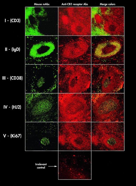
The preferential expression of CB2 receptors in B cells led us to investigate the in situ distribution of this molecule in the B-cell zones of secondary lymphoid organs. For this purpose, dual-immunofluorescence studies were performed on tonsil tissue sections by combining anti-CB2 receptor Abs together with several MoAbs identifying the different compartments of the B-cell follicles. In this tissue, the absence of CB2 receptor staining in interfollicular T-cell areas (CD3+) was observed as well as a marked homogeneous labeling of the mantle zones containing the resting IgD+ B cells (Fig 4, lines I and II, respectively). By contrast, germinal center (GC) areas displayed a heterogeneous labeling. In some cases, they appeared labeled with the same intensity as the mantle zone (Fig 4, lines II and IV), and in other cases they displayed a slight decreased CB2 receptor staining (Fig 4, lines I and V). Variations of intensity of CB2 receptor labeling observed in some GC may be associated with the presence of a particular cell subset in these areas. Indeed, when GC were full of follicular dendritic cells (FDC), as shown after labeling with anti-FDC HJ2 MoAbs, GC were entirely stained by anti-CB2 receptor Abs (Fig 4, line IV), indicating that FDC expressed the CB2 receptor. By contrast, we found that weak expression of CB2 receptors was frequently associated with the presence of Ki67+ proliferating cells that are localized in the dark zones of GC in human tonsils (Fig 4, line V). These observations, which were reproducibly repeated in three different tonsils, showed that CB2 receptors appeared preferentially expressed in both follicular mantles and GC of B-cell zones. In GC, FDC expressed CB2 receptors, whereas Ki67+ cells displayed substantial lower levels of these receptors.
In situ localization of CB2 receptors on tonsil tissue sections. Frozen tissue sections were simultaneously labeled with MoAbs characterizing different anatomical compartments in tonsils and with anti-CB2 receptor Abs. The MoAb labeling are displayed in the first column and are green-colored: CD3 (I); anti-IgD (II); CD38 (III); anti-follicular dendritic cells (IV); anti-Ki67 (V). The anti-CB2 receptor Ab labelings are displayed in the second column and are red-colored. The last line shows an irrelevant control performed with the specific peptide as described in Fig 2. The third column shows the merge colors. These data are representative of three different tonsils.
In situ localization of CB2 receptors on tonsil tissue sections. Frozen tissue sections were simultaneously labeled with MoAbs characterizing different anatomical compartments in tonsils and with anti-CB2 receptor Abs. The MoAb labeling are displayed in the first column and are green-colored: CD3 (I); anti-IgD (II); CD38 (III); anti-follicular dendritic cells (IV); anti-Ki67 (V). The anti-CB2 receptor Ab labelings are displayed in the second column and are red-colored. The last line shows an irrelevant control performed with the specific peptide as described in Fig 2. The third column shows the merge colors. These data are representative of three different tonsils.
Regulation of the expression of CB2 receptors during B-cell differentiation.
The low CB2 receptor expression associated with proliferating B cells suggested a subtle regulation of this receptor during B-cell differentiation. To confirm the above-noted histological observations, expression of CB2 receptors was studied in purified B cells from three different human tonsils, using anti-CB2 receptor Abs and flow cytometry.
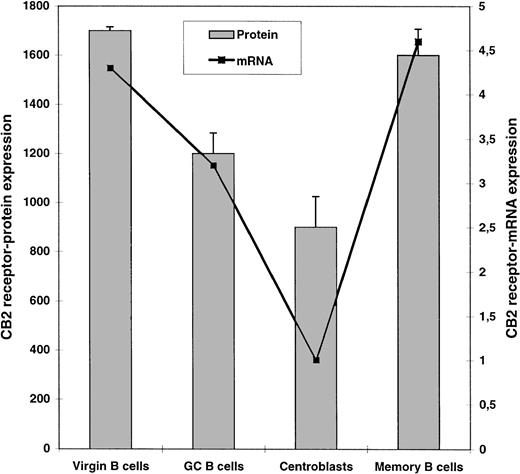
The expression of CB2 receptors was found to be modulated during B-cell differentiation (Fig 5). In CD38+ GC B cells, a decrease of CB2 receptor expression was observed and confirmed when B lymphocytes acquired CD77, corresponding to their differentiation in centroblasts. Finally, CB2 receptor expression was restored when B cells reached their terminal stages of differentiation to become memory B cells (IgD−CD44+CD38−).
Analysis of the regulation of expression of CB2 receptors during B-cell differentiation. Human tonsillar B-cell subsets were analyzed by flow cytometry (□) and by RT-PCR (▪) to quantitate CB2 receptor expression at the protein and transcript levels, respectively. For flow cytometric study, phenotypes of virgin B cells (IgD+ CD38−), GC B cells (IgD− CD38+), centroblasts (IgD− CD77+), and memory B cells (IgD− CD38− CD44+) were performed on tonsillar B cells as indicated in Materials and Methods. Results are the mean ± SD from three different donors and were calculated as described in Fig 3. For RT-PCR, the level of mRNA in each subset was quantitated from 2 × 10⁵ virgin B cells, GC B cells, centroblasts, or memory B cells sorted by FACS. CB2-receptor mRNA contents were normalized with that of β2-microglobulin and are expressed in arbitrary units. This experiment was repeated from two different donors with similar results.
Analysis of the regulation of expression of CB2 receptors during B-cell differentiation. Human tonsillar B-cell subsets were analyzed by flow cytometry (□) and by RT-PCR (▪) to quantitate CB2 receptor expression at the protein and transcript levels, respectively. For flow cytometric study, phenotypes of virgin B cells (IgD+ CD38−), GC B cells (IgD− CD38+), centroblasts (IgD− CD77+), and memory B cells (IgD− CD38− CD44+) were performed on tonsillar B cells as indicated in Materials and Methods. Results are the mean ± SD from three different donors and were calculated as described in Fig 3. For RT-PCR, the level of mRNA in each subset was quantitated from 2 × 10⁵ virgin B cells, GC B cells, centroblasts, or memory B cells sorted by FACS. CB2-receptor mRNA contents were normalized with that of β2-microglobulin and are expressed in arbitrary units. This experiment was repeated from two different donors with similar results.
These results were confirmed when the modulation of CB2 receptors was studied at the level of mRNA content during B-cell differentiation. A quantitative RT-PCR–based method was performed on 2 × 10⁵ highly purified B cells belonging to different B-cell subsets. Two analyses performed on B-cell subsets from two different donors showed that centroblasts (CD77+) displayed a fourfold loss in their CB2 receptor-mRNA content compared with virgin B cells (IgD+; Fig 5).
Involvement of CB2 receptors in the proliferation of virgin and GC B cells induced by CD40 MoAbs.
To study the function of CB2 receptors during B-cell differentiation, tonsillar B cells were separated into two subsets corresponding to virgin (IgD+) and GC (CD38+) B cells. We first examined the effects of the CP55,940 cannabinoid agonist on the proliferation of both subsets triggered with optimal concentrations of CD40 MoAbs in the presence of CD32+-L cells. CP55,940 induced a dose-dependent increase of proliferation on both subsets (Fig 6). The thymidine uptake mediated by CD40 MoAbs was increased by 27% for the virgin B cells and 20% for the GC B cells in the presence of 10 nmol/L of the cannabinoid ligand. These effects were optimal at 72 hours, although they can be detected as soon as 24 hours. They were not inhibited by the CB1 receptor antagonist SR 141716α22 and were totally affected by the CB2 receptor antagonist SR 144528α (Fig7). Furthermore, we observed that CP55,940 was not able to induce by itself any proliferation of B-cell subsets in the absence of CD40 MoAbs (data not shown).
Effect of CP55,940 on the proliferation of B-cell subsets. Virgin (A) and GC (B) B-cell subsets were induced to proliferate in the presence of different concentrations of CP55,940 for 72 hours after ligation of CD40 antigen using CD32+ L cells. Data shown are representative of two different experiments performed from two different donors. *P ≤ .05.
Effect of CP55,940 on the proliferation of B-cell subsets. Virgin (A) and GC (B) B-cell subsets were induced to proliferate in the presence of different concentrations of CP55,940 for 72 hours after ligation of CD40 antigen using CD32+ L cells. Data shown are representative of two different experiments performed from two different donors. *P ≤ .05.
CB2 receptors accounted for the CP 55,940-increased B-cell proliferation. The CB1 receptor antagonist SR 141716 (▪) and the CB2 receptor antagonist SR 144528 (•) were preincubated at indicated concentrations for 30 minutes with purified tonsillar B cells before the addition of 10 nmol/L CP 55,940. B cells were induced to proliferate for 72 hours after ligation of CD40 antigen. Data are expressed taking as 100% the difference between [3H] thymidine uptakes (which corresponded to 15,200 cpm) into B cells activated with CD40 MoAbs with and without 10 nmol/L CP 55,940. *P ≤ .05.
CB2 receptors accounted for the CP 55,940-increased B-cell proliferation. The CB1 receptor antagonist SR 141716 (▪) and the CB2 receptor antagonist SR 144528 (•) were preincubated at indicated concentrations for 30 minutes with purified tonsillar B cells before the addition of 10 nmol/L CP 55,940. B cells were induced to proliferate for 72 hours after ligation of CD40 antigen. Data are expressed taking as 100% the difference between [3H] thymidine uptakes (which corresponded to 15,200 cpm) into B cells activated with CD40 MoAbs with and without 10 nmol/L CP 55,940. *P ≤ .05.
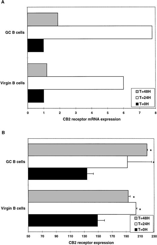
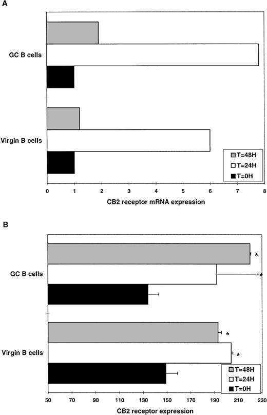
We then examined the regulation of CB2 receptors in virgin and GC B cells triggered by CD40 ligation. Transcripts of CB2 receptors were dramatically upregulated in both B-cell subsets. CB2 receptor mRNA content was maximum around 24 hours and returned to its basal level within 48 hours (Fig 8A). This upregulation of CB2 receptors was confirmed at the protein level using anti-CB2 receptor Abs and flow cytometry. CB2 receptors increased during the first 24 hours and was maintained at 48 hours in virgin and GC B cells (Fig 8B). This might indicate that CD40 ligation induces the transcription of CB2 receptors.
Regulation of CB2 receptor expression during CD40-mediated activation. Virgin and GC B-cell subsets were triggered by CD40 ligation in the presence of CD32+-L cells. Expression of CB2 receptors were quantitated at 24 and 48 hours by RT-PCR (A) and flow cytometry (B) as described in Figs 3 and 5. Data are representative of two different experiments performed from two different donors. *P ≤ .05.
Regulation of CB2 receptor expression during CD40-mediated activation. Virgin and GC B-cell subsets were triggered by CD40 ligation in the presence of CD32+-L cells. Expression of CB2 receptors were quantitated at 24 and 48 hours by RT-PCR (A) and flow cytometry (B) as described in Figs 3 and 5. Data are representative of two different experiments performed from two different donors. *P ≤ .05.
DISCUSSION
Both Δ9-THC and the chemical analog CP55,940 exert their psychoactive effects through the brain cannabinoid receptor.1,32 They activate different signaling pathways5-8 that are all inhibited by the potent and selective SR 141716α antagonist.22 Both cannabinoid compounds also recognize another receptor, CB2, which is mainly localized in cells of the immune system.20 These two receptors belong to the seven-transmembrane G-protein–coupled receptor family. Several receptors of this family, such as the chemokine receptors, display major functions in the immune system through their involvement in the traffic and activation of leukocytes33,34 as well as their implication in the infection of immune cells by the human immunodeficiency virus.35-37 It was thus relevant to postulate that the large range of pharmacological effects of cannabinoid compounds reported to date on the immune system9-19 25 could be mediated through the CB2 receptor.
CB2 cannabinoid receptors are rather considered as orphan receptors, because only derivatives of arachidonic acid are able to bind these receptors as putative endogenous ligands with low affinities.38,39 Signaling induced by cannabinoid receptors has extensively been studied in transfected or gene reporter-transformed cell lines.23,24,40,41 To contribute to the understanding of the function of CB2 receptors in the immune system, we decided to accurately study the expression of CB2 receptors in lymphocytes. We raised polyclonal Abs highly specific for the C-terminal part of this receptor. Using a semiquantitative flow cytometric assay, we found that, in human peripheral blood, the rank order of CB2 receptor expressions was B cells > NK cells > T8 cells > T4 cells, confirming our previously reported results of determination of CB2 receptor mRNA expression level in these cells.26 27 Moreover, the evaluation of the number of secondary Abs bound per cell made it possible to estimate the CB2 receptor quantity to 2,000 receptors per B cell (data not shown).
The preferential expression of CB2 receptors in the B-cell lineage led us to study their regulation during B-cell differentiation. We thus examined CB2 receptors in tonsil tissue sections by dual-color confocal microscopy and found that CB2 receptors were restricted to B-cell areas in accordance with a previous autoradiographic study of the binding of [3H]-CP55,940 to rat immune tissues.21 In secondary follicles, labeling by anti-CB2 receptor Abs was clearly observed in the follicular mantle, whereas in GC, heterogeneous staining was observed. In GC, CB2 receptor less staining was found to be associated with the presence of proliferating cells (Ki67+), whereas expression of CB2 receptors was found to be associated with the presence of FDC.
Identification of different B-cell subsets along the B-cell differentiation pathway in tonsils has recently been described using flow cytometric technique.42 We used this technique to characterize virgin B cells, GC B cells, centroblasts, and memory B cells. During B-cell differentiation process, a dramatic downregulation of CB2 receptor labeling was observed when B cells left the virgin B-cell stage to become centroblasts. CB2 receptor expression was restored at the end of differentiation when memory B cells appeared. The polyclonal anti-CB2 receptor Abs target the intracytoplasmic CB2 receptor C-terminal tail. Therefore, the decrease in CB2 receptor staining in centroblasts may be explained either by a decrease in CB2 receptor transcription or a receptor modification at the Ab recognition site after intracellular signaling. To exclude the latter, CB2 receptor transcripts were quantitated in highly purified B-cell subsets. A decrease in CB2 receptor mRNA level was also observed in centroblasts, confirming the downregulation of CB2 receptors at the protein level during B-cell differentiation.
The original distribution of CB2 receptors among cells of the immune system and their fine modulation during B-cell differentiation suggested that these receptors may exert their function on immune cells depending on their lineages, their stages of differentiation, and their partitioning at different locations within secondary lymphoid organs. Our previous observations that cannabinoid ligands enhanced human B-cell proliferation mediated by cross-linking of surface Igs, whereas no effect was noticeable on human T-cell proliferation mediated by phytohemagglutinin (PHA), argued in favor of a B-cell lineage-specific expression of CB2 receptors.25 Moreover, cannabinoid agonists effects were totally inhibited by pertusis toxin, demonstrating that cannabinoid receptors are coupled to a Gi protein in B cells. To examine the pattern of expression in the mature B-cell compartment, we compared the proliferative response of virgin and GC B cells to the cannabinoid agonist, CP55,940, under CD40-MoAb challenge. Low concentrations of CP55,940 enhanced the proliferation of both subsets in the presence of CD40 MoAbs. This enhancement was mediated by CB2 receptors, because the selective CB2 receptor antagonist SR 144528A inhibited CP 55,940 effects in a dose-dependent manner, whereas the CB1 receptor antagonist SR 141716A was without any effect. Moreover, the putative endogenous CB1 cannabinoid ligand anandamide (arachidonylethanolamide)38 was not able to enhance the proliferation of B-cell subsets induced by CD40 MoAbs (data not shown), confirming its lack of activity on the peripheral cannabinoid receptor transfected in CHO cell line.43 Furthermore, stimulation of CB2 receptors by CP55,940 in the absence of CD40 MoAbs was not sufficient to induce a proliferation of B cells, indicating that CB2 receptors may act as coreceptors in the CD40-transduction pathway. The CB2 receptor-mediated enhancement of the proliferation of B cells at various stages of differentiation suggested a regulation of its expression after exposition to CD40 MoAbs. Indeed, a strong upregulation of CB2 receptors was observed in virgin and GC B cells stimulated with CD40 MoAbs. We have recently shown that CP55,940 induces the activation of p42/p44 MAPK and the expression of the growth-related gene krox-24 in a CB2 receptor-transfected cell line.23 The activation of MAPK after ligation of the B-cell receptor is also associated to the induction of krox-24 through the activation of p21ras pathway.44 Ligation of CD40 by MoAbs leads to the activation of another signaling pathway that is the stress-activated protein kinase pathway (SAPK).45The fact that CB2 receptors may act as coreceptors in both signaling pathways, MAPK via surface IgM and SAPK via CD40 antigen, suggests that CB2 receptor signals may converge at a Gi protein regulating the activity of p21ras, which is an effector shared by the two important pathways of B-cell differentiation.46
ACKNOWLEDGMENT
The authors thank Pierre Gros for a critical review of the manuscript and Catherine Carayon for her secretarial assistance.
The publication costs of this article were defrayed in part by page charge payment. This article must therefore be hereby marked “advertisement” in accordance with 18 U.S.C. section 1734 solely to indicate this fact.
REFERENCES
Author notes
Address reprint requests to Pierre Carayon, PhD, Immunology Department, Sanofi Recherche, 371, rue du Professeur Joseph-Blayac, 34184 Montpellier Cedex 04, France; e-mail:catherine.carayon@tls1.elfsanofi.fr.



![Fig. 7. CB2 receptors accounted for the CP 55,940-increased B-cell proliferation. The CB1 receptor antagonist SR 141716 (▪) and the CB2 receptor antagonist SR 144528 (•) were preincubated at indicated concentrations for 30 minutes with purified tonsillar B cells before the addition of 10 nmol/L CP 55,940. B cells were induced to proliferate for 72 hours after ligation of CD40 antigen. Data are expressed taking as 100% the difference between [3H] thymidine uptakes (which corresponded to 15,200 cpm) into B cells activated with CD40 MoAbs with and without 10 nmol/L CP 55,940. *P ≤ .05.](https://ash.silverchair-cdn.com/ash/content_public/journal/blood/92/10/10.1182_blood.v92.10.3605/4/m_blod42205007x.jpeg?Expires=1767812837&Signature=I6hh1Qz-PVgjANWqb1VRQ9k0Aon3bQ6Dddw7qUBhB8JKnJ6xOYVW6S2nmDKihznt4W~ayT6hoCQOpOfqp3TH9G4I18DX2ZS03ZrHxo6goIK2Hh7kFCtXcjsTHjWIXFrFs91tUsQuFzRVYK4P3sVMRX6LOCMyJAeTKSEmTaJ7Fz6f1gIAaiRgR83djY-bBeXbx9Y4RN2n3Uso2wE-8vE7fWArahJEzxejAYGkM9kdI~0Lbof2B5lcqYTeQV9ha1DvLxfoUzspshONNStUtf8KQIbFj1niZ64NaY1ffyfrm2phcN5wNAuOMrg3umHMdO9OE~WKqaczHRzHldZbW~s7qw__&Key-Pair-Id=APKAIE5G5CRDK6RD3PGA)



![Fig. 7. CB2 receptors accounted for the CP 55,940-increased B-cell proliferation. The CB1 receptor antagonist SR 141716 (▪) and the CB2 receptor antagonist SR 144528 (•) were preincubated at indicated concentrations for 30 minutes with purified tonsillar B cells before the addition of 10 nmol/L CP 55,940. B cells were induced to proliferate for 72 hours after ligation of CD40 antigen. Data are expressed taking as 100% the difference between [3H] thymidine uptakes (which corresponded to 15,200 cpm) into B cells activated with CD40 MoAbs with and without 10 nmol/L CP 55,940. *P ≤ .05.](https://ash.silverchair-cdn.com/ash/content_public/journal/blood/92/10/10.1182_blood.v92.10.3605/4/m_blod42205007x.jpeg?Expires=1767881338&Signature=Jn540gzYpjpCthHl5GVyoLolRwe78OsgY9nu4U~h6Db6-xCtJSEhAFgjv0oA6-USL12UgJ8ZTwfR3C8YIfo28VnKD3OHeONInG6U5eAQ6~l66he0VeWStklzDjqwUX3ptPPP~r3-W0gkUzWnCNQ3dqE0WZVen8SWOWjBq5aOjs6jjKfYikP7T7vv7AqO1dnLtHsqIQZwE5P78erD5v-zFVdG-5gLZFmrzpoz7T3t5gsG5BIO-5sLkc4n6S7D68-kpu8ZN9Z1ur6tgUvdTEfQebm7QADUeDrZBklobaHgtE2KuM~l9vNM6-z3NtzSOGDWHwBZHO3yDEm8SPY4Zdq9CA__&Key-Pair-Id=APKAIE5G5CRDK6RD3PGA)
