To the editor:
Detection of vascular endothelial growth factor in AIDS-related primary effusion lymphomas
Primary effusion lymphoma (PEL), an infrequent type of AIDS-related non-Hodgkin's lymphoma involving B lymphocytes infected with Kaposi's sarcoma-associated herpesvirus (KSHV) and often Epstein-Barr virus, is characterized by lymphomatous effusions in the pleural or abdominal cavity, usually without an identifiable tumor mass.1,2 Recently, liquid growth of PEL-derived cell lines in the peritoneal cavity of immunodeficient mice was found to require the presence of vascular endothelial growth factor (VEGF),3an angiogenesis factor that is also a potent inducer of vascular permeability.4 5 Based on these results suggesting a role for VEGF in the pathogenesis of PEL, we measured levels of VEGF in PEL effusions from patients with AIDS and compared them to those in other malignant and nonmalignant effusions.
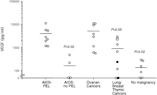
Samples from 8 AIDS-PEL effusions (2 pleural, 1 pericardial and 5 peritoneal) were obtained from the AIDS Malignancy Bank (National Cancer Institute, Bethesda, MD) and from clinical samples collected in our institutes. Control pleural, peritoneal, or pericardial fluids were obtained from 4 AIDS patients with inflammatory effusions without PEL, 5 HIV-negative patients with nonmalignant effusions, 8 patients with ovarian cancer, 5 patients with lung cancer, 1 patient with breast cancer, and 2 patients with thymic cancer. The concentrations of VEGF were measured by enzyme-linked immunosorbent assay (R&D Systems, Minneapolis, MN). All AIDS-PEL effusions had measurable VEGF (Figure). In AIDS PEL effusions, VEGF levels (median 3977 pg/mL; range 1133-11417 pg/ml) were higher than in AIDS effusions without PEL (median 175 pg/mL; range <15-484 pg/mL;P < .02, Student t test), other nonmalignant effusions (median 137 pg/mL; range <15-303 pg/mL;P < .02), and in lung/breast/thymic malignant effusions (median 969 pg/mL; range <15-2993 pg/mL; P < .05). VEGF levels in malignant ovarian effusions (median 5098 pg/mL; range 574-11091 pg/mL) were comparable to those in AIDS PEL effusions (P = .595).
VEGF levels in effusions from patients with malignant and nonmalignant disease.
AIDS-PEL (n = 8), nonmalignant AIDS, no PEL (n = 4), ovarian cancer (n = 8), lung cancer (n = 5; open circles), breast cancer (n = 1; open square), thymic cancer (n = 2; closed circles) and nonmalignant samples (n = 5). Bars indicate the average of each group. Statistical significance versus AIDS-PEL was determined by Student t test analysis.
It was previously reported that levels of VEGF in malignant ascites are generally higher than in nonmalignant effusions, with wide variations noted depending on the nature of the malignancy.4,5 Tumors of sarcoma and carcinoma origin have been reported to produce substantially more VEGF than hematological tumors.4Malignant ascites from ovarian cancers were found to contain some of the highest levels of VEGF, on the average 45-fold higher than in nonmalignant controls.5 Here we show that levels of VEGF in PEL effusions are comparable to those in malignant ovarian effusions.
Compared to normal or other malignant lymphocytes, PEL cells have markedly reduced expression of several adhesion molecules.1This defect may prevent PEL cell anchorage and subsequent development of solid tumors. By promoting vascular leakage, VEGF may contribute to PEL liquid growth in body cavities. In mice, VEGF neutralization prevented the establishment of PEL ascites. Patients with PEL, usually with advanced AIDS, have displayed a median survival of only 3 months even with administration of chemotherapy.3 Neutralization of VEGF in PEL effusions may represent a rational and novel therapeutic approach not likely to further compromise patient immune status.


