Peripheral blood mononuclear cells (PBMCs) from patients with human T-cell lymphotropic virus type I (HTLV-I)–associated myelopathy/tropical spastic paraparesis (HAM/TSP) proliferate spontaneously in vitro. This spontaneous lymphoproliferation (SP) is one of the immunologic hallmarks of HAM/TSP and is considered to be an important factor related to the pathogenesis of HAM/TSP. However, the cell populations involved in this phenomenon have not yet been definitively identified. To address this issue, the study directly evaluated proliferating cell subsets in SP with a flow cytometric method using bromodeoxyuridine and Ki-67. Although both CD4+ and CD8+ T cells proliferated spontaneously, the percentage of proliferating CD8+ T cells was 2 to 5 times higher than that of CD4+ T cells. In addition, more than 40% of HTLV-I Tax11-19–specific CD8+T cells as detected by an HLA-A*0201/Tax11-19 tetramer proliferated in culture. In spite of this expansion of HTLV-I–specific CD8+ T cells, HTLV-I proviral load did not decrease. This finding will help elucidate the dynamics of in vivo virus-host immunologic interactions that permit the coexistence of high HTLV-I–specific CD8+ cytotoxic T-lymphocyte responses and high HTLV-I proviral load in HAM/TSP.
Introduction
Human T-cell lymphotropic virus type I (HTLV-I) is an exogenous human retrovirus that has been demonstrated to be the etiologic agent in adult T-cell leukemia and a progressive neurologic disease called HTLV-I–associated myelopathy/tropical spastic paraparesis (HAM/TSP).1 2 HAM/TSP is caused by preferential damage of the thoracic spinal cord and is characterized clinically by muscle weakness, hyperreflexia, spasticity in the lower extremities, and urinary disturbance. Although development of this disease is not completely understood, virus-host immunologic interactions have been suggested to play a role in the pathogenesis of this disorder.
Although several immunologic parameters have been shown to be elevated in patients with HAM/TSP, including high frequencies of circulating HTLV-I–specific CD8+ cytotoxic T lymphocytes (CTL), high antibody titers against HTLV-I antigens are found in both peripheral blood and cerebrospinal fluid, as well as an increased expression of cytokine and chemokines.3-7 The immunologic hallmark of HTLV-I–infected individuals is the capacity of peripheral blood mononuclear cells (PBMCs) to spontaneously proliferate in vitro,8-11 that is, extraordinarily high uptake of3H-thymidine (typically >100 000 cpm after 4 days in culture) in the absence of exogenous antigens or stimulants. Moreover, the magnitude of this spontaneous lymphoproliferation (SP) is more pronounced in HAM/TSP patients than in asymptomatic HTLV-I carriers8-11 and has been used as an evaluation of treatment for this disorder.12 13 However, it is still unknown if this spontaneous proliferation is solely a consequence of activation of CD4+ HTLV-I–infected T-cells or may in part be due to expansion of CD8+ virus-specific lymphocytes.
HTLV-I has a preferential tropism for CD4+ T cells in vivo,14 and a high amount of HTLV-I proviral load has been reported in patients with HAM/TSP.15 The taxregion of HTLV-I encodes the Tax protein, a strong transactivator of both viral and host genes, including interleukin 2 (IL-2) and IL-2 receptor (IL2r).16,17 Although HTLV-I Tax protein could not be detected in fresh PBMCs ex vivo, Tax protein was found after several hours of culture in vitro.18 These observations suggested that the high SP particularly demonstrated from PBMCs of patients with HAM/TSP may be due to HTLV-I–infected CD4+ T cells proliferating by an IL2–IL2r autocrine loop in vitro.19 Additionally, SP of specific cell subsets has been examined directly from PBMCs of patients with HAM/TSP. With the use of purified cell separation techniques, SP was observed in T cells but not in B cells or monocytes.11 Moreover, CD4+ cells were found to proliferate spontaneously in vitro and to express HTLV-I antigens, whereas purified CD8+ cells did not.20 However, purified CD8+ cells were still capable of proliferating when cultured with irradiated autologous HTLV-I–infected CD4+ cells in the presence of exogenous IL-2.20 In addition, expansion of CD8+ cells in SP in HAM/TSP has also been reported on the basis of a proportional change of cell surface markers.21 These studies suggest that both CD4+ and CD8+ cells proliferate in SP. However, the methods used did not allow identification or quantification of specific phenotypic cell subsets in SP.
In this study, we describe a flow cytometric method for directly identifying proliferating cell subsets in SP. Bromodeoxyuridine (BrdU) is a thymidine analog that is incorporated into DNA during the S phase of the cell cycle.22 Ki-67 is a nuclear cell proliferation–associated antigen expressed in proliferating cells during the late G1 to M phase of the cell cycle.23 BrdU incorporation and Ki-67 expression can be detected and quantified by using fluorescence-conjugated monoclonal antibodies (mAbs). We also examined the proliferation of HTLV-I-Tax–specific CD8+ T cells during SP by using a combination of BrdU or Ki-67 and an HLA-A*0201/Tax11-19 tetramer. Using these methods, we demonstrated predominant expansion of CD8+ T cells, including HTLV-I-Tax–specific CD8+ T cells, during SP. This report is the first direct evidence of the proliferation of specific cell subtypes in SP in HAM/TSP. In addition, we investigated the amount of HTLV-I provirus and HTLV-I Tax protein expression during SP. The predominant involvement of CD8+ cells in SP may underlie the pathogenesis and progression of HAM/TSP.
Patients, materials, and methods
Subjects
PBMCs were obtained from patients with HAM/TSP and from HTLV-I seronegative healthy donors. All samples were taken with informed consent. The characteristics of patients are summarized in Table 1. PBMCs were isolated from peripheral blood on Ficoll density gradients. All cells were resuspended in CRPMI medium (RPMI 1640 supplemented with L-glutamine, penicillin/streptomycin, and HEPES buffer) containing 20% fetal calf serum (FCS) and 20% dimethyl sulfoxide, and frozen in liquid nitrogen until use.
Proliferation assays
For cell proliferation assays, cells were suspended in CRPMI supplemented with 5% human AB serum and cultured in 96-well U-bottom plates at 3 × 10⁵ per well. Cells were either unstimulated or stimulated with phytohemagglutinin (PHA; Sigma, St Louis, MO). Proliferating cells were identified in several different ways. For 3H-thymidine assays, 1 μCi3H-thymidine was added to each well for the last 4 hours of incubation. Incorporated 3H-thymidine was measured by using a β-plate counter (Wallal, Gaithersburg, MD).
Because 3H-thymidine assays do not allow distinction of phenotypes of proliferating cells, proliferation was also measured by using a BrdU Flow Kit (Pharmingen, San Diego, CA). This procedure uses a fluorescence-conjugated anti-BrdU mAb to label BrdU that has been incorporated into newly synthesized DNA within dividing cells. Concomitant staining with mAbs against surface markers permits identification of phenotypic subsets of proliferating cell populations by using flow cytometry. BrdU (10 μM) was added to the cell culture for 16 hours before staining and fixation. Cells were washed with phosphate-buffered saline containing 1% FCS and 0.1% NaN3and incubated with anti–human CD4-phycoerythrin (PE; Caltag Laboratories, Burlingame, CA) and antihuman CD8-RPE/Cy-5 (Dako Laboratories, Denmark) or antihuman CD8-PE (Caltag Laboratories) and antihuman CD4-Tri-Color (Caltag Laboratories) for 20 minutes at 4°C. Cells were then fixed with a paraformaldehyde solution for 30 minutes at 4°C, permeabilized with saponin for 10 minutes at 4°C, and re-fixed with paraformaldehyde for 5 minutes at 4°C. Following fixation, cells were treated with 300 μg/mL DNase for 1 hour at 37°C, then incubated with anti-BrdU–fluorescein isothiocyanate (FITC) or a matched isotype control mAb for 20 minutes at room temperature, as per kit instructions. Flow cytometric analyses were performed by using a FACS Calibur (Becton Dickinson, Mountain View, CA).
Proliferation of T-cell subsets identified with the BrdU assay was further confirmed by measuring the expression of a nuclear-proliferating cell antigen, Ki-67. Cultured cells were washed and labeled with antihuman CD4-PE (Caltag Laboratories) and antihuman CD8-RPE/Cy-5 (Dako) for 30 minutes at 4°C. They were then fixed and permeabilized with a paraformaldehyde and saponin solution from a Cytoperm/Cytofix kit (Pharmingen) for 20 minutes at 4°C. Fixed cells were labeled with either antihuman Ki-67-FITC mAb or a matched isotype control antibody for 30 minutes at 4°C and analyzed by flow cytometry.
Identification of Tax-specific CD8+ cells
The HTLV-I Tax 11-19 peptide is a dominant epitope that is recognized by HLA-A2–restricted CD8+ CTLs in patients with HAM/TSP.7 24 Analysis of Tax-11-19–specific CD8+ cells was performed by using a PE-conjugated HLA-A*0201 tetramer (provided by NIAID MHC Tetramer Core Facility, Atlanta, GA; and NIH AIDS Research and Reference Reagent Program). The tetramer was loaded with HTLV-I Tax 11-19 peptide (LLFGYPVYV), which was synthesized and 95% purified by high-performance liquid chromatography (New England Peptide, Fitchburg, MA). Tetramer loaded with HIV Gag77-85 peptide (SLYNTVATL) was used as a negative control. Cultured cells were washed and incubated with HLA-A2 tetramer, antihuman CD4-FITC mAb (Caltag Laboratories), and antihuman CD8-RPE/Cy5 mAb (Dako) for 30 minutes at 4°C, then analyzed by flow cytometry. Tetramer-labeled cells were also fixed and permeabilized as described earlier and stained for BrdU incorporation or Ki-67 expression.
Identification of HTLV-I Tax–expressing cells
PBMCs (5 × 10⁵) were placed on a culture well (round bottom 96-well plate) in 200 μL RPMI-1640 supplemented with L-glutamine, penicillin, streptomycin, and 5% human AB serum. Harvested cells were washed with phosphate-buffered saline containing 1% FCS and 0.1% NaN3 and incubated with antihuman CD4-PE mAb (Caltag Laboratories) and antihuman CD8-RPE/Cy5 mAb (Dako) for 20 minutes at 4°C. Cells were then fixed and permeabilized with 4% formaldehyde and 0.1% saponin (CytoFix/Cytoperm Kits; Pharmingen) for 20 minutes at 4°C. After washing with 0.1% saponin buffer (Perm/Wash solution; Pharmingen), the cells were incubated with anti–HTLV-I Tax mAb (the reagent was obtained through the AIDS Research and Reference Reagent Program, Division of AIDS, NIAID, NIH; HTLV-1 tax hybridoma [168A51-42] from Dr Beatrice Langton) for 30 minutes at 4°C. Followed by washing, FITC-conjugated goat F(ab′)2antimouse immunoglobulin G2a mAb (Southern Biotechnology Associates, Birmingham, AL) was used as a second antibody for labeling anti–HTLV-I Tax mAb. Flow cytometric analyses were performed by using a FACS Calibur (Becton Dickinson).
Quantitative polymerase chain reaction
HTLV-I proviral load was measured by using an ABI PRISM 7700 Sequence Detector (Applied Biosystems, Foster City, CA) as previously described.15 DNA was extracted from 1 × 10⁶cells by using Puregene DNA Isolation Kit (Gentra, Minneapolis, MN) and was adjusted to 10 ng/μL. Polymerase chain reaction (PCR) conditions were as follows: 10 μL DNA solution was added to 40 μL reaction mixture containing 10 mM Tris-HCl; 50 mM KCl; 10 mM EDTA; 60 nM ROX (passive reference dye); 5.5 mM MgCl2; 0.2 μM of each primer; 0.1 μM TaqMan probe; 200 μM each of dATP, dGTP, and dCTP; 400 μM dUTP; 0.5 U uracil-N-glycosylase; and 1.25 U Taq polymerase (AmpliTaq Gold; Applied Biosystems).
The primer set for HTLV-I pX region was 5′-ACAAAGTTAACCATGCTTATTATCAGC-3′ positioned at 7276-7302 and 5′-ACACGTAGACTGGGTATCCGAA-3′ positioned at 7355-7334. The primer set for β-actin was 5′-CACACTGTGCCCATCTACGA-3′ positioned at 2146-2165 and 5′-CTCAGTGAGGATCTTCATGAGGTAGT-3′ positioned at 2250-2225. The TaqMan fluorescent probe for HTLV-I pX region was 5′-TTCCCAGGGTTTGGACAGAGTCTTCT-3′ positioned at 7307-7332 and for β-actin was 5′-ATGCCCTCCCCCATGCCATCCTGCGT-3′ positioned at 2171-2196. The thermocycler conditions were as follows: 50°C for 2 minutes, 95°C for 10 minutes, then 45 cycles of 95°C for 15 seconds, and 60°C for 1 minute.
The amount of HTLV-I proviral DNA was calculated by the following formula: copy number of HTLV-I (pX) per 100 cells = [(copy number of pX) / (copy number of β-actin / 2)] × 100.
Results and discussion
Cultured PBMCs from patients with HAM/TSP contain 2 cell populations distinguished by size (large cells and small cells).25 These populations can be identified with a flow cytometer by plotting forward scatter and side scatter of the cells. The phenotypic composition of each cell population was analyzed by using mAbs against CD3 (T lymphocytes), CD16 (natural killer cells), CD14 (monocytes), and CD19 (B cells). Before culture, the smaller cell population consisted of lymphocytes and the larger cell population consisted of monocytes. After SP of HAM/TSP PBMCs, the large cell population contained lymphocytes, shown by an increased percentage of CD3+ cells (data not shown).25 In contrast, the percentage of CD3+ cells in the large cell population did not change in an unstimulated culture of healthy PBMCs. This finding supports a morphologic change on blasting and suggests that the lymphocytes in the large cell population may be preferentially activated.25
PBMCs from patients with HAM/TSP were cultured for 6 days and assayed for proliferation and lymphocyte phenotypic composition. Proliferation observed using the 3H-thymidine assay was consistent with earlier descriptions of SP in HAM/TSP.8-11During SP of HAM/TSP PBMCs, 3H-thymidine counts increased over time in culture, reaching maximal levels of 30 000 to 130 000 cpm on day 4 or day 5 (data not shown). Unstimulated PBMCs from healthy donors incorporated less then 1000 cpm at the same time points, whereas PHA-stimulated PBMCs from the same donors typically peaked around 80 000 to 100 000 on day 3 or day 4 (data not shown).
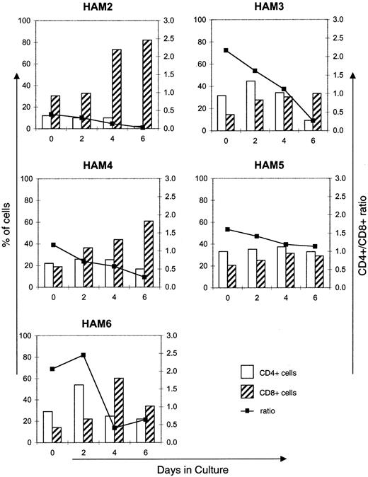
On days 0, 2, 4, and 6 of the proliferation, PBMCs were harvested and stained with CD4-FITC and CD8-RPE/Cy5 and analyzed by flow cytometry. CD4+ and CD8+ subsets were quantified and used to calculate a ratio of CD4+ cells to CD8+ cells. Data from 5 patients with HAM/TSP are summarized in Figure 1. As shown, the ratio of CD4+ cells to CD8+ cells decreased for each patient over 6 days. Surprisingly, this change in ratio appeared to be driven by the increasing CD8+ population more than by a decrease in CD4+ percentages. Occasional drops in CD8+ and/or CD4+ cells by day 6 are likely a consequence of maintaining cell cultures for prolonged periods without supplementing the media and are consistent with decreases in proliferation observed in 3H-thymidine incorporation assays after day 4, as described earlier.
Alteration of CD4+ and CD8+populations in HAM/TSP in vitro.
CD4+ and CD8+ subsets from 5 patients with HAM/TSP are graphed as a percentage of gated cells (see text for details). Ratio of CD4+ cells to CD8+ cells decreased in all 5 patients over 6 days in culture.
Alteration of CD4+ and CD8+populations in HAM/TSP in vitro.
CD4+ and CD8+ subsets from 5 patients with HAM/TSP are graphed as a percentage of gated cells (see text for details). Ratio of CD4+ cells to CD8+ cells decreased in all 5 patients over 6 days in culture.
The decreasing CD4+/CD8+ ratio is simple numerical evidence, suggesting that CD8+ cells are involved in in vitro proliferation. However, because the3H-thymidine method of measuring proliferation is not capable of identifying phenotypic subsets of cells, we used 2 nonradioisotopic reagents, BrdU and Ki-67, coupled with flow cytometry. Proliferation of each T-cell subset was determined by the percentage of CD4+ or CD8+ cells also labeled with anti-BrdU or anti–Ki-67. Representative data from the BrdU incorporation assay are shown in Figure 2. The histogram cells are based on their expression of CD4 or CD8 and BrdU. Both CD4+ and CD8+ cells gated by the small cell population did not show proliferation. However, proliferation is clearly seen in both CD4+ and CD8+ populations gated by the large cell population on days 3 and 4. The percentage of proliferating CD4+ (or CD8+) cells in total CD4+ (or CD8+) cells is graphed in Figure3A (see legend about calculation). The CD8+ subset proliferated to a greater degree than the CD4+ subset. Some CD4− and CD8−cells in the large cell population were also BrdU positive. However, most of these double-negative cells were in the small cell population and were BrdU negative. An unstimulated culture of PBMCs from a healthy donor showed no proliferation in either CD4+ or CD8+ cells. When PBMCs from a healthy donor were stimulated with PHA, equal percentages of both CD4+ and CD8+ populations were labeled with BrdU (data not shown). These results corroborate the proliferation seen with the3H-thymidine assay and suggest that the BrdU assay is an accurate representation of cellular proliferation. Combined proliferation data from 4 patients with HAM/TSP are summarized in Figure 3B. All patients with HAM/TSP showed predominant proliferation of CD8+ cells in SP in which the percentage of BrdU-labeled CD8+ cells by day 4 was 2 to 5 times greater than BrdU-labeled CD4+ cells. Staining with anti–Ki-67 correlated with BrdU labeling in both stimulated and unstimulated healthy donor cells and in cells from patients with HAM/TSP (data not shown). Thus, the results with both BrdU and Ki-67 staining provide the first direct evidence for predominant proliferation in vitro of CD8+ cells from HAM/TSP PBMCs.
Spontaneous proliferation of CD4+ cells and CD8+ cells.
In vitro proliferation of CD4+ and CD8+ cells, measured by incorporation of BrdU, is shown for a representative patient with HAM/TSP (HAM-4). The data are representative of 4 patients with HAM/TSP tested in the same way. Histograms demonstrate that both CD4+ and CD8+ cells that were gated by small cell population (R1) or large cell population (R2) incorporated BrdU. Few cells in R1 were labeled with BrdU during culture. Both CD4+ and CD8+ cells in R2 showed BrdU as being positive during culture. And a larger percentage of CD8+cells proliferated than did CD4+ cells. The number in each histogram represents the percentage of BrdU-positive cells.
Spontaneous proliferation in HAM/TSP.
(A) This graph presents the percentage of proliferating cells of HAM-4 (whose histogram is shown in Figure 2) that was calculated as the following: % BrdU+CD8+ cells = [number of BrdU+CD8+ cells in R1 + number of BrdU+CD8+ cells in R2] / [number of total CD8+ cells in R1 + R2]. % BrdU+CD4+ cells = [number of BrdU+CD4+ cells in R1 + number of BrdU+CD4+ cells in R2] / [number of total CD4+ cells in R1 + R2]. (B) In vitro proliferation of CD4+ and CD8+ cells was quantified for 4 patients with HAM/TSP as shown in Figure 3A. This graph shows the mean percentage of proliferating cells in each subset for 4 patients. Error bars indicate SEM. As seen in individual patients, the combined data indicate a higher degree of proliferation in CD8+ cells than in CD4+ cells.
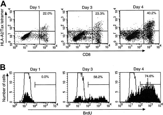
CD8+ cells specific for HTLV-I antigens are prominent in the peripheral blood of patients with HAM/TSP and may be important in progression of the disease.5,26 Proliferation of HTLV-I antigen-specific CD8+ cells might, therefore, be expected to contribute to CD8+ cell expansion. To address this issue, CD8+ cells specific for the HTLV-I Tax 11-19 peptide were identified in 3 HLA-A2 HAM/TSP patients, using an HLA-A*0201/Tax 11-19 peptide tetramer. Tetramer-positive cells were measured in both the small cell and large cell populations. Results from one representative patient with HAM/TSP (HAM-4) are shown in Figure4A. On day 0 and day 1, most Tax-specific CD8+ cells were found in the gate small cell population, and some Tax-specific CD8+ cells were BrdU negative in the gated large cell population. Over 4 days in culture, the Tax-specific CD8+ cells increased in the large cell population from 22.0% to 40.2% of total CD8+ cells in the large cell population. Interestingly, Tax-specific CD8+ cells in the small cell population decreased from day 1 to day 4 in the same patient (data not shown), suggesting that these cells became activated in vitro and underwent morphologic changes that shifted them into the population of larger cells. To identify proliferation of these Tax-specific CD8+ cells, PBMCs were concomitantly labeled with Tax tetramer and either BrdU or Ki-67. After gating on the tetramer-positive CD8+ population, histograms were generated to demonstrate the increase in the percentage of cells positive for BrdU (Figure 4B showed representative data of HAM-4). After 4 days in culture, 40.2% (HAM-2), 70.0% (HAM-3), and 74.6% (HAM-4) of Tax-specific CD8+ cells were labeled with BrdU in 3 patients with HAM/TSP, respectively. Similar Tax-specific CD8+ cell proliferation was measured by Ki-67 expression (data not shown). The expansion of Tax-specific CD8+ cells in SP supports the view that HTLV-I–specific CD8+ T lymphocytes may play a critical role in the inflammatory process of HAM/TSP. This view is further supported by evidence of the elevated production of proinflammatory cytokines, such as interferon γ and tumor necrosis factor α, by HTLV-I–specific CD8+ T lymphocytes in HAM/TSP.3
Proliferation of Tax-specific CD8+ cells in HAM/TSP in vitro.
(A) Tax-specific CD8+ cells of HAM-4 were identified and quantified, using an HLA-A*0201/Tax tetramer. Dot plots gated on the large cell population (R2) are presented. Percentage of HTLV-I Tax-specific CD8+ cells was increased in culture, as indicated in the upper-right quadrants. The number indicates the percentage of Tax-specific CD8+ cells in R2 per total CD8+ cells in R2. (B) Gating on Tax-specific CD8+ cells allowed measurement of proliferation. Numbers indicate the percentage of Tax-specific CD8+ cells that incorporated BrdU. These cells showed a very high rate of proliferation in vitro.
The observed expansion of Tax-specific CD8+ cells in SP might also be expected to reduce the amount of HTLV-I present. In support of this hypothesis, an increased frequency of CD8+cells has been associated with reduced Tax expression in vivo. Similarly, the frequency of Tax-specific CTL was negatively correlated with the percentage of CD4+ cells in fresh peripheral blood leukocytes from patients with HAM/TSP.18 To investigate a possible relationship between HTLV-I load and Tax-specific CD8+ cell proliferation more directly, proviral load was determined in patients with HAM/TSP during SP by using quantitative real-time PCR. Results are summarized in Figure5. HTLV-I proviral load did not significantly change during culture. Although the CD4/CD8 ratio at day 4 in Figure 1 decreased compared with that at day 0, HTLV-I proviral load did not change (Figure 5). If HTLV-I were to infect only CD4+ cells, then the HTLV-I proviral load might have decreased at day 4 as the percentage of CD4+ also appeared to be decreasing over time (Figure 1). However, because the HTLV-I proviral load was relatively stable during this 4-day culture period when the percentage of CD4+ cells was decreased, it suggests that other cells (possibly CD8+) may be HTLV-I infected. This finding is consistent with the recent report that CD8+ T cells may also be infected with HTLV-I.27 Therefore, expansion of HTLV-I–infected CD8+ T cells may contribute to the proviral load in SP.
HTLV-I proviral load in HAM/TSP.
HTLV-I proviral load was measured at each time point in culture as the number of copies of provirus per 100 cells. No significant change was observed in proviral load in culture.
HTLV-I proviral load in HAM/TSP.
HTLV-I proviral load was measured at each time point in culture as the number of copies of provirus per 100 cells. No significant change was observed in proviral load in culture.
It was unexpected that HTLV-I proviral load was stable in spite of Tax-specific CD8+ cell expansion. Therefore, we investigated HTLV-I Tax protein expression by using an intracellular protein detection technique. We chose to study the HTLV-I Tax protein because it is a major target of the HTLV-I–specific CTLs that pivotally control viral infection. As shown in Figure6, HTLV-I Tax protein-expressing cells could not be detected in uncultured PBMCs, but were detectable during culture, and reached a maximum at 12 hours. After 72 hours, HTLV-I Tax-expressing cells were no longer detected. This result is consistent with published data.18 It has been also demonstrated that CD4+ cells naturally infected with HTLV-I were killed in vitro by CD8+ lymphocytes by a perforin-dependent cytotoxic pathway, and CD8+ CTLs controlled HTLV-I antigen-expressing cells.18 However, the stable proviral load in culture while Tax-specific CD8+ cells were still expanding suggests the involvement of an additional mechanism to escape from CD8+ CTLs. One possible mechanism involves the expansion of HTLV-I–infected (CD4+ and CD8+) cells by mitotic proliferation (infected cells do not express HTLV-I antigens) rather than infectious transmission (infected cells express HTLV-I antigens and produce HTLV-I virions). This first type of propagation might result in an increase in integrated HTLV-I without increased HTLV-I antigen expression. Without expressing HTLV-I Tax, these infected cells could not be targeted by Tax-specific CD8+CTLs. As Tax-expressing cells were killed by Tax-specific CD8+ CTLs, mitotic proliferation (silent provirus) would selectively predominate. Following the reduction of Tax expression, Tax-specific CD8+ cell proliferation might be supported by cytokines, such as IL-15, which is an important factor for maintaining CD8+ cells.28 Other mechanisms associated with SP have been previously reported that include interaction between HTLV-I–infected T cells and noninfected T cells mediated by CD2/LFA-3 and LFA-1/ICAM.29 Further study of this phenomenon may help us to understand the mechanism that permits coexistence of high HTLV-I–specific CD8+ proliferative and CTL responses and high HTLV-I proviral load in HAM/TSP in vivo.30,31 In a recent HIV study, it has been reported that even after effective combination therapy that reduced plasma HIV RNA to undetectable levels, HIV can persist in a latent form in resting CD4+ T cells.32
Alteration of HTLV-I Tax expression and proviral load.
PBMCs from 4 patients with HAM/TSP were cultured for 0 hour, 12 hours, 24 hours, 48 hours, 72 hours, and 96 hours. HTLV-I Tax protein expression of the cultured cells and proviral load were measured at each time point. The closed triangle (dotted line) indicates the percentage of Tax-expressing cells. The open circle (solid line) indicates the copy number of HTLV-I provirus (DNA) per 100 cells. (A) HAM-3. (B) HAM-4.
Spontaneous proliferation of CD4+ and CD8+ cells may be an important phenomenon not only in vitro but also in situ in HAM/TSP. Ijichi et al33 have proposed that an autoaggressive process against bystander tissue plays a crucial role in the pathogenesis of this disease. In this hypothesis, lymphocytes that include both HTLV-I–infected CD4+ cells and HTLV-I antigen-specific CD8+ cells enter spinal cord lesions and proliferate in situ in a manner similar to in vitro SP. The in situ immune response propagated against viral antigens may act as an autoaggressive effector for the development of central nervous system tissue damages in HAM/TSP. In support of this hypothesis, an immunohistochemical phenotypic analysis of infiltrated mononuclear cells in a spinal cord lesion in a patient with HAM/TSP has showed the presence of both CD8+ cells and CD4+cells.34 35
This report provides direct evidence that CD8+ cells in vitro predominantly expand in SP of PBMCs from HAM/TSP. Although there is no direct in vivo correlate for spontaneous lymphoproliferating PBMCs, the observations in this study may be used as a surrogate marker for virus-host immunologic interactions that may occur in vivo. Thus, these findings support the ex vivo observations demonstrating expansion of CD8+ cells, including HTLV-I Tax-specific CD8+ lymphocytes that have been suggested to play a critical role in the pathogenesis of HAM/TSP. The elucidation of the role of these and other viral antigen-specific CD8+cells in HAM/TSP may reflect similar mechanisms in other diseases with viral etiologies.
J.A.S. and M.N. contributed equally to this study.
The publication costs of this article were defrayed in part by page charge payment. Therefore, and solely to indicate this fact, this article is hereby marked “advertisement” in accordance with 18 U.S.C. section 1734.
References
Author notes
Steven Jacobson, Viral Immunology Section, Neuroimmunology Branch, National Institute of Neurological Disorders and Stroke, National Institutes of Health, Building 10, Room 5B-16, 9000 Rockville Pike, Bethesda, MD 20892; e-mail:jacobsons@ninds.nih.gov.


![Fig. 3. Spontaneous proliferation in HAM/TSP. / (A) This graph presents the percentage of proliferating cells of HAM-4 (whose histogram is shown in Figure 2) that was calculated as the following: % BrdU+CD8+ cells = [number of BrdU+CD8+ cells in R1 + number of BrdU+CD8+ cells in R2] / [number of total CD8+ cells in R1 + R2]. % BrdU+CD4+ cells = [number of BrdU+CD4+ cells in R1 + number of BrdU+CD4+ cells in R2] / [number of total CD4+ cells in R1 + R2]. (B) In vitro proliferation of CD4+ and CD8+ cells was quantified for 4 patients with HAM/TSP as shown in Figure 3A. This graph shows the mean percentage of proliferating cells in each subset for 4 patients. Error bars indicate SEM. As seen in individual patients, the combined data indicate a higher degree of proliferation in CD8+ cells than in CD4+ cells.](https://ash.silverchair-cdn.com/ash/content_public/journal/blood/98/5/10.1182_blood.v98.5.1506/5/m_h81711469003.jpeg?Expires=1766597624&Signature=q9m0unH3~hgL7QxWcFbCZ~~2miXRQa4hqaw0j4D6SfrQg8B9IgVGqA~uCSIgpq1JJcssBs2HNsfke9575yxMFSvR5Ev81SPyPc6~Cd~eImqdTRYnrtNNuM6-FwRlmIAfIAMhHa7LGkHJ5xdKRUNOALxrT3xwPMKkEblLhpz9mG34sVdLpTtLkVLx59j2lSixU3P9bPB5MebgkMWeLNGNRXCz~tmnFb9uYhZc8cxt3FOHvHuV5X8UEVFkUHsFlT5gc3dtvd0hhzFfpm84dMQ83n1xElltlQMtu2juphhsRRjmWhW0t6bJFaDCc7tq8ci8arSbC32FvWoqAXowYxxg0w__&Key-Pair-Id=APKAIE5G5CRDK6RD3PGA)


