Epstein-Barr virus (EBV) is a tumorigenic human herpesvirus that persists for life in healthy immunocompetent carriers. The viral strategies that prevent its clearance and allow reactivation in the face of persistent immunity are not well understood. Here we demonstrate that EBV infection of monocytes inhibits their development into dendritic cells (DCs), leading to an abnormal cellular response to granulocyte macrophage–colony-stimulating factor (GM-CSF) and interleukin-4 (IL-4) and to apoptotic death. This proapoptotic activity was not affected by UV inactivation and was neutralized by EBV antibody-positive human sera, indicating that binding of the virus to monocytes is sufficient to alter their response to the cytokines. Experiments with the relevant blocking antibodies or with mutated EBV strains lacking either the EBV envelope glycoprotein gp42 or gp85 demonstrated that interaction of the trimolecular gp25–gp42–gp85 complex with the monocyte membrane is required for the effect. Our data provide the first evidence that EBV can prevent the development of DCs through a mechanism that appears to bypass the requirement for viral gene expression, and they suggest a new strategy for interference with the function of DCs during the initiation and maintenance of virus-specific immune responses.
Introduction
Viruses that establish persistent infection develop multiple strategies to evade host immune responses. Striking examples are provided by human herpesviruses that block antigen presentation and produce homologues of cytokines and chemokines that can subvert inflammatory responses (reviewed in Ploegh1). EBV is an oncogenic herpesvirus that infects most adults and persists for life in immunocompetent hosts. It is associated with several malignancies of hematopoietic and nonhematopoietic origin. Several lines of evidence indicate that major histocompatibility complex (MHC) class I-restricted CD8+ cytotoxic T lymphocytes (CTLs) are critical for the control of EBV infection. The symptomatic primary infection, infectious mononucleosis, and the asymptomatic carrier-state are characterized by vigorous CTL responses to viral proteins expressed in latently and productively infected cells (reviewed in Rickinson and Moss2). Lack of CTL responses correlates with uncontrolled proliferation of EBV-carrying B-cell lymphomas in immunocompromised patients, and these immunodeficiency-associated malignancies can be prevented or even cured by adoptive transfer of EBV-specific CTLs.3 Viral escape strategies that allow persistence and reactivation in the face of this effective immunity are largely unknown.
DCs play a pivotal role in the initiation and maintenance of immune responses because of their ability to capture exogenous antigens and present them to T lymphocytes in the form of MHC class I-associated peptides, a phenomenon known as cross-priming (reviewed in Banchereau et al4). It is well established that interference with the function of DCs regulates the life cycle and pathogenesis of many viruses,5-11 but its contribution to EBV immunity is as yet unexplored. Previous studies have focused on the ability of EBV-transformed lymphoblastoid cell lines (LCLs) to reactivate EBV-specific cytotoxic T-lymphocyte responses in vitro from autologous peripheral blood.12 LCLs coexpress viral antigens and high levels of MHC class I and class II adhesion and costimulatory molecules, and it is generally assumed that this immunogenic phenotype is responsible for the extensive expansion of reactive T cells that characterize infectious mononucleosis. However, LCLs cannot activate EBV-specific CD8+ T cells from naive donors. This is consistent with a number of human and animal studies that demonstrated a poor capacity of B cells to activate primary CTL responses.13-16 In contrast to B cells, DCs are efficient in triggering primary T-cell responses in vitro and in vivo. It has been recently shown that monocyte-derived DCs can efficiently take up EBV antigens from apoptotic or necrotic LCLs.17-19 These antigens were processed and presented in association with MHC class I molecules and were recognized by specific CTLs in vitro. Notably, the EBV nuclear antigen (EBNA) 1, which is not presented to MHC class I-restricted CTLs because of a cis-actingblockade of ubiquitin–proteasome-dependent proteolysis,20,21 is still capable of inducing strong CTL responses in HLA-A2 and -B35–positive individuals and requires cross-priming for its presentation to CTLs.22 Thus, specialized antigen-presenting cells are likely to be involved in the induction of at least some EBV-specific responses in vivo.
Triggering of Th1-type immune responses and development of strong cytotoxic activity usually require DCs derived from myeloid precursors, such as blood monocytes.23 EBV can bind to monocytes, modulate cytokine production, and inhibit phagocytic activity of these cells.24-28 Here we show that EBV infection prevents the development of DCs promoting the apoptosis of monocytes cultured in the presence of granulocyte macrophage–colony-stimulating factor (GM-CSF) and interleukin-4 (IL-4). Binding of the EBV envelope gp42–gp85–gp25 fusion complex to the monocyte surface appears to play a critical role in the proapoptotic activity of the virus.
Materials and methods
Preparation of blood dendritic cells
Blood DCs were generated from peripheral blood mononuclear cells (PBMCs) as decribed.29 Mononuclear cells, isolated from the blood of healthy donors by density gradient centrifugation, were resuspended at a density of 1.5 to 2 × 10⁶ cells/mL in culture medium (RPMI 1640 medium supplemented with 10% heat-inactivated fetal calf serum, 2 mM L-glutamine, 100 U/mL penicillin, 100 μg/mL streptomycin) and were allowed to adhere to the surface of a plastic flask for 2 hours at 37°C in a 5% CO2 incubator. Confluent monolayers were washed 3 times in phosphate-buffered saline (PBS) to remove the nonadherent cells and then were cultured for indicated times in medium supplemented with 1000 U/mL recombinant GM-CSF and 1000 U/mL recombinant IL-4 (a kind gift of Schering-Plough Research Institute, Kenilworth, NJ). Cytokines were replenished by exchanging one third of the culture supernatant every third day, at which time cell viability was also estimated by trypan blue dye exclusion. Changes in cell morphology were monitored daily by visual inspection using an inverted microscope. Where indicated, CD14+ monocytes were purified using the monocyte-negative isolation kit (Dynal AS, Oslo, Norway) according to the manufacturer's instructions. PBMCs (10 × 106) were reacted with a mixture of antibodies specific for various T-, B-, and NK-cell markers, and the positive cells were depleted by incubation with antimouse antibody-conjugated Dynabeads, followed by capture on an MPC-1 magnetic particle concentrator. The negatively selected population contained more than 98% CD14+ cells as detected by staining and fluorescence-activated cell sorter (FACS) analysis.
EBV infection
Infectious EBV was obtained from 10-day-old cell-free supernatant of the virus producer B95.8 cell line. The virus was concentrated from filtered supernatants (0.45 μM) by centrifugation at 45 000g for 90 minutes at 4°C. Pellets were resuspended in PBS. B95.8 supernatants were depleted of infectious virus either by 6 consecutive rounds of absorption on CD21+Raji cells (1 × 10⁸ cells in 20 mL B95.8 supernatant for 1 hour at 37°C). Alternatively, virus absorption was performed on tosylactivated magnetic beads (Dynabeads M-280; Dynal) conjugated with purified gp350-specific monoclonal antibody (mAb) 2L10 (2 mg/mL beads). Beads treated with blocking reagent or conjugated with CD3-specific mAb OKT3 were used as controls. Antibody conjugation and blocking of active sites were performed according to the manufacturer's procedures. Virus titers were determined by immortalization of freshly separated B lymphocytes and by expression of the EBNAs in the EBV-negative B-lymphoma line Bjab. Anticomplement-enhanced immunofluorescence staining was performed 48 hours after infection using a previously characterized EBV antibody–positive human serum. The virus was inactivated by exposure of B95.8 supernatant to UV light (254 nm) for 5 minutes (UV source model UVG-11; Upland, CA) from a distance of 10 cm, which resulted in a complete loss of transforming activity and EBNA induction in Bjab cells. Confluent monolayers of adherent PBMCs or purified CD14+ cells were infected by incubation for 2 hours at 37°C. The monolayers were then washed and cultured in medium containing IL-4 and GM-CSF to induce DC development. Where indicated, the virus preparations were preincubated for 30 minutes at room temperature with 1:20 dilution of the indicated human sera or 100 μg/mL purified antibodies before they were used for the infection of monocytes. The expression of EBV mRNA was detected by reverse transcription–polymerase chain reaction (RT-PCR) using primers specific for the EBNA1, EBNA2, LMP1, and BZLF1 messages, as previously described.30
Analysis of cell surface marker expression and [3H]-thymidine incorporation assay
Surface markers were detected using a panel of mAbs specific for HLA class I, HLA-DR, CD3, CD19, CD14, CD80, CD86, CD83, CD11C, and CD1a. Phycoerythrin and fluorescein isothiocyanate (FITC)–conjugated antibodies used for direct immunofluorescence and isotype-matched controls were described elsewhere.29 Fluorescence intensity was monitored with a FACScan flow cytometer (Becton Dickinson, San Jose, CA) using the CellQuest software.
Stimulatory capacity of DC preparations was assessed by [3H]-thymidine incorporation assays. DCs were collected, irradiated with 40 Gy, and mixed with allogeneic PBMCs in complete medium at the indicated responder-stimulator ratios. The cell suspension was adjusted to a density of 3 × 10⁵ cells/mL and was distributed in triplicate to 96-well, U-bottom microtiter plates (100μL/3 × 104 T cells/well). Cells were cultured in a CO2 incubator for 5 to 7 days and were pulsed with [3H]-thymidine (0.037 MBq/well) during the last 8 hours of incubation. Plates were then harvested to glass filters on a Tomtec harvester 96 (Orange, CT), and filters were counted on a Wallac 1450 Microbeta liquid scintillation counter (Wallac, Turku, Finland).
Mutated viral strains, EBV-specific antibodies, and purified gp350/220
Recombinant viruses were constructed in the Akata strain by homologous recombination with appropriate DNA fragments in which the gp42 or gp85 open-reading frames were disrupted by the insertion of a neomycin resistance cassette (gp42) or a cassette expressing the neomycin resistance gene and a gene for green fluorescence protein (gp85), respectively.31,32 Wild-type and recombinant viruses were produced by inducing EBV lytic cycle in Akata cells with anti–human IgG as previously described.31 Viral stocks were equalized for EBV content, which was assessed by binding to Raji cells followed by indirect immunostaining with the gp350/220-specific mAb 2L10 and FACS analysis.
A truncated EBV gp350 lacking the membrane anchor was produced in the mouse fibroblast cell line C127 using a bovine papilloma virus-based expression vector and purified as previously described.33EBV antibody-positive and -negative human sera were obtained from healthy laboratory personnel and from patients with nasopharyngeal carcinoma. Antibody titers to the EBV nuclear antigens and viral capsid antigen were determined by anticomplement-enhanced immunofluorescence staining or indirect immunofluorescence, respectively. Mouse antibodies F-2-134 (specific to the EBV envelope glycoprotein gp42),35 E1D136 (reacting with the EBV gp85–gp25 complex),35 72A137(specific to the EBV gp350/220 glycoprotein), and OKT338(specific to CD3) were purified from culture supernatants of the corresponding hybridoma cell lines.
Detection of apoptosis
Cytospins of EBV-infected and control cells were fixed with freshly prepared paraformaldehyde (4% in PBS), and the cells were permeabilized (0.1% Triton X-100 in 0.1% sodium citrate) and stained with Hoechst 33258 (Sigma-Aldrich, Stockholm, Sweden) to detect apoptotic nuclei. The samples were examined with a Leitz-BMRB fluorescence microscope (Leica, Heidelberg, Germany). Images were captured with a Hamamatsu (Osaka, Japan) 4800 cooled CCD camera and were processed with Adobe Photoshop software. Efficiency of Annexin V binding to the surfaces of EBV-infected and control cells was measured using Annexin V–FITC Apoptosis Detection Kit I (PharMingen, San Diego, CA). Monoclonal antibodies specific to Bim (StressGen Biotechnologies, Victoria, BC, Canada), Bcl-2 (Zymed Laboratories, Carlton Court, CA), Bad and Bax (R&D Systems, Oxon, United Kingdom), and polyclonal rabbit anti–Bcl-XL and anti–caspase-3 antibodies (PharMingen) were used to analyze the expression of Bcl-2 family members and caspase-3 cleavage by immunoblotting.
Results
EBV infection leads to apoptosis of developing DCs
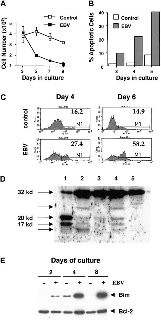
To determine whether EBV can interfere with the development of DCs, confluent monolayers of adherent mononuclear cells were infected with the prototype B95.8 virus and then cultured for 7 days in the presence of GM-CSF and IL-4. Cells expressing typical DC morphology and cell surface markers, including high levels of MHC class II and CD86, were recovered from the infected cultures, but a dramatic decrease in cell number was observed compared with uninfected controls (Table1). Major differences were revealed on examination of the cultures by light microscopy and monitoring of cell recovery over time. Floating cells appeared in the uninfected cultures within 2 to 3 days; after that, the cultures appeared as single-cell suspensions, containing occasional loose clumps of large, viable, dendritic-shaped cells. The number of cells remained fairly constant during the first 7 days and started to decrease only after prolonged culture (Figure 1A). In the infected cultures, cell detachment had started already after 1 day, and the cultures were extensive formations of dense clumps containing dark and irregularly shaped cells. A significant decrease in cell viability began from days 3 to 5 (Figure 1A). Similar differences were observed when the effect of EBV infection was tested on purified CD14+ monocytes isolated by negative selection, and these cells were therefore used in subsequent experiments. To investigate whether the induction of apoptosis may explain the lower recovery caused by EBV infection, the cultures were examined by Hoechst staining. Typical apoptotic changes, including membrane ruffling and nuclear condensation, were detected in uninfected and EBV-infected cultures. However, the percentage of apoptotic cells was significantly higher in the infected cultures already at day 3 and steadily increased in parallel with the progressive drop of cell recovery (compare panels A and B in Figure 1). In independent experiments, comparable numbers of apoptotic cells were revealed by their capacity to bind Annexin V with a relatively high efficiency. The percentage of such cells also increased progressively from day 4 to day 6 in EBV-infected cultures, whereas it remained fairly constant in mock-infected populations (Figure 1C). Caspase-3 cleavage, a point of convergence for different apoptotic pathways,40 was monitored by immunoblotting of total cell lysates. Enzymatically active 20-kd and 17-kd forms of caspase-3 were revealed in cell lysates of EBV-infected DCs collected at day 4 and day 6 of culture but were less evident in lysates of mock-infected cells (Figure 1D). Consistent with the relatively slow kinetics of apoptotic death, prolonged exposure of filters was required to visualize these cleavage products. Induction of apoptosis was accompanied by strong up-regulation of Bim, a proapoptotic member of the Bcl-2 family (Figure 1E). In accordance with the notion that Bim induces apoptosis by binding to Bcl-2 and neutralizing its antiapoptotic activity,42 43 Bcl-2 expression was not affected or was only slightly increased in some experiments, resulting in a significant decrease of the Bcl-2–Bim ratio. Expression levels of Bcl-XL were low and appeared to be unchanged on EBV infection (data not shown). Expression of proapoptotic members of the Bcl-2 family, Bad and Bax, was, respectively, either not affected by EBV infection or not detectable in control or infected cells (data not shown).
EBV infection induces apoptosis of monocytes cultured in GM-CSF and IL-4.
Monolayers of adherent PBMCs or purified CD14+monocytes were infected with supernatants from the EBV producer B95.8 cell line for 2 hours at 37°C and were cultured in medium with or without GM-CSF and IL-4. Each culture condition was tested in triplicate in every experiment. (A) Time kinetics of cell recovery in control (open symbols) and EBV-infected cultures (closed symbols) containing GM-CSF and IL-4. Mean ± SD of triplicates. Results are from 1 of 20 representative experiments. (B) Cytospins of cells cultured for 5 days in GM-CSF and IL-4 were examined by phase-contrast microscopy and Hoechst staining. From day 3, the percentage of apoptotic cells was significantly higher in EBV-infected cultures (gray bars) than in the uninfected control (white bars) and increased in parallel with the decrease of cell recovery. Results are from 1 of 2 representative experiments. (C) FACS analysis of FITC-conjugated Annexin V binding to EBV-infected and control cells collected at day 4 or day 6 of culture. Gated populations of propidium iodide-negative cells are shown. Values inside the histogram plots represent the percentage of gated cells in the M1 region. Results are from 1 of 2 representative experiments. (D) Caspase-3 cleavage in EBV-infected cells. Total cell lysates of EBV peptide-specific antigen-activated cytotoxic T-cell clone BK289 (lane 1), EBV-infected (lanes 2 and 4), and control (lanes 3 and 5) monocytes collected at day 4 (lanes 2 and 3) or day 6 (lanes 4 and 5) of culture with the lymphokines were analyzed by immunoblotting with caspase-3–specific rabbit polyclonal antibody. The 20-kd and 17-kd products, corresponding to enzymatically active forms of caspase-3 and generated by cleavage of 32-kd proenzyme, are easily detectable in the T-cell lysate because of Fas triggering.39 The same products are seen in the lysates of EBV-infected cells but not in control cells. The band of lower molecular weight most likely represents the enzymatically inactive 12-kd small subunit of caspase-3.40 41 Identity of the fragment with the highest molecular weight remains uncertain. (E) Expression of Bim and Bcl-2 proteins in control (EBV−) or EBV-infected (EBV+) cultures was analyzed by immunoblotting after the indicated periods of time.
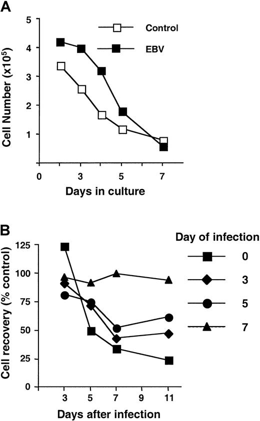
The activity of apoptosis-inducing agents is often determined by the stage of cell differentiation or activation. We asked, therefore, whether the status of monocyte–DC differentiation affects the sensitivity of cells to apoptotic death on EBV infection. First, cell morphology and viability were monitored during culture in the absence of cytokines. As illustrated by the representative experiment shown in Figure 2A, EBV infection did not induce apoptosis in monocytes. Indeed, the recovery of viable cells was slightly higher in EBV-infected cultures than in uninfected controls. This was partly explained by the early detachment of EBV-infected cells already noted in the cytokine-containing cultures. To analyze the sensitivity of cells to EBV-induced apoptosis at different stages of differentiation toward DC phenotype, cells cultured in the presence of IL-4 and GM-CSF were infected with EBV at different time points, as indicated in Figure 2B. In agreement with the data presented in Figure1A, EBV infection at the initiation of the culture resulted in the loss of approximately 70% of cells relative to uninfected controls. Cells infected at day 3 or day 5 of culture also died in response to the infection; however, the level of cell loss gradually decreased along with lymphokine-induced differentiation. Thus, only 50% of cells were lost in cultures infected at day 5. By day 7, virtually all cells cultured in GM-CSF and IL-4 acquired an immature DC phenotype characterized by the expression of CD86, CD80, and high levels of MHC class I and II molecules (Figure 3A and data not shown). In some cases, DCs that developed in EBV-infected cultures showed an up-regulation of CD86, CD83, and more heterogeneous expression of CD11c compared with mock-infected controls (Figure 3A and data not shown). However, these changes were not observed with every DC preparation and did not lead to any detectable differences in the stimulatory capacity of these cells, as assessed by mixed-lymphocyte reactions with allogeneic PBMCs (Figure 3B). At this stage, DCs in control cultures appeared to be unresponsive to EBV, as assessed by cell death (Figure 2B). These results indicated that apoptotic death is triggered by EBV infection only in cells undergoing differentiation toward the DC phenotype and may reflect an abnormal cellular response to the differentiation-inducing lymphokines.
EBV infection selectively affects cells undergoing differentiation into DCs.
(A) Recovery of uninfected and EBV-infected cells cultured in the absence of GM-CSF and IL-4. Results are from 1 of 4 representative experiments. (B) Complete differentiation into immature DCs is associated with resistance to the inhibitory effect of the virus. Cells cultured in the presence of GM-CSF and IL-4 were infected with EBV at different days of culture. Cell recovery was monitored at indicated time points after infection and was expressed as a percentage relative to uninfected controls to compensate for cell loss always observed in DC cultures with prolonged incubation. Mean values obtained from 3 independent experiments are shown.
Phenotype and T-cell stimulatory capacity of DCs developing from EBV- or mock-infected monocytes after 7 days of culture with GM-SCF and IL-4.
(A) Cells were recovered from mock (control) or EBV-infected cultures on day 7, and surface expression of indicated markers was analyzed by immunostaining and FACS analysis. (B) Uninfected (control), mock-infected (adsorbed EBV), or EBV-infected monocytes were cultured for 7 days in the presence of the lymphokines, harvested, irradiated, and used as stimulators at the indicated stimulator-responder ratios in allogeneic mixed-lymphocyte cultures. [3H]-Thymidine incorporation by stimulated T cells was measured after 7 days, as described in “Materials and methods.” In this experiment, the loss of DCs in the EBV-infected cultures was approximately 65% compared with the control cells. Results from 1 of 8 representative experiments are shown.
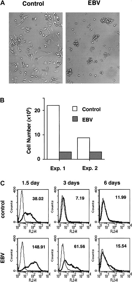
To explore this phenomenon further, cell morphology and viability were compared in cultures containing either GM-CSF or IL-4 alone. A dramatic difference was observed in the GM-CSF cultures. Although uninfected cells formed monolayers of firmly adherent macrophages that remained viable during the entire observation period, EBV-infected cells detached rapidly and started to die at a rate only slightly slower than that observed in the presence of GM-CSF and IL-4 (Figure4A and not shown). Independently of EBV infection, cells cultured in IL-4 alone died within 4 to 6 days, as expected. However, a significantly higher percentage of small cells showing morphologic signs of apoptosis was observed in the EBV-infected cultures starting from day 2, and the recovery of viable cells was significantly decreased by day 4 (Figure 4B). The effect of virus infection on the response to cytokine stimulation was also confirmed by analysis of surface marker expression. CD14 is rapidly down-regulated upon culture of blood monocytes in GM-CSF and IL-4 because of the differentiating activity of IL-4.44 45 This loss of expression was significantly delayed in EBV-infected cultures, where high levels of CD14 were still detected in most of the cells after 3 days (Figure 4C). Collectively, these findings demonstrate that EBV infection blocks the differentiation of blood monocytes into DCs, altering their response to GM-CSF and IL-4 and promoting apoptosis in the presence of the cytokines.
Response of monocytes to GM-CSF and IL-4 is altered by EBV infection.
(A) Light microscopy images of purified monocytes cultured for 6 days in medium containing 1000 U/mL recombinant GM-CSF. Most of the cells in the control cultures adhered firmly to the plastic, whereas virtually all the EBV-infected cells detached and formed tight, medium-sized clumps. Two of 10 representative cultures initiated with monocytes from 2 donors are shown. (B) Cell recovery of purified monocytes cultured with 1000 U/mL recombinant IL-4. Cells cultured in IL-4 alone died within 5 to 6 days, as expected. The recovery of viable cells at day 4 was 5- to 10-fold lower in EBV-infected cultures than in uninfected controls. Results from 2 experiments performed in parallel are shown. (C) Surface expression of CD14 was compared on control or EBV-infected cells by immunostaining and FACS analysis. Down-regulation of CD14 was delayed in EBV-infected DCs. Results from 1 of 3 experiments are shown.
Inhibition of DC development is independent of viral gene expression
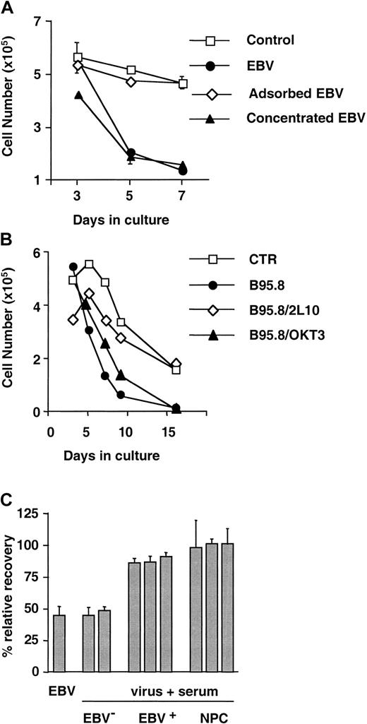
The finding that EBV infection inhibits the development of DCs from purified monocytes and the failure to reproduce the phenomenon by coculture of uninfected monocytes with infected mononuclear cells in a 2-chamber system (not shown) suggest that contact with the virus is required for the effect. The following sets of experiments were performed to confirm this observation. Monocytes were infected with B95.8 supernatants depleted of infectious virus by absorption on EBV receptor–positive Raji cells or with concentrated virus obtained by ultracentrifugation. The virus-depleted supernatant did not affect recovery, whereas the yield of DCs was further decreased by infection with concentrated virus (Figure 5A). To ensure the specificity of virus absorption, magnetic beads were conjugated with either the gp350-specific mAb 2L10 or the CD3-specific mAb OKT3 as a control. Incubation of B95.8 supernatant with OKT3 beads or beads with blocked binding sites resulted in the unspecific decrease of EBV titers by 30% to 50%, as assessed by EBNA staining or EBV binding assay (data not shown). Therefore, prolonged incubations were required to clearly reveal the inhibitory activity of the virus. Nevertheless, only absorption with 2L10 mAb, but not with OKT3 mAb-conjugated beads, significantly diminished the ability of the B95.8 supernatant to induce apoptosis in developing DCs, confirming that the inhibitory effect requires the presence of the virus (Figure 5B). In line with this conclusion, the activity of the virus was abolished by preincubation with sera from healthy EBV carriers or from patients with nasopharyngeal carcinoma that contained high titers of neutralizing antibodies, whereas sera from EBV antibody–negative donors had no effect (Figure 5C).
Binding of the virus is required for the inhibition of DC development.
(A) The effect of the virus was abolished by adsorption on EBV receptor–positive cell lines and was enhanced by concentration. EBV was concentrated from B95.8 supernatant by ultracentrifugation, and EBV-depleted supernatant was obtained by adsorption on EBV receptor–positive Raji cells. Purified CD14+ monocytes were infected with untreated, absorbed, or concentrated B95.8 supernatant for 2 hours at 37°C and then were cultured in the presence of GM-CSF and IL-4. Results are from 1 of 3 to 5 representative experiments performed with each type of virus preparation. Mean ± SD of triplicates. (B) Virus absorption was performed using magnetic beads conjugated with 2L10 mAb. Beads conjugated with OKT3 mAb were used as control. Results are from 1 of 3 representative experiments. (C) Effect of the virus was neutralized by preincubation with EBV antibody–positive sera. B95.8 supernatant was preincubated for 30 minutes at room temperature with 1:20 dilution of sera from EBV antibody-negative (EBV−) or -positive (EBV+) healthy donors or from patients with nasopharyngeal carcinoma before it was used for infection of purified monocytes. The percentage recovery relative to uninfected controls is shown. Mean ± SD of 4 experiments.
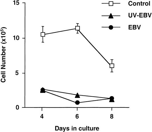
Recent reports suggest that EBV can replicate in monocytes, though with low efficiency.26 We tested, therefore, whether viral gene expression was required for the proapoptotic effect. Monocytes were exposed to replication-competent or UV-inactivated virus, and cell recovery was monitored in cultures containing GM-CSF and IL-4. As shown in Figure 6, inactivation of the virus with doses of UV light sufficient to prevent the transformation of freshly isolated B lymphocytes and the expression of EBNAs in the EBV-negative Bjab cell line (not shown) did not affect its ability to inhibit the development of DCs. Analysis of viral gene expression by RT-PCR failed to detect transcripts of the latent viral genesEBNA1, EBNA2, LMP1, andLMP2a and the lytic gene BZLF1 in cells collected at different times after infection (data not shown).
UV-inactivated EBV retains its inhibitory activity.
Purified CD14+ monocytes were infected with replication-competent or inactivated virus. B95.8 supernatants were inactivated by exposure to a 254-nm UV light source for 5 minutes from a distance of 10 cm. The dose corresponded to double the amount required to abolish the transformation of freshly isolated B lymphocytes and the induction of EBNA expression in the EBV-negative B-lymphoma line Bjab. The efficiency of inactivation was confirmed by the failure to detect EBV-specific mRNAs by RT-PCR at different times after infection (not shown). Results from 1 of 5 representative experiments are shown. Mean ± SD of triplicates.
Effect of the virus requires engagement of the membrane-fusion complex
The inhibitory activity of UV-inactivated virus suggests that binding of EBV to monocytes may be sufficient to alter their response to GM-CSF and IL-4. The major EBV envelope glycoprotein, gp350/220, was shown to interact with monocytes and to activate the expression of several cellular genes.27,28 We asked whether the effect of the virus could be reproduced by exposure to purified recombinant gp350. As shown in Figure 7A, culturing of monocytes in the presence of increasing concentrations of gp350 or on gp350-coated plates (not shown) had no effect on, or even slightly increased, the recovery of DCs. Hence, we turned to other components of the viral envelope. In addition to binding to CD21, infection of B lymphocytes requires the engagement of a trimolecular complex composed of the EBV glycoproteins gp42, gp85, and gp25.35,46,47Gp42 interacts with the β chain of HLA-DR, -DQ, and -DP48,49 and promotes viral penetration, which is mediated by the homologue of the herpesvirus membrane-fusion complex gH/gL, gp85, and gp25. The gp85/gp25 heterodimer alone acts as an alternative EBV ligand in CD21−, MHC class II-negative epithelial cells by interacting with an as-yet-unidentified membrane molecule.32,50 Preincubation of replication-competent (Figure 7B) or UV-inactivated virus (not shown) with antibodies to gp350/220 did not prevent cell death, further confirming that gp350/220 ligation is not involved in this viral effect. An isotype-matched CD3-specific antibody was also inactive, whereas DCs were rescued by preincubation with the mAb F-2-1—which blocks the interaction of gp42 with MHC class II51—and E1D1—which blocks penetration mediated by gp85/2535,46 and inhibits binding to epithelial cells in the absence of CD21.32 Involvement of the tripartite complex was further confirmed using mutant EBV strains deficient in the expression of the gp42 or gp85 proteins. Purified CD14+ monocytes were infected with wild-type Akata virus, gp42- or gp85-deficient virus, and B95.8 virus as a control and then were cultured for 7 days in the presence of IL-4 and GM-CSF. The wild-type Akata and B95.8 viruses exhibited a comparable ability to suppress DC development, whereas the inhibitory activity of viruses lacking either gp42 or gp85 was significantly impaired (Figure 7C). Thus, binding of EBV to the proposed coreceptor expressed on monocytes appeared to be sufficient to modify their response to GM-CSF and IL-4 and to inhibit the development of DCs.
Engagement of the EBV membrane-fusion complex is required for inhibition of DC development.
(A) Culture of monocytes in the presence of recombinant gp350/220 did not inhibit the development of DCs. Purified monocytes were cultured for 7 days in medium containing GM-CSF, IL-4, and the indicated concentration of purified recombinant gp350. Results are from 1 of 4 representative experiments. (B) CD14+ monocytes were infected with B95.8 virus or virus preincubated for 30 minutes at room temperature with 100 μg/mL mAb, as indicated. The inhibitory activity of the virus was blocked by mAb F-2-1, which interferes with the interaction of gp42 with HLA class II, and by E1D1, which prevents gp85–gp25-mediated membrane fusion and inhibits binding to epithelial cells in the absence of gp42. The 72A1 antibody to gp350 and OKT3 antibody to CD3 had no effect. Results from 1 of 2 experiments giving comparable results are shown. (C) Recombinant EBV lacking gp42 or gp85 showed a significantly impaired inhibitory activity compared to wild-type viruses derived from Akata or B95.8. Mean ± SD of 3 experiments.
Discussion
The data presented here provide the first demonstration that EBV infection inhibits the development of myeloid DCs from blood monocyte precursors. We show that EBV-infected monocytes die by apoptosis in the presence of GM-CSF and IL-4. This correlates with an aberrant response to both cytokines because the infected cells failed to differentiate into adherent macrophages in response to GM-CSF alone, and their death was accelerated in the presence of IL-4 compared with uninfected control cells. Viral interference with the cytokine effect is further illustrated by the early blockade of CD14 down-regulation (Figure 4), which is normally observed as a rapid response to IL-4. Strong up-regulation of Bim expression is likely to account for cell death induced by EBV infection and is also consistent with inability of IL-4 and GM-CSF to trigger survival signals known to prevent Bim up-regulation in other models.52 53
Interference with the function of DCs is a common feature of viral pathogenesis and is usually achieved by the expression of viral genes and virus replication in mature DCs or their precursors.6,9,10,54,55 We have found that the proapoptotic activity of EBV is not affected by UV inactivation, excluding a major contribution of de novo synthesis of viral proteins or mRNAs. Thus, EBV appears to be unique in its ability to suppress the development of DCs without the need for productive infection. Experiments with blocking antibodies and recombinant virus strains indicate that binding of EBV to monocytes through the viral membrane-fusion complex may be sufficient to trigger events that will eventually lead to cell death. Two components of the fusion complex, gp42 and the gp85–gp25 heterodimer, appear to be involved. Interestingly, the corresponding members of the human cytomegalovirus fusion complex were shown to activate signal transduction in infected cells but were not implicated in the induction of apoptosis.56,57 Purified gp42 has been shown to suppress lymphocyte proliferation in allogeneic mixed-lymphocyte cultures, but the mechanism of this effect has not been elucidated.49Our data suggest that gp42 may exert its suppressive effect through the inhibition of DC development. The blocking activity of antibodies that interfere with the interaction of gp42 with HLA-DR and the impaired activity of the gp42-deficient virus indicate that ligation of HLA class II may be required for the effect. Indeed, HLA-DR mediates signals that may result in cell proliferation, cell–cell adhesion, or cell death. Triggering of MHC class II molecules with a subset of specific mAbs initiates apoptosis in DCs. Mature DCs are sensitive to HLA-DR–mediated apoptosis, whereas immature DCs and macrophages are less sensitive and monocytes appear to be resistant.58 In contrast, the most prominent proapoptotic activity of EBV is observed with the infection of monocytes at the early stages of their differentiation into DCs (Figure 2). Sensitivity of cells to the viral effect decreases along with monocyte progression to the DC phenotype, and the virus does not affect completely differentiated mature DCs. This suggests that gp42 mediates DC apoptosis through a mechanism that is distinct from antibody-mediated MHC class II ligation. The inhibitory effect of gp85-specific blocking antibodies and diminished proapoptotic activity of gp85-deficient virus are also consistent with an independent role of the gp85–gp25 heterodimer in the inhibition of DC development. If so, the interaction of gp42 with HLA class II may be required to bring the heterodimer in proximity with the cell membrane. In addition, other virion-associated components, such as tegument proteins that may be delivered to the cytosol after viral fusion, may also contribute to the induction of apoptosis.
The mechanisms through which interaction with the EBV fusion complex alters the response of monocytes to signals that usually promote survival and differentiation remain unknown. Analysis of GM-CSF and IL-4 receptor expression by staining with specific antibodies failed to demonstrate significant changes in the infected cells (not shown), but this does not exclude a receptor-mediated effect. Site-specific phosphorylation of the IL-4 receptor by receptor-associated kinases results in differential triggering of cell growth, gene activation, differentiation, or resistance to apoptosis (reviewed in Nelms et al59). Thus, EBV infection might affect the signaling capacity of the receptors by altering their pattern of phosphorylation. It is noteworthy that interaction of the most abundant EBV envelope protein, gp350–gp220, with a CD21-like moiety on the monocyte membrane, up-regulates tumor necrosis factor–α (TNF-α) through activation of protein kinase C, phosphatidylinositol 3 kinase, and the NF-κB family of transcription factors,27,28 whereas exposure to intact virions inhibits TNF-α production.60It remains to be seen whether specific combinations of kinases or phosphatases are recruited by binding of EBV through the membrane-fusion complex.
Our findings indicate that EBV may interfere with the development and maintenance of specific immune responses. Prolonged contact between antigen-presenting DCs and naive T cells is required for the initiation of primary immune responses. According to different models of T-cell activation, the number of antigen-loaded DCs available to specific T cells represents a critical parameter for the induction of immunity, and whether the DC supply is efficient may largely determine the outcome of the immune response.61,62 At least a proportion of DCs develops directly from monocyte precursors.63 By inhibiting the differentiation of monocytes into mature DCs, EBV may temporarily halt the onset of immune responses during primary infection, creating a time window for efficient viral replication. This could permit the accumulation of a sufficiently large pool of virus-infected B lymphocytes and could allow their access to the memory B-cell compartment. Supporting this scenario, absolute monocytopenia was observed during the acute phase of infectious mononucleosis in the only reported case that was followed from the time of first encounter with the virus to the resolution of clinical symptoms.64Notably, infectious mononucleosis represents a rare outcome of primary EBV infection. The clinical picture of the disease develops as a result of an abnormally intensive immune response and can be explained, in the context of the proposed model, as a failure of the virus to exert sufficiently strong suppression on the immune system. The usual success of this viral strategy may account for the generally asymptomatic course of primary EBV infection.
Many viruses are known to act as potent activators of DC maturation, thereby promoting the development of specific antiviral immunity.65-68 However, several viruses block DC development or deregulate the process of DC maturation. Such activity has been reported for human herpes simplex10,69 and has been suggested for human cytomegalovirus,11 and it appears to be associated with the ability of herpesviruses to establish persistence in infected hosts. Constant viral shedding is a characteristic of healthy EBV carriers, who constitute most of the human population. DC activation by EBV virions could provide a constantly acting endogenous danger signal, resulting in a break of tolerance. It is tempting to propose that the inhibitory activity of EBV on DC development may represent an essential mechanism in the establishment of EBV persistence, working as a silencer for potentially deleterious immunologic consequences of viral reactivation. In agreement with this idea, analysis of surface marker expression did not reveal any significant phenotypic differences between control and EBV-infected cells surviving 7 days of culture in the presence of lymphokines. The 2 cell populations were also comparable in their functional activity, as determined by allogeneic mixed-lymphocyte reactions (Figure 3). Although we cannot exclude that some subtle differences between DCs developing in virus-infected and control cultures could be revealed on analysis of their capacity to stimulate or polarize antigen-specific T-cell responses, the available data indicate that neither exposure to the virus nor uptake of apoptotic material from EBV-infected, monocyte-derived cells results in DC activation. It should be noted, however, that in vivo the DC population is composed of phenotypically and functionally distinct subsets. Results presented in this study suggest that EBV could interact with and affect the development of DCs and DC precursors of different origins, and detailed characterization of such interactions should be forthcoming.
EBV-infected memory B cells appear to be the primary site of virus reactivation in healthy virus carriers (reviewed in Masucci and Ernberg70). These cells are probably unable to boost immune responses because the presentation of antigen on resting B cells has a tolerizing effect on T lymphocytes.13 Their capacity to transfer the virus to adjacent monocytes may not be affected by the presence of neutralizing antibodies because of the ability of herpesviruses to spread by cell-to-cell contact in vivo. Notably, high titers of neutralizing antibodies do not protect against superinfection with EBV in immunocompromised patients.71 Thus, impairment of cross-priming by DCs may delay the reactivation of EBV-specific immune responses during persistent infection, allowing efficient spread to new hosts. The high capacity of developing DC to migrate from the periphery to the lymphatic tissue is well established. It is conceivable, therefore, that the death of EBV-infected developing DCs could also limit the transport of the virus to lymph nodes, where it can propagate through lytic replication and proliferation of latently infected cells.72 Finally, interference with the development of DCs may offer a new explanation for the role of virus replication in the pathogenesis of EBV-associated malignancies. Enhanced virus production has been shown to precede the outgrowth of EBV-associated large-cell lymphomas in immunosuppressed patients,73 and elevated titers of antibodies to antigens of the productive cycle correlate with increased risk and relapse of endemic Burkitt lymphoma and nasopharyngeal carcinoma.74 75 The significance of this prodromal burst of virus replication has remained obscure because EBV is strictly latent in the malignant cells. Our findings suggest that, by diminishing the activity of certain types of DCs, virus production could be an important factor in tumor progression.
Supported by grants from the Swedish Cancer Society, the Swedish Paediatric Cancer Foundation, the Swedish Foundation of Strategic Research, the Petrus and Augusta Hedlund Foundation, and the Karolinska Institutet, Stockholm, Sweden.
The publication costs of this article were defrayed in part by page charge payment. Therefore, and solely to indicate this fact, this article is hereby marked “advertisement” in accordance with 18 U.S.C. section 1734.
References
Author notes
Victor Levitsky, Microbiology and Tumorbiology Center, Karolinska Institutet, Nobels väg 16, S-17177 Stockholm, Sweden; e-mail: victor.levitsky@mtc.ki.se.

![Fig. 3. Phenotype and T-cell stimulatory capacity of DCs developing from EBV- or mock-infected monocytes after 7 days of culture with GM-SCF and IL-4. / (A) Cells were recovered from mock (control) or EBV-infected cultures on day 7, and surface expression of indicated markers was analyzed by immunostaining and FACS analysis. (B) Uninfected (control), mock-infected (adsorbed EBV), or EBV-infected monocytes were cultured for 7 days in the presence of the lymphokines, harvested, irradiated, and used as stimulators at the indicated stimulator-responder ratios in allogeneic mixed-lymphocyte cultures. [3H]-Thymidine incorporation by stimulated T cells was measured after 7 days, as described in “Materials and methods.” In this experiment, the loss of DCs in the EBV-infected cultures was approximately 65% compared with the control cells. Results from 1 of 8 representative experiments are shown.](https://ash.silverchair-cdn.com/ash/content_public/journal/blood/99/10/10.1182_blood.v99.10.3725/6/m_h81022532003.jpeg?Expires=1769589421&Signature=L8IVMUdRhTYlHLWfo4ixCbLb2xvg0yS5V0WsGfm6CsYrm3inYdeMZGD~67uM5xdcalSeudUyq-vV-H-Ia6n4aYXQesLWlHa8R1Vy9p-3X5T6ICaD3SH8UFXAo4v7wn7JEaYZV~6JSB7VKaPrb9Hi-C7aVFqwAQe8GR~wtXZERAtCsZy3M5f3qy2pgjFyLE9NvDdD0Yvm~VVwr6z2yFbFuNqoMtB65xjdPJTULGaL3a6Xp4rHGFpEODUz8GLwQrcU6Qj5rrA2B3qlxN1H1~VYnJlq14Z3Qr~H0tl0SyZYCkt5SG~QyJNiI5Bp3QScE3Yv6VLx49ENlgQ7bdCOdLMdpg__&Key-Pair-Id=APKAIE5G5CRDK6RD3PGA)
