HLA class I expression depends on the formation of a peptide-loading complex composed of class I heavy chain; β2-microglobulin; the transporter associated with antigen processing (TAP); and tapasin, which links TAP to the heavy chain. Defects in TAP result in a class I deficiency called the type I bare lymphocyte syndrome (BLS). In the present study, we examined a subject with a novel type I BLS who does not exhibit apparent TAP abnormalities but who has a tapasin defect. The subject's TAPASIN gene has a 7.4-kilobase deletion between introns 3 and 7; an Alu repeat–mediated unequal homologous recombination may be the cause of the deletion. No tapasin polypeptide was detected in the subject's cells. The cell surface class I expression level in tapasin-deficient cells was markedly reduced but the reduction was not as profound as in TAP-deficient cells. These results suggest that tapasin deficiency is another cause of type I BLS.
Introduction
Type I bare lymphocyte syndrome (BLS) is characterized by the lack of cell surface HLA class I.1,2 Patients with this syndrome showed a reduced number of CD8+ T cells and the lack of natural killer (NK) activities.3,4 Type I BLS is caused by a deficiency in the transporter associated with antigen processing (TAP).4-7 TAP transports peptides from the cytoplasm into the inner lumen of the endoplasmic reticulum (ER), and defects in TAP induce poor peptide loading on class I heavy chains (HCs), resulting in a reduction in the number of mature class I molecules on the cell surface. Peptide binding of HCs occurs on the ER by the formation of a complex that is composed of HCs, β2-microglobulin, TAP, and tapasin.8 Tapasin is a 48-kd transmembrane protein that has 2 immunoglobulinlike domains and the ER-retention motif.9,10 The TAPASINgene is located at the HLA region.11 Tapasin binds with TAP, forms a bridge between TAP and HCs, enhances peptide loading, and retains immature HCs in the ER.12,13 The importance of tapasin in the class I assembly has been indicated by studies using a human mutant cell line, 721.220, which has a truncated tapasin by aberrant splicing.9,14TAPASIN-gene–targeted mice had an impaired class I assembly, reduced level of cell surface class I expression, and impaired immune responses.15 16
Here we analyzed a subject with a novel type I BLS who did not manifest the symptoms commonly observed in TAP-deficient subjects and found that the subject has tapasin deficiency.
Study design
Antibodies
Antitapasin (peptides at 313-330) and anti-TAP1 rabbit antibodies5 were provided by K. Tanaka (Tokyo Metropolitan Institute of Medical Sciences, Japan); Rgp48N (antitapasin, N-terminal peptides at 2-20)17 by P. Cresswell (Yale University, New Haven, CT); the anti-TAP2 monoclonal antibody 429.3 by P. M.van Endert18 (INSERM, Paris, France); HC10 (anti-HLA class I) by H. L. Ploegh (Harvard Medical School, Boston, MA); and w6/32 (anti-HLA class I) by Serotec (Kidlington, United Kingdom).
Cell preparations
Peripheral blood mononuclear cells (PBMCs) were isolated from a subject S.M. The B-cell line SM-LCL was established from the patient's PBMCs. The TAP1-deficient KMW-B2 cells have been described previously.5
Polymerase chain reaction amplification and sequencing
Genomic DNA and complementary DNA (cDNA) were prepared as described previously.5 DNA sequencing was performed by means of ABI 310 (PE Applied Biosystems, Foster City, CA).
Flow cytometric analysis
Cells were stained by fluorescein isothiocyanate (FITC)–conjugated antibodies and analyzed by means of EPICS Elite flow cytometer (Beckman Coulter, Fullerton, CA)
Results and discussion
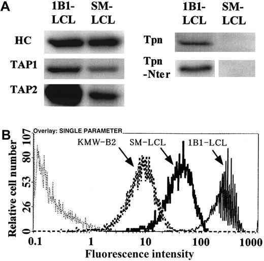
S.M. was a 54-year-old woman suffering from a primary chronic glomerulonephritis for 10 years. She has been receiving hemodynamic dialysis for 5 years. She had a history of herpes zoster and polyps of stomach and colon. Because kidney transplantation has been considered, S.M. was subjected to serological HLA typing. Her class II antigens could be typed, but class I typing was not successful (data not shown), indicating that she has type I BLS. HLA-DNA typing identified her HLA genotype to be homozygous for A*2601, B*1501, Cw*0303, and DRB1*1501, although she is not an offspring of a consanguineous marriage. Western blotting showed that the cell line SM-LCL has TAP polypeptides of correct sizes with slightly smaller amounts than those of the control cells (1B1-LCL) and that SM-LCL lacks tapasin polypeptides (Figure1A). The tapasin polypeptide was also not detected in the patient's PBMCs (data not shown), indicating that she has tapasin deficiency. Tapasin increases the TAP expression level by stabilizing it,10 and this may explain the reduction in the amounts of TAP. Figure 2A shows polymerase chain reaction (PCR) analysis of S.M.'s TAPASINgene. A fragment encompassing exons 1 to 3 was amplified (lane 2), and the exon 8 fragment was also amplified (lane 6). On the other hand, a fragment encompassing exons 4 to 7 was not amplified (lane 4), suggesting the existence of a homozygous deletion including exons 4 to 7 in the S.M. TAPASIN gene. To determine the breakpoint of the deletion, a region corresponding to introns 3 and 7 was analyzed in detail (Figure 2B). Both introns of the intact TAPASIN gene contain many Alu-repetitive family sequences.19 Several specific primers for the intron portions with unique sequences were designed, and PCRs using various primer combinations were performed. Representative results are shown in Figure 2A. No amplified fragments were obtained from S.M. DNA with the use of primers downstream of the AluSp subfamily sequence of intron 3 or primers upstream of the AluSx subfamily sequence of intron 7 (lanes 8 and 10, respectively). On the contrary, an approximately 0.9-kilobase (kb) fragment was amplified with primers in introns 3 and 7 from the subject (lane 12). These results indicate that the breakpoints were located near these primers, and the deletion was about 7.4 kb (Figure 2C). Sequencing analysis of the fragment further revealed that putative recombination sites were in 14-base stretches (Figure 2D). This kind of an unequal homologous recombination between Alu repeats has been reported for several genes.20 Reverse transcription–PCR and cDNA sequencing analyses showed that S.M. tapasin cDNA lacks a fragment corresponding to exons 4 to 7 and encodes a frameshifted and truncated polypeptide (data not shown). The putative truncated polypeptide could not be detected by an N-terminal tapasin-specific antibody17 (Figure 1A), suggesting the absence of translation or rapid degradation of the protein. Results of flow cytometry of SM-LCL are shown in Figure 1B. The cell surface class I expression level of the subject's cells was markedly reduced. A similar reduction in class I expression level was observed in peripheral blood T cells, B cells, and monocytes (data not shown). Since tapasin is indispensable for optimum peptide loading, most class I molecules may be empty or loaded with a low-affinity peptide and remained in the ER or degraded.13 The reduction in the expression level of class I in tapasin-deficient cells was not as great as that in TAP-deficient cells. Tapasin retains immature HCs in the ER via its retention motif in the cytoplasmic portion.10 Therefore, more immature HCs might be transported to the cell surface in tapasin-deficient cells than in TAP-deficient cells.15 Alternatively, these cell surface class I molecules in tapasin-deficient cells may load peptides via a tapasin-independent pathway.21 22
Western blot and flow cytometric analysis of SM-LCL.
(A) Western blot analysis of SM-LCL. Cell lysates of 1B1-LCL (healthy control) and SM-LCL were put into reactions with antibodies as indicated. Tpn indicates tapasin; Tpn-Nter, tapasin molecules that were detected by the anti–N-terminal tapasin antibody, Rgp48N. In the Tpn-Nter lane, portions around 48 kd (normal tapasin) and 18 kd (putative truncated tapasin) were shown in 1B1-LCL and SM-LCL, respectively. (B) Flow cytometric analysis. Cell surface HLA class I expression was detected by monoclonal antibody FITC-w6/32. Cells were SM-LCL (Tpn-deficient), KMW-B2 (TAP1-deficient), and 1B1-LCL (healthy control). Thin dotted line indicates control FITC–immunoglobulin G.
Analysis of normal and S.M.'s
TAPASIN genes. (A) Genomic polymerase chain reaction (PCR) of a TAPASIN gene. PCR products were analyzed by 1.5% agarose gel electrophoresis. Odd lanes show 1B1-LCL (healthy control). Even lanes show SM-LCL. PCR primers used were as follows: exon 1-3; 5′-AGGAAGAGGAGGCTTCATGG-3′(forward), 5′-CCTTAGAATCTACCCACCCTT-3′(reverse); exons 4-7, 5′-TCATGCCCCTTCCCAACCC-3′(forward), 5′-ATAGGATGAGGAGATAAAGTGA-3′(reverse); exon 8, 5′-TTAAGAAGATGCAGGAGATACA-3′(forward), 5′-CACTCAGTGGAGAGAGATTG-3′(reverse); intron 3, 5′-CAGGCCTATACTTTCTTAAT-3′ (forward), 5′-ACCCCTTTCTTGTGCCTAGT-3′(reverse); intron 7, 5′-AACAATATAAGAAATTACAGGC-3′(forward), 5′-TGTATCTCCTGCATCTTCTTAA-3′(reverse). PCR products sizes are as follows: exons 1-3, 908 base pairs (bp); exons 4-7, 1529 bp; exon 8, 305 bp; intron 3, 1711 bp; intron 7, 1038 bp. (B) Schematic of normal and S.M.'s TAPASIN gene structure. Upper line shows normalTAPASIN gene; lower line, S.M.'s TAPASIN gene. Boxes indicate exons. PCR primer positions are indicated by arrowheads (exon primers) or arrows (intron primers; black arrows, intron 3; white arrows, intron 7, respectively). A 7.4-kilobase (kb) portion indicated by doted lines was deleted in S.M.'s TAPASIN gene. Scales of exons and introns are approximations. (C) Intron 3 structure of S.M.'s TAPASIN gene. Upper line shows normal tapasin intron 3 region; middle line, S.M.'s intron 3 region; lower line, normal tapasin intron 7 region. Arrows indicate putative breakpoints. Boxes indicate exons and Alu-repetitive sequences. In SM-tpn, open boxes correspond to the exon 3 to exon 4 region; shaded boxes correspond to the exon 7 to exon 8 region of normal TAPASIN gene. A hatched box (marked with arrows) indicates a putative recombination region. (D) Nucleotide sequence of putative recombination junction. Upper sequence shows normal tapasin intron 3 AluSp region; middle sequence, S.M.'s tapasin; lower sequence, normal tapasin intron 7 AluSx region. Arrows indicate differences in nucleotide sequence positions between S.M.'s and normal intron 3 or intron 7. Box indicates a putative recombination junction area.
TAP-deficient subjects have respiratory inflammations and skin ulcers.2,7,23 The pathological mechanisms have not been clarified, but the involvement of autoreactive cytotoxic lymphocytes has been suggested.7,24,25 However, our tapasin-deficient subject, S.M., did not show any of these symptoms to date. The reasons for these differences have not yet been clarified. Cell surface expression levels or the quality of class I molecules may be critical; alternatively, defects in TAP itself, but not tapasin, may contribute to the pathology. Tapasin-deficient mice showed poor CD8+ T-cell development, defects in antigen presentation, and reduced T-cell and NK-cell responses.15 16 Although the numbers of peripheral blood CD8 T cells and NK cells were reduced (6.4% and 1% of PBMCs, respectively, in 2 × 10⁶ PBMCs per milliliter blood), S.M. has not shown apparent immune deficiency and has not been suffering from particular virus infections except herpes zoster to date. Further study of S.M. is required for better understanding of the biological and physiological functions of tapasin in the immune system.
We thank Peter Cresswell, Peter M. van Endert, Keiji Tanaka, and Hidde L. Ploegh for kindly providing the antibodies. We also thank Kazuhiro Shimizu, Natalia Lapteva, and Yasuhiko Nagasaka for technical assistance and Takeshi Araki, Hiroshi Furukawa, Motoko Nishimura, Mie Nieda, Hirohiko Hohjoh, Ayako Kobayashi, Masatomo Maeda, and Eisei Noiri for constructive discussions.
Prepublished online as Blood First Edition Paper, May 13, 2002; DOI 10.1182/blood-2001-12-0252.
Partly supported by Grants-in-Aid for Scientific Research (B) from the Ministry of Education, Science, Sports and Culture of Japan.
The publication costs of this article were defrayed in part by page charge payment. Therefore, and solely to indicate this fact, this article is hereby marked “advertisement” in accordance with 18 U.S.C. section 1734.
References
Author notes
Toshio Yabe, Department of Research, Tokyo Metropolitan Red Cross Blood Center, Hiroo 4-1-31, Shibuya-ku, Tokyo 150-0012, Japan; e-mail: to-yabe@tokyo.bc.jrc.or.jp.


This feature is available to Subscribers Only
Sign In or Create an Account Close Modal