Abstract
Coupling of ovalbumin (OVA) to anti–DEC-205 monoclonal antibody (mAb) (αDEC) induced the proliferation of OVA-specific T cells in vivo. Expansion was short-lived, caused by dendritic cells (DCs), and rendered T cells anergic thereafter. Phenotypic analysis revealed the induction of CD25+/CTLA-4+ T cells suppressing proliferation and interleukin-2 (IL-2) production of effector CD4+ T cells. The findings were supported by 2 disease models: (1) CD4+ T-cell–mediated hypersensitivity reactions were suppressed by the injection of αDEC-OVA and (2) the application of hapten-coupled αDEC-205 reduced CD8+ T-cell–mediated allergic reactions. Thus, targeting of antigens to immature DCs through αDEC antibodies led to the induction of regulatory T cells, providing the basis for novel strategies to induce regulatory T cells in vivo.
Introduction
Dendritic cells (DCs) are highly efficient antigen-presenting cells that collect antigens in the periphery of the body and transport them to lymphoid organs. Peptides are produced and bound to major histocompatibility complexes (MHC), resulting in T-cell proliferation and in the initiation of an immune response.1 In general, DCs use pinocytosis, phagocytosis, and receptor-mediated endocytosis for antigen uptake; the latter is the most effective way to charge DC with antigens. To facilitate receptor-mediated endocytosis, DCs express a variety of receptors such as Fc receptors, macrophage mannose receptor (MMR), and a novel antigen receptor, DEC-205.2 DEC-205 is a decalectin, which, along with MMR and phospholipase receptor A2, belongs to the type 4 lectin family. All these receptors are involved in mediating endocytosis, but DEC-205 differs functionally from its sister molecules. It orchestrates a novel pathway of antigen uptake by recycling through late endosomal compartments rich in MHC class 2 products and enhancing antigen presentation to CD4+ T cells up to 100-fold compared with the homologous MMR.3 In vivo DEC-205 is nearly exclusively expressed by DCs in lymphoid organs,4 whereas MMR is expressed by subcapsular macrophages, Kupffer cells, and some epithelial cells. In this study, we were aiming to exploit the specific expression pattern of DEC-205 for antigen targeting to DCs in vivo.
The delivery of antigens to defined subsets of leukocytes by means of antibodies has long been hampered by the fact that target surface molecules were not specific for a single cell type or that cell-type–restricted surface markers failed to effectively mediate the delivery of antibody-antigen conjugates to an antigen-presenting compartment. Thus far, different antibodies directed against antigen-receptors, such as the Fc receptor and the B-cell receptor (BCR), have been used for targeting purposes. These receptors are known to mediate endocytosis and targeting of antigens to FcϵRII in the mouse or to MHC class 2 molecules in the rat, and they did indeed increase the immune response against the respective antigens.5,6 However, all the receptors used so far are expressed by different cells, such as monocytes, DCs, and granulocytes. In this report we show that antibodies directed against DEC-205 (αDEC) can target antigens exclusively to DCs in vivo. In comparison to other methods that use in vitro generated—and therefore mature—DCs, DEC targeting allowed the loading of antigen to immature DCs. We found that the presentation of antigens by DCs in this steady state led only to short-lived proliferation of antigen-specific T cells and that 8 days after injection, CD4+/CD25+ T cells with regulatory properties were induced. This CD25+ population was able to suppress interleukin-2 (IL-2) production and proliferation of conventional CD4+ T cells. The significance of this immune-suppressive effect could further be demonstrated in CD8- and CD4-dependent disease models. Here, the injection of αDEC-antigen conjugates suppressed the footpad swelling reaction (DTH) or allergic contact sensitivity reaction. Thus, these results represent a novel approach to induce regulatory T cells in vivo and emphasize the role of immature DCs as inducers of tolerance in vivo.
Materials and methods
Cell culture and mice
Balb/c and DO11.10 mice were bred and housed in the animal facility at the University of Mainz, Germany. The hydridomas NLDC-145 (anti-DEC), TIB120 (anti-MHC class 2), and Gl117 (β-galactosidase, isotype control) were cultured in R5 (RPMI 1640 supplemented with 5% fetal calf serum [FCS], penicillin-streptomycin [Pen-Strep], and glutamine; all from PAA Biochemicals, Linz, Austria), and antibodies were purified by ammonium chloride precipitation followed by protein A-G columns (Pierce, Rockville, MD). DCs derived from bone marrow were prepared according to standard methods. Briefly, bone marrow cells were cultured in R5 supplemented with granulocyte macrophage–colony-stimulating factor (GM-CSF) for 6 days. Thereafter, nonadherent cells were harvested and transferred to 6-well dishes. To some samples ovalbumin protein (100 μg/mL; Sigma, St Louis, MO) was added, and maturation was induced by the addition of lipopolysaccharide (5 ng/mL; Sigma). After 48 hours mature bone marrow dendritic cells (BMDCs) were harvested and subjected to proliferation assays.
Conjugation of ovalbumin to antibodies
Purified hybridoma supernatant of NLDC-145 (anti–DEC-205), TIB-120 (anti-MHC class 2), and Gl117 (isotype control) antibodies were labeled with activated ovalbumin (Pierce) using the heterobifunctional cross-linker succinimidyl 4-(N-maleimidomethyl) cyclohexane-1-carboxylate (SMCC; Pierce) according to the manufacturer's protocol. Briefly, the antibodies were reduced using 2-mercaptoethylamine HCl (MEA) and were separated from the MEA by gel filtration (Pierce). Thereafter, a mixture of cross-linker–activated ovalbumin and reduced antibodies were incubated overnight. Unreacted cross-linker was separated by gel filtration from the proteins, and the antibody-OVA conjugates were further enriched using spin columns (Millipore). To remove unbound OVA, the antibody-OVA conjugate was passed over a protein A/G column (Pierce) and was analyzed by sodium dodecyl sulfate–polyacrylamide gel electrophoresis (SDS-PAGE). For trinitrochlorobenzene (TNCB) coupling, antibodies were incubated with TNCB for 30 minutes at 37°C; afterward, unreacted TNCB was separated by gel filtration from the antibodies.
Immunohistology
Antibody conjugates were injected at 1 μg per footpad, popliteal lymph nodes (LNs) were removed, and 4-μm–thin cryosections were prepared. Specimens were fixed in 4% paraformaldehyde (PFA) and stained in a moist chamber. Injected αDEC-OVA conjugates were detected by incubating the sections with phycoerythrin (PE)–labeled goat anti-rat antibodies (Dianova, Germany). Then the specimens were incubated with rat immunoglobulin G (IgG) to block free secondary antibodies, followed by incubation with fluorescein isothiocyanate (FITC)–conjugated αMHC class 2 (clone M114; BD Bioscience, Heidelberg) antibodies. Specimens were examined by fluorescence microscopy.
Isolation of CD11c+/– cells and in vitro antigen presentation to DO11.10 cells
Different antibody-OVA conjugates were injected into the footpads of Balb/c mice (1 μg/footpad). After indicated time points, LNs were removed and single-cell suspensions were prepared using collagenase (Roche, Mannheim, Germany). Cells were incubated with antimouse CD11c MACS microbeads for 30 minutes on ice, and CD11c+ and the remaining CD11+ cells were separated on a magnetic field. This procedure yielded 95% CD11c+ cells in the positive fraction and approximately 5% CD11c+ cells in the negative fraction. Graded doses, as indicated, were dispensed in 96-well plates. To obtain OVA-specific responder cells, T cells were prepared from LNs and spleens from DO11.10 transgenic mice. Cells were incubated with anti-CD4 MACS beads (Miltenyi, Bergisch Gladbach, Germany), and, after a 30-minute incubation period, CD4+ T cells were enriched through a magnetic field to a purity of more than 92% CD4+ cells. OVA-specific responder T cells (1 × 10⁵ per well) were added to graded doses of CD11+ and CD11c+ stimulator cells. After 2 to 3 days of culture in 96-well plates, 3H-thymidine (1 mCi [37 KBq]-well; Amersham, Freiburg, Germany) was added for the last 16 hours before harvesting. Incorporation of radioactivity was determined by scintillation counting. Data shown are the means of triplicate cultures where the standard deviation was less than 10% of the mean cpm.
Labeling of DO11.10 T cells with CFSE and in vivo proliferation
DO11.10 T cells were purified with beads as previously described, and aliquots of cells were incubated with CFSE (carboxyfluorescein diacetate, succinimidyl ester; Molecular Probes, Göttingen, Germany) for 30 minutes. After that, 5 × 10⁶ labeled cells were injected intravenously into Balb/c recipients, and 24 hours later antibody-OVA conjugates were injected. After 2 days LNs and spleens were removed, and cells were incubated with PE-labeled anti-CD4 antibodies (BD Bioscience) followed by incubation with anti-PE MACS microbeads. Cells were enriched on a magnetic column, and CFSE labeling was measured in a FACScan by gating on PE-positive cells. Proliferation is indicated by a decrease in fluorescence intensity—that is, after each round of T-cell division, the CFSE dye is equally distributed among the dividing cells.
Isolation of T cells, FACS analysis, and restimulation in vitro
Balb/c cells were seeded with 5 × 10⁶ DO11.10 T cells. After 24 hours, antibody-OVA conjugates (1 μg/foot) were injected into the footpads. Mice were killed at indicated time points, and fluorescence-activated cell sorter (FACS) analysis and restimulation assays were performed.
To study survival of the injected cells, suspensions of spleens and LNs were stained with FITC-labeled KJ1-26 antibody (Caltag, Hamburg, Germany) and PE-labeled anti-CD4 antibodies. In some experiments, T cells were enriched. Cell suspensions were incubated with tissue-culture supernatants of the following hybridomas (all from ATCC): TIB120 (anti-MHC class 2), RA3-6B2 (anti-B cell), and F4/80 (anti-macrophage). After 2 washes, cells were incubated with sheep antirat Dynabead (Dynal, Oslo, Norway) for 30 minutes. Cell suspensions were placed in front of a magnetic field, and nonadherent cells were recovered. These cells contained more than 90% CD4+ and CD8+ T cells, as determined by FACS analysis. To obtain CD4+ cells, these precleared T-cell suspensions were incubated with antimouse CD4 MACS beads (Miltenyi), and CD4+ T cells were isolated by running the cell suspension through a magnetic column. This procedure resulted in more than 95% pure CD4+ T cells. Similarly, DO11.10 T cells were isolated from these pre-enriched cultures. Briefly, T cells were incubated with PE-labeled KJ1-26 antibody, followed by incubation with anti-PE MACS beads. KJ1-26+ T cells were then separated by MACS columns. For OVA-specific restimulation experiments, 5 × 10⁴ T cells per well were incubated with indicated numbers of ovalbumin-pulsed BMDCs in 96-well plates (Becton Dickinson, Heidelberg, Germany). After 2 to 3 days, T-cell proliferation was assayed by 3H-thymidine. In some experiments CD25+ cells were depleted from the Dynabead-enriched T cells before purification of the CD4+ subset. Briefly, cells were incubated with PE-labeled anti-CD25 antibodies (Chemicon, Hofheim, Germany) followed by anti-PE MACS beads. CD25+ cells were then separated by a magnetic field and kept separately. To access the regulatory effect of CD25+ T cells, cells were added to antigen presentation assays and mixed lymphocyte reactions (MLRs). For MLR, 5 × 10⁴ CD4+ T cells were cocultured with 2 × 10⁴ allogeneic BMDCs, and 25 000, 12 500, or 6250 CD25+ T cells were added to these cultures. T-cell proliferation was determined by 3H-thymidine incorporation. In some assays, isolated CD25+ T cells were cultured together with T-cell–depleted spleen cells as accessory cells (APCs), 0.5 μg/mL anti-CD3 antibodies, and equal numbers of isolated CD4+ responder T cells. T-cell proliferation was determined by 3H-thymidine incorporation. For FACS analysis, CD4+ T cells were incubated with PE-labeled KJ1-26 antibody, and FITC-labeled anti-CD25 (Chemicon). Before anti–CTLA-4 (BD Bioscience) staining, cells were fixed/permeabilized, and samples were analyzed using FACScan (Becton Dickinson). For intracellular IL-2 detection, T cells were prepared and restimulated in vitro as described. 5 × 10⁶ LN cells were pulsed in 24-well dishes with OVA peptide overnight and for the last 4 hours in the presence of brefeldin A (10 μg/mL; Sigma). After washing and fixation/permeabilization, cells were stained with FITC-labeled anti-CD4 antibodies and PE-labeled anti–IL-2 antibodies (Becton Dickinson) and were analyzed using FACScan.
Delayed-type hypersensitivity and contact hypersensitivity reactions
For OVA-specific delayed-type hypersensitivity (DTH) reactions, DO11.10 cells were purified and injected into Balb/c mice (5 × 10⁶ cells). After 48 hours, αDEC-OVA, Gl117-OVA, or soluble OVA was injected. Five days later mice were sensitized by subcutaneous injection of 30 μg OVA 323-339 peptide in IFA (Difco, Detroit, MI). After 7 days, 30 μg OVA peptide in incomplete Freund adjuvant (IFA) was injected into the right footpad, and IFA only was injected into the left footpad. For analysis of DTH reactions, swelling was measured after 24 hours using a caliper rule. Results are displayed as mean difference ± standard deviation in footpad thickness of left versus right footpad. Significance was tested using the Student t test. For in vitro proliferation, LN cells were prepared as described earlier, and 2 × 10⁵ cells were incubated with graded doses of OVA. Proliferation was measured after 72 hours. For contact hypersensitivity (CHS) reactions, abdomens of mice were shaved and painted with 0.5% TNCB (Sigma) dissolved in acetone olive oil. After 3 days, 10 μg TNCB-derivatized αDEC-205 or respective controls were injected subcutaneously into mice, followed by challenge at the right ear after 3 days. Ear swelling was measured after 24 hours using a caliper rule. Results are displayed as mean difference ± standard deviation in ear thickness of left versus right. Significance was tested by Student t test. For in vitro proliferation, LN cells were prepared as described earlier, and 5 × 10⁵ cells were incubated with graded doses of TNBS. Proliferation was measured after 72 hours.
Results
Coupling of OVA to αDEC-205 antibodies enhances targeting and antigen presentation in vivo
We have recently shown that injected αDEC antibodies target peptides to LNs in vivo.7 However, in these experiments, a peptide was genetically engineered into the Fc region of the antibody, and these recombinant antibodies were prepared from cell culture supernatant. Because this approach is tedious, we wanted to test whether whole proteins can be cross-linked to αDEC and to determine how this coupling affects in vivo antigen presentation. In initial experiments, the model antigen OVA was coupled to αDEC antibodies, and conjugates were further purified yielding a preparation that contained antibody-OVA conjugates and unconjugated antibodies but no free OVA (data not shown).
Because cross-linking of antibodies to OVA might affect their capacity to target their specific antigen, we tested targeting properties of the antibody-OVA conjugates in vivo by injecting mice subcutaneously with αDEC-OVA or respective isotype control OVA (Gl117-OVA) conjugates. Twenty-four hours later, injected antibodies were visualized in tissue sections. We found αDEC-OVA conjugates scattered in the T-cell area of the draining LN and demonstrate extensive colabeling with MHC class 2+ cells (Figure 1A). In contrast, when sections were double-labeled with the macrophage-specific antibody SER-4, no colocalization could be observed (Figure 1B), suggesting that only small and undetectable amounts of αDEC-OVA antibodies were captured by Fc receptors expressed by macrophages. When isotype controls were injected, no targeting was detectable (data not shown). The strong colocalization with MHC class 2 suggested that OVA coupled to αDEC-205 gains access to an active antigen-processing compartment in DCs in vivo. To test for antigen presentation, different antibody-OVA conjugates (as indicated) were injected subcutaneously into mice, and CD11+ LN DCs from draining LN were isolated 48 hours later and incubated with OVA-specific T cells (DO11.10). Figure 1C shows that low numbers of LN DCs isolated from αDEC-OVA–injected mice are able to stimulate the proliferation of DO11.10 T cells. In contrast, much higher numbers of DCs were required from αMHC class 2-OVA– or Gl117-OVA–injected mice to obtain T-cell proliferation. Moreover, this stimulatory activity was restricted to CD11c+ LN cells because the CD11c+ fraction did not stimulate T-cell proliferation above background levels (Figure 1C).
αDEC-205 antibodies target OVA to MHC class 2+ cells and induce T-cell proliferation. (A-B) αDEC-OVA conjugates were injected into the footpads of mice. Twenty-four hours later tissue sections from LNs were stained with PE-labeled goat anti-rat antibodies (to detect injected αDEC-OVA [red]). Specimens were further double labeled with FITC-αMHC class 2 antibodies (A) or FITC–SER-4 antibodies (B), respectively (green). Colabeling is indicated by yellow. Original magnification, ×200. (C) Twenty-four hours after the injection of 10 μg antibody-OVA conjugates, CD11c+ and CD11c– cells were prepared and cocultured with OVA-specific DO11.10 T cells. T-cell proliferation was assayed by 3H-thymidine incorporation. (D) A mixture of αDEC and OVA and of αDEC or OVA alone was injected, and CD11c+ cells were tested for antigen presentation as in panel C. (E) Mice were reconstituted with CFSE-labeled, OVA-specific DO11.10 T cells and injected with different antibody-OVA conjugates as indicated. Forty-eight hours later, LN cells were prepared and stained with PE-labeled αCD4 antibody. CFSE labeling was assayed by FACS after gating on CD4+ cells, and T-cell division is indicated by dilution of the dye (ie, decrease of fluorescence).
αDEC-205 antibodies target OVA to MHC class 2+ cells and induce T-cell proliferation. (A-B) αDEC-OVA conjugates were injected into the footpads of mice. Twenty-four hours later tissue sections from LNs were stained with PE-labeled goat anti-rat antibodies (to detect injected αDEC-OVA [red]). Specimens were further double labeled with FITC-αMHC class 2 antibodies (A) or FITC–SER-4 antibodies (B), respectively (green). Colabeling is indicated by yellow. Original magnification, ×200. (C) Twenty-four hours after the injection of 10 μg antibody-OVA conjugates, CD11c+ and CD11c– cells were prepared and cocultured with OVA-specific DO11.10 T cells. T-cell proliferation was assayed by 3H-thymidine incorporation. (D) A mixture of αDEC and OVA and of αDEC or OVA alone was injected, and CD11c+ cells were tested for antigen presentation as in panel C. (E) Mice were reconstituted with CFSE-labeled, OVA-specific DO11.10 T cells and injected with different antibody-OVA conjugates as indicated. Forty-eight hours later, LN cells were prepared and stained with PE-labeled αCD4 antibody. CFSE labeling was assayed by FACS after gating on CD4+ cells, and T-cell division is indicated by dilution of the dye (ie, decrease of fluorescence).
To verify that αDEC injection does not enhance antigen presentation by an unknown mechanism independently from its uptake by the DEC-205 receptor, we injected a mixture of OVA and uncoupled αDEC-205 antibodies. In these experiments (Figure 1D), T-cell proliferation remained at background levels, indicating that the injection of αDEC monoclonal antibody (mAb) alone does not enhance the antigen presentation simply by binding to CD11c+ LN cells. In addition to these in vitro stimulation experiments, we wanted to further demonstrate that T-cell proliferation is induced in vivo by αDEC-OVA injection. Therefore, we reconstituted Balb/c mice with CFSE-labeled, OVA-specific DO11.10 T cells 24 hours before injection of αDEC-OVA conjugates; 48 hours after injection, CFSE fluorescence of isolated T cells was analyzed by FACS. Figure 1E shows that the injection of αDEC-OVA induced strong T-cell proliferation (as indicated by the dilution of the dye), whereas in mice injected with controls only sparse proliferation was detectable. The major population of CFSE-negative cells derives from CD4+ host T cells and remains unchanged on the injection of antibody-OVA conjugates.
We next asked whether the injection of αDEC-OVA induces long-lasting T-cell activation in vivo. To test for T-cell activation, we isolated OVA-specific T cells from reconstituted Balb/c mice 8 days after injection of the respective antibody-OVA conjugates and cocultured these T cells with OVA-pulsed BMDCs in vitro. In these assays, T cells isolated from αDEC-OVA–injected mice became anergic and failed to proliferate in response to OVA-pulsed BMDCs (Figure 2A). In contrast, T cells isolated from αMHC class 2-OVA– or Gl117-OVA–injected mice proliferated normally. In these initial experiments, we observed that proliferation on restimulation in vitro was normal when αDEC-OVA was injected with an activating stimulus such as complete Freund adjuvant (CFA) (Figure 2A, open triangles), suggesting that activation of the DCs was necessary to prevent T-cell anergy. This effect was indeed caused by DC maturation, as was demonstrated by the coinjection of αCD40 antibodies together with αDEC-OVA. It has been shown that αCD40 antibodies lead to DC maturation in vivo.8 Accordingly, our experiments showed that after application of this DC maturation stimulus, T cells were rescued from anergy and proliferated normally when restimulated in vitro (Figure 2A, open circles). Taking previous results into account showing that injected transgenic T cells undergo apoptosis over time, we next tested whether the injection of αDEC-OVA or respective controls affects the survival of the seeded DO11.10 T cells in vivo. We showed (Figure 2B) that the total number of seeded T cells gradually declines over time; however, though marginally higher numbers of DO11.10 T cells were detectable in Gl117-OVA–injected mice, we could not detect any significant difference of DO11.10 T-cell survival between αDEC-OVA and isotype-treated mice 8 days after injection. Transgenic DO11.10+ T cells were detectable until day 20 after injection. Therefore, we conclude that the deletion of the OVA-specific T cell from the periphery occurs at similar rates. To further investigate the means by which T-cell proliferation was suppressed after αDEC-OVA injection, the phenotype of recovered T cells was analyzed by FACS. As shown in Figure 2C, the IL-2 receptor α-chain (CD25), a marker for activated T cells and for regulatory T cells, was up-regulated 2 days after the injection of αDEC-OVA conjugates regardless of whether αCD40 was coinjected. These results are in agreement with the data presented in Figure 1E, showing initial proliferation of the DO11.10 T cells after injection of αDEC-OVA conjugates. However, in Gl117-OVA–injected mice, the up-regulation of CD25 was less marked than in other groups, possibly reflecting the poor presentation of OVA and activation of T cells by this antibody conjugate. Interestingly, when T cells were analyzed 8 days after the injection of OVA conjugates (which corresponds to when restimulation assays were performed), elevated CD25 expression was detectable only in αDEC-OVA–injected mice (0.4% of all CD4+ T cells and approximately 20% of the recovered DO11.10 T cells). In T cells recovered from mice that had received αDEC-OVA together with the DC-maturation stimulus αCD40, CD25 expression had returned to background levels (0.1% of all CD4+ T cells and approximately 5% of recovered DO11.10 T cells). This effect was OVA specific and restricted to DO11.10 T cells because CD25 expression of the CD4+ host T cells was not affected by injection of the respective conjugates (approximately 5% of the cells expressed CD25). In addition, CTLA-4 was exclusively up-regulated in αDEC-OVA–injected mice after 8 days, and double-labeling revealed that CTLA-4 is coexpressed by CD25+ T cells (Figure 2D). These results could be repeated several times (Table 1), and they revealed a significant (P < .02) difference in the percentage of CD25+/CTLA+ cells in mice injected with αDEC-OVA compared with Gl117-OVA–injected controls.
OVA-specific T cells do not proliferate on restimulation after presentation of OVA by DCs in the steady state and show up-regulation of CD25 and CTLA-4. (A) Mice were reconstituted with OVA-specificT cells and were injected with different antibody-OVA conjugates as indicated. Eight days later, KJ1-26+ OVA-specific T cells were purified by MACS and cocultured with OVA-pulsed BMDCs. T-cell proliferation was assayed by 3H-thymidine incorporation. (B) To assess survival of the injected DO11.10 T cells, mice were killed after reconstitution and injection of αDEC-OVA conjugates at the time points indicated. Total T-cell suspensions were prepared using magneto-beads and were stained with FITC-labeled clonotypic antibody KJ1-26 and PE-labeled αCD4. Thereafter, FACS was used to analyze cells. Numbers in the diagrams display the percentage of KJ1-26+ cells. (C) For phenotypic analysis, 2 or 8 days after injection of the respective antibody-OVA conjugates, CD4+ T cells were purified by MACS and double labeled with FITC-conjugated anticlonotypic TCR antibodies (KJ1-26) and PE-labeled anti-CD25 or anti–CTLA-4 antibodies, respectively. Thereafter, total cell suspensions were analyzed using FACS without further gating, and numbers shown are the percentages of cells within the designated quadrant. (D) Mice were treated as in panel C, and T cells were stained with FITC-labeled anti-CD25 and PE-labeled anti–CTLA-4 antibodies and respective isotype controls. Thereafter, cells were analyzed using FACS.
OVA-specific T cells do not proliferate on restimulation after presentation of OVA by DCs in the steady state and show up-regulation of CD25 and CTLA-4. (A) Mice were reconstituted with OVA-specificT cells and were injected with different antibody-OVA conjugates as indicated. Eight days later, KJ1-26+ OVA-specific T cells were purified by MACS and cocultured with OVA-pulsed BMDCs. T-cell proliferation was assayed by 3H-thymidine incorporation. (B) To assess survival of the injected DO11.10 T cells, mice were killed after reconstitution and injection of αDEC-OVA conjugates at the time points indicated. Total T-cell suspensions were prepared using magneto-beads and were stained with FITC-labeled clonotypic antibody KJ1-26 and PE-labeled αCD4. Thereafter, FACS was used to analyze cells. Numbers in the diagrams display the percentage of KJ1-26+ cells. (C) For phenotypic analysis, 2 or 8 days after injection of the respective antibody-OVA conjugates, CD4+ T cells were purified by MACS and double labeled with FITC-conjugated anticlonotypic TCR antibodies (KJ1-26) and PE-labeled anti-CD25 or anti–CTLA-4 antibodies, respectively. Thereafter, total cell suspensions were analyzed using FACS without further gating, and numbers shown are the percentages of cells within the designated quadrant. (D) Mice were treated as in panel C, and T cells were stained with FITC-labeled anti-CD25 and PE-labeled anti–CTLA-4 antibodies and respective isotype controls. Thereafter, cells were analyzed using FACS.
Percentages of KJ1-26+/CD25+ and KJ1-26+/CTLA-4+ cells among CD4+T cells 8 days after injection of αDEC-OVA and G1117-OVA, respectively
. | αDEC-OVA—injected mice . | . | G1117-OVA—injected mice . | . | ||
|---|---|---|---|---|---|---|
| Experiment no. . | % CD25 . | % CTLA-4 . | % CD25 . | % CTLA-4 . | ||
| 1 | 0.40 | 0.30 | 0.10 | 0.10 | ||
| 2 | 0.50 | 0.30 | 0.20 | 0.10 | ||
| 3 | 0.80 | 0.60 | 0.10 | 0.00 | ||
| 4 | 0.30 | 0.30 | 0.00 | 0.00 | ||
| Mean ± SD | 0.50 ± 0.19 | 0.38 ± 13 | 0.10 ± 0.07 | 0.05 ± 0.05 | ||
Data represent mean ± SD.
Depletion of CD25+ T cells restores proliferation and IL-2 production
Neither marker, CD25 nor CTLA-4, is specific for regulatory T cells, and functional assays must be performed to demonstrate the presence of genuine Treg. Therefore, we next tested whether the depletion of potentially regulatory CD25+ T cells would restore T-cell proliferation in in vitro assays. T cells from αDEC-OVA– and Gl117-OVA–injected mice, respectively, were subsequently depleted of CD25+ T cells and subjected to in vitro restimulation assays as described.
As shown in Figure 3A, T-cell proliferation returned to normal levels in samples derived from αDEC-OVA–injected mice after removal of the CD25+ T cells. With CD25+ cells present, T-cell proliferation was suppressed compared with Gl117-OVA–injected mice. In turn, when isolated CD25+ T cells were added to cocultures of OVA-pulsed DCs and T cells isolated from Gl117-OVA–injected mice, the suppression of T-cell proliferation could be noted. As expected, isolated CD25+ T cells alone did not proliferate when cocultured with OVA-pulsed DCs. To further exclude the possibility that the injection of αGl117-OVA selectively blocks the ability of CD25+ T cells to suppress T-cell proliferation, we purified Treg cells from αDEC-OVA– and Gl117-OVA–injected mice, incubated these cells with nontransgenic CD4+ T cells (5 × 10⁴ cells each), and stimulated these cocultures with a combination of anti-CD3/APCs. In these experiments, Gl117-OVA–derived CD25+ T cells were able to block the proliferation of conventional T cells (Figure 3B), indicating that the injection of antibody-OVA conjugates does not block the regulatory function of CD25+ T cells. It has been shown that regulatory T cells reduce the IL-2 production of conventional CD4+ T cells.9 Accordingly, we could not detect IL-2 by intracellular staining of T cells isolated from αDEC-OVA–injected mice (Figure 3C), whereas IL-2 production was evident in controls and in αDEC-OVA–injected mice that received a DC maturation stimulus by αCD40 injection. As in T-cell proliferation, IL-2 production could be restored after the depletion of CD25+ T cells. Therefore, we conclude that the presentation of OVA by immature DCs in vivo leads to the activation of Treg, suppressing the proliferation and limiting the IL-2 production of conventional CD4+ T cells.
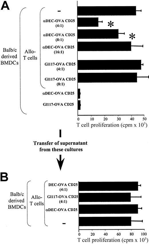
Depletion of CD25+ T cells restores proliferation and IL-2 production. (A) Mice were reconstituted with OVA-specific T cells and injected with αDEC-OVA or αGl117-OVA (isotype). Eight days later, OVA-specific T cells (αDEC-OVA T cells and Gl117-OVA T cells) were prepared and cocultured with OVA-pulsed BMDCs. In some experiments, as indicated, CD25+ T cells were depleted, and isolated CD25+ cells were added to T-cell preparations (1:4 ratio) derived from Gl117-OVA–injected mice. T-cell proliferation was assayed by H3-thymidine incorporation. The asterisk indicates a significant difference (α < 0.05) from proliferation obtained in samples with isotype-control–injected mice (Gl117-OVA T cells). (B) Mice were treated as in panel A, and CD25+ T cells were isolated after 8 days and incubated with a mixture of anti-CD3 antibodies, spleen cells, and CD4+ responder T cells. In controls either CD25+ or CD4+ T cells were stimulated. T-cell proliferation was determined by 3H-thymidine incorporation. The asterisk indicates a significant difference (α < 0.05) from proliferation obtained with stimulated CD4+ T cells only. Error bars indicate mean ± SD. (C) Mice were injected, and cells were isolated and cocultured as in panel A. Two days after coculture, cells were fixed/permeabilized and stained with FITC-labeled αCD4 and PE-labeled αIL-2 antibodies and were analyzed by FACS. Numbers shown are the percentage of cells within the designated quadrant.
Depletion of CD25+ T cells restores proliferation and IL-2 production. (A) Mice were reconstituted with OVA-specific T cells and injected with αDEC-OVA or αGl117-OVA (isotype). Eight days later, OVA-specific T cells (αDEC-OVA T cells and Gl117-OVA T cells) were prepared and cocultured with OVA-pulsed BMDCs. In some experiments, as indicated, CD25+ T cells were depleted, and isolated CD25+ cells were added to T-cell preparations (1:4 ratio) derived from Gl117-OVA–injected mice. T-cell proliferation was assayed by H3-thymidine incorporation. The asterisk indicates a significant difference (α < 0.05) from proliferation obtained in samples with isotype-control–injected mice (Gl117-OVA T cells). (B) Mice were treated as in panel A, and CD25+ T cells were isolated after 8 days and incubated with a mixture of anti-CD3 antibodies, spleen cells, and CD4+ responder T cells. In controls either CD25+ or CD4+ T cells were stimulated. T-cell proliferation was determined by 3H-thymidine incorporation. The asterisk indicates a significant difference (α < 0.05) from proliferation obtained with stimulated CD4+ T cells only. Error bars indicate mean ± SD. (C) Mice were injected, and cells were isolated and cocultured as in panel A. Two days after coculture, cells were fixed/permeabilized and stained with FITC-labeled αCD4 and PE-labeled αIL-2 antibodies and were analyzed by FACS. Numbers shown are the percentage of cells within the designated quadrant.
Injection of αDEC-OVA activates Treg in vivo
CD25+ Treg cells have to be activated through the TCR to exert their suppressive effects. Once activated, they exert their regulatory effect antigen-nonspecifically.10 In experiments shown so far, T cells isolated from injected mice were restimulated with OVA-pulsed DCs that could activate Treg in an antigen-specific manner and might, therefore, not reflect their in vivo activation status. To test whether CD25+ Treg isolated from αDEC-OVA–injected mice was already activated in vivo, MLRs were performed using Balb/c-derived BMDCs as stimulator cells and B6 T cells as responders. Freshly isolated CD25+ Treg derived from αDEC-OVA–injected mice were added in graded doses to these cultures. In these experimental settings, the CD25+ T cells isolated from αDEC-OVA–injected, DO11.10–reconstituted Balb/c mice are syngeneic to the respective stimulator cells, and no ex vivo activation occurs. Here we show (Figure 4A) that CD25+ T cells derived from αDEC-OVA–injected mice significantly suppress T-cell proliferation in an MLR in a dose-dependent manner. In contrast, when CD25+ T cells isolated from Gl117-OVA–injected mice were added to the MLR, no suppressive effect could be recorded. Finally, CD25+ T cells themselves did not proliferate under these conditions. Thus, these data indicate that CD25+ Treg derived from αDEC-OVA–injected mice are activated in vivo and exert their suppressive effects in an antigen-nonspecific manner.
Injection of αDEC-OVA activates Treg in vivo. (A) CD25+ T cells from reconstituted mice that were injected with αDEC-OVA or Gl117-OVA were isolated and mixed with B6-derived CD4+ T cells at different ratios. Cells were then stimulated with Balb/c-derived BMDCs, and T-cell proliferation was assayed by 3H-thymidine incorporation. Pure B6 T cells served as positive control. The asterisk indicates a significant difference (experiments were carried out 5 times; mean of triplicate wells; α < 0.05) from proliferation obtained with T cells only. (B) Tissue-culture supernatants from cocultures of T cells described in panel A (as indicated) were added to conventional MLR, and T-cell proliferation was assayed by 3H-thymidine incorporation (mean of triplicate wells ± SD is shown).
Injection of αDEC-OVA activates Treg in vivo. (A) CD25+ T cells from reconstituted mice that were injected with αDEC-OVA or Gl117-OVA were isolated and mixed with B6-derived CD4+ T cells at different ratios. Cells were then stimulated with Balb/c-derived BMDCs, and T-cell proliferation was assayed by 3H-thymidine incorporation. Pure B6 T cells served as positive control. The asterisk indicates a significant difference (experiments were carried out 5 times; mean of triplicate wells; α < 0.05) from proliferation obtained with T cells only. (B) Tissue-culture supernatants from cocultures of T cells described in panel A (as indicated) were added to conventional MLR, and T-cell proliferation was assayed by 3H-thymidine incorporation (mean of triplicate wells ± SD is shown).
To further demonstrate that the suppressive effects observed in cocultures with DEC-OVA–derived T cells is not caused by soluble mediators such as IL-10 or transforming growth factor-β (TGF-β), we transferred the tissue-culture supernatant from these αDEC-OVA–derived “suppressed” cultures to cultures containing T cells isolated from control mice (Figure 4B). Here we show that no suppressive activity was transferred by the tissue-culture supernatants, indicating that direct cell contact of CD25+ T cells is necessary for Treg to exert suppressive activity.
Injection of αDEC conjugates suppresses delayed-type hypersensitivity reactions in vivo
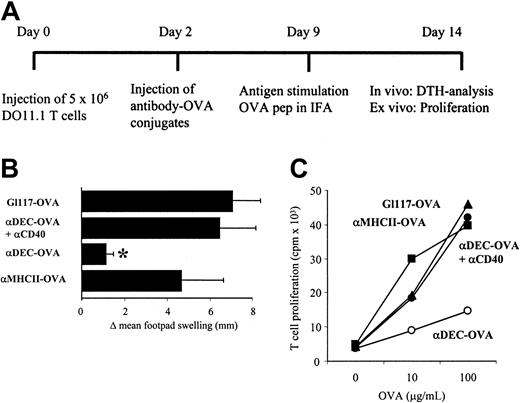
To corroborate our in vitro observations with in vivo results, we tested the effect of αDEC-OVA injections in a CD4+ T-cell–driven, delayed-type hypersensitivity (DTH) model. In this model, we reconstituted Balb/c mice with DO11.10 T cells and injected the respective antibody-OVA conjugates. Thereafter, a T-cell response was induced by the injection of OVA into footpads, and the swelling reaction was determined as a measure for T-cell activation. Figure 5 shows that the injection of αDEC-OVA before challenge with OVA reduced footpad swelling significantly. In contrast, the injection of Gl117-OVA had no effect on DTH reaction. When αMHC class 2-OVA conjugates were injected into mice, a minimal reduction of footpad swelling could be observed that was not considered significant (Student t test). Similar to results obtained in in vitro restimulation assays, the coadministration of DC-activating αCD40 antibodies abolished the regulatory effects, and footpad thickness was increased as in untreated samples. These data could further be confirmed by restimulation assays in vivo. We show (Figure 5C) that in vitro proliferation of T cells isolated from αDEC-OVA–injected mice was suppressed compared with untreated or isotype-OVA–injected mice.
Injection of αDEC conjugates inhibits T-cell proliferation in DTH reactions in vivo. (A) Mice were treated as described in the scheme. (B) After challenge with 30 μg OVA peptide in IFA into the right footpad, footpad thickness was measured after 24 hours using a caliper rule. The asterisk indicates a significant difference (mean of 5 mice/group; α < 0.05) from antibody control (G1117-OVA). Results from 1 of 3 typical experiments are shown. (C) LN cells obtained from mice described in panel B were prepared and cultured in the presence of OVA, as indicated. ▴ indicates GI117-OVA; •, α MHCII-OVA; ▪, αDEC-OVA + αCD40; and ○, αDEC-OVA. T-cell proliferation was assayed by 3H-thymidine uptake.
Injection of αDEC conjugates inhibits T-cell proliferation in DTH reactions in vivo. (A) Mice were treated as described in the scheme. (B) After challenge with 30 μg OVA peptide in IFA into the right footpad, footpad thickness was measured after 24 hours using a caliper rule. The asterisk indicates a significant difference (mean of 5 mice/group; α < 0.05) from antibody control (G1117-OVA). Results from 1 of 3 typical experiments are shown. (C) LN cells obtained from mice described in panel B were prepared and cultured in the presence of OVA, as indicated. ▴ indicates GI117-OVA; •, α MHCII-OVA; ▪, αDEC-OVA + αCD40; and ○, αDEC-OVA. T-cell proliferation was assayed by 3H-thymidine uptake.
Injection of αDEC conjugates suppresses contact hypersensitivity reactions in vivo
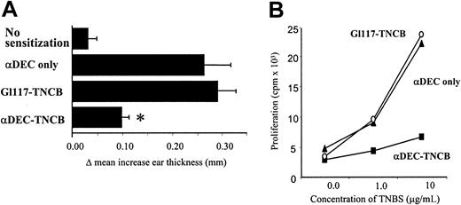
Our previous results have shown that coupling of a protein antigen to αDEC antibodies results in the induction of anergy in the CD4+ T-cell compartment. To extend these findings in a different CD8-driven disease model, we tested the effects of αDEC injection in a CD8+ T-cell–dependent contact hypersensitivity (CHS) reaction using the contact sensitizer trinitrochlorobenzene (TNCB). In a therapeutic setting, groups of 8 mice were sensitized against TNCB and treated by injection of αDEC-205 or isotype control antibodies (Gl117) that were derivatized with TNCB. Respective controls were left untreated or received αDEC antibody without TNCB. Thereafter, mice were challenged by the application of TNCB onto one ear, and ear-swelling reactions were measured 24 hours later. In these experiments (Figure 6), the injection of TNCB-derivatized αDEC antibodies reduced ear swelling significantly. Injection of the respective isotype control (Gl117) had no effect on the CHS. Similar results could be obtained in LN restimulation assays—that is, T cells isolated from mice that received derivatized αDEC antibodies showed reduced proliferation compared with respective control groups (Figure 6B). Therefore, we conclude that antigens given to immature DCs in the steady state induce T-cell unresponsiveness in vitro and in vivo.
Injection of αDEC conjugates inhibits T-cell proliferation in CHS reactions in vivo. (A) For CHS reactions, mice were sensitized with TNCB followed by injection of derivatized αDEC-205 or Gl117 antibodies. Five days later, mice were challenged with TNCB at the right ear, and ear thickness was measured after 24 hours using a caliper rule. The asterisk indicates a significant difference (mean of 5 mice/group; α < 0.05) from antibody control (αDEC only). Results from 1 of 4 typical experiments are shown. (B) Mice were treated as in panel A, except that LN cells were prepared, 5 × 10⁵ cells were incubated with TNBS, and T-cell proliferation was determined 3 days later.
Injection of αDEC conjugates inhibits T-cell proliferation in CHS reactions in vivo. (A) For CHS reactions, mice were sensitized with TNCB followed by injection of derivatized αDEC-205 or Gl117 antibodies. Five days later, mice were challenged with TNCB at the right ear, and ear thickness was measured after 24 hours using a caliper rule. The asterisk indicates a significant difference (mean of 5 mice/group; α < 0.05) from antibody control (αDEC only). Results from 1 of 4 typical experiments are shown. (B) Mice were treated as in panel A, except that LN cells were prepared, 5 × 10⁵ cells were incubated with TNBS, and T-cell proliferation was determined 3 days later.
Discussion
Our study demonstrates that the specific targeting of immature DCs with the mAb αDEC-205 coupled to various antigens results in the presentation of the respective antigen in a tolerizing fashion. We show in an OVA-specific system that after the initial expansion of antigen-specific T cells, these specific T cells are anergized and express CD25 and CTLA-4 antigen, the phenotypic hallmarks of regulatory T cells. Functional analysis of this T-cell population revealed that CD25+ T cells from αDEC-OVA–injected animals dose-dependently suppressed proliferation and IL-2 production of conventional CD4+ T cells in a cell-contact–dependent way. Depletion of CD25+ T cells from bulk T-cell cultures restored T-cell proliferation. These phenotypic and functional aspects qualify T cells induced by αDEC-OVA as genuine regulatory T cells.11 In further CD4- and CD8-driven disease models, we demonstrate that the induction of regulatory T cells by αDEC results in the suppression of immune responses in vivo. Thus, in vivo targeting of antigens to immature DCs by αDEC mAb results in the control of immune responses by the induction of regulatory T cells.
Regulatory T cells (Treg) were first described by Sakaguchi et al.12 Originally, Treg cells were extensively characterized in the murine system as a subpopulation of 5% to 10% of all peripheral CD4+ T cells. Development of Treg cells occurs from the thymus at approximately postnatal day 3, and their maintenance depends possibly on the recognition of autoantigens in the periphery in combination with the triggering of CD28 and CD154 coreceptors.13,14 Eradication of Treg cells results in the development of different autoimmune diseases, among them gastritis and thyroiditis, indicating that the main function of the CD25+ T regulatory cells is the suppression of otherwise autoreactive T cells.15-18 The suppressive mechanisms by which CD25+ Treg cells exert their inhibitory effects in vitro were found to be dependent on a direct T-regulatory/T-effector cell contact. The inhibitory effect itself is antigen nonspecific but requires antigen-dependent activation of the regulatory T cells through their T-cell receptor.9,10 In this regard, antigen-presenting cells play a crucial role in activation of Treg, and in the human system it has been shown that immature DCs in vitro can induce alloantigen-reactive Treg cells. Repetitive stimulation of naive CD4+ T cells with allogeneic immature DC, in the absence of immune-modulating agents, results in the differentiation of naive T cells into nonexpanding CD4+/CD25+ T cells.19 This T-cell population induced by immature DCs in vitro, produces high amounts of IL-10 but no IL-4, IL-5, or IL-2.20 They can act directly on activated TH1 cells and inhibit their antigen-specific proliferation and cytokine production in a cell-contact–dependent manner. The suppressive activity of these regulatory T cells is antigen-nonspecific and can be partially inhibited by the addition of exogenous IL-2. Thus, the functional activities of these human regulatory T cells are similar to the murine CD4+/CD25+ Treg cells described earlier. In the aggregate, these data indicate that immature DCs, through the induction of regulatory T cells, are critically involved in the process of immunoregulation.
To specifically target immature DCs in vivo, the DEC-205 receptor is an ideal candidate molecule. DEC-205 is a decalectin originally characterized by the monoclonal antibody NLDC-145. As a receptor, for purposes of antigen targeting to DCs in situ, it offers several advantages. DEC-205 is expressed in abundance on DCs in the T-cell areas,4 and antibodies bound to DEC-205 are efficiently internalized and delivered to antigen-processing compartments.3 In vivo, αDEC-205 mAb targets to DCs efficiently, with a dose of less than 1 μg antibody leading to presentation of the coupled antigen by DCs. It also appears that antigens internalized by DEC-205 are stored in the DCs for prolonged periods of time, further increasing the efficiency of antigen presentation by DCs. Most important, internalization of the antigen by DEC-205 does not induce maturation of the DCs. Immature DCs had been shown to induce anergic, regulatory T cells in vitro,19,21 and αDEC-205 targeting of antigens to immature DCs led to anergic T cells in vivo. These anergic T cells have regulatory capacities and can be used to suppress CD4-or CD8-mediated immune responses in vivo. This notion is further supported by results showing that subtypes of DEC-205+ DCs are involved in conveying tolerance.22 A DEC-205–expressing subset of lymphoid DCs has been shown to mediate deletion of potentially autoreactive T cells through Fas-mediated killing,23 and a DEC-205+ subset of liver DC exerts tolerogenic functions.24 Therefore, our data finally prove the hypothesis proposed by Steinman et al25,26 that DCs in the steady state are prone to induce tolerance in vivo and that the DEC-205 receptor is a crucial molecule in this process. In this concept the function of DCs and finally the outcome of an immune response is determined by the maturation status of the DCs. Using αDEC-mediated targeting, we have indeed demonstrated that the immature, tolerogenic DCs could be converted to potent T-cell stimulatory cells after the induction of maturation— that is, when αDEC conjugates were given together with DC-activating αCD40 antibodies,8 T cells were fully activated and not anergized. These results might reflect an in vivo situation in which tissue damage or inflammation delivers signals to DCs resulting in a mature and fully activated phenotype.27,28 Only these mature DCs are potent T-cell stimulators that activate T cells for the induction of an immune response.8,27,29 Beyond several in vitro approaches for the induction of Treg, our findings also demonstrate that CD4+/CD25+ regulatory T cells can be induced by immature DCs in vivo and that these cells are involved in the control of diseases such as allergic contact sensitivity. CD8+ T cells mainly drive this hypersensitivity reaction, and our results reflect an in vivo example for the suppressive effect of Treg on CD8+ effector cells, a mechanism recently proposed by Piccirillo and Shevach.30 Initially, the tolerogenic effect of immature DCs in vivo has been observed in liver transplantation or in a model of experimental allergic encephalomyelitis.31-33 In addition, human volunteers vaccinated with immature DCs in vivo showed induction of IL-10–producing T cells with regulatory activity.34,35 The major problem with the direct use of immature DCs for in vivo therapeutic approaches in mice and humans, however, is that the fate of the injected DCs is impossible to predict. They may die before they have triggered the appropriate T cells, or, what is worse, they might be driven to maturation under the influence of proinflammatory cytokines and immunize instead of tolerize. These problems can be circumvented by the direct in vivo targeting of immature DCs with αDEC-205 mAb.
Our data open the field for novel therapeutic strategies targeting immature DCs in vivo by αDEC-205. We show that the coupling of antigen to αDEC-205 mAb leads to the induction of regulatory T cells that are able to down-modulate immune responses in vivo. The results in murine DTH footpad swelling reactions and ACD ear swelling reactions further emphasize that CD4- and CD8-mediated immune responses can be affected by this novel therapeutic approach. Although the effect observed is antigen nonspecific, the immunosuppression is not long-lasting because Treg will die in vivo shortly after activation. Thus, coupling of known autoantigens or allergens to αDEC-205 mAb might provide a novel means to cure allergic and autoimmune diseases.
Prepublished online as Blood First Edition Paper, January 23, 2003; DOI 10.1182/blood-2002-10-3229.
Supported by Deutsche Forschungsgemeinschaft grants EN209-7.1 and SFB548-A3 and by a Mainz-Forschungsfonds (MAIFOR) grant (K.M.).
The publication costs of this article were defrayed in part by page charge payment. Therefore, and solely to indicate this fact, this article is hereby marked “advertisement” in accordance with 18 U.S.C. section 1734.
We thank A. Meinl and M. Rivera for excellent technical assistance and Kerstin Steinbrink and Steven Oliver for critical reading of the manuscript.

![Figure 1. αDEC-205 antibodies target OVA to MHC class 2+ cells and induce T-cell proliferation. (A-B) αDEC-OVA conjugates were injected into the footpads of mice. Twenty-four hours later tissue sections from LNs were stained with PE-labeled goat anti-rat antibodies (to detect injected αDEC-OVA [red]). Specimens were further double labeled with FITC-αMHC class 2 antibodies (A) or FITC–SER-4 antibodies (B), respectively (green). Colabeling is indicated by yellow. Original magnification, ×200. (C) Twenty-four hours after the injection of 10 μg antibody-OVA conjugates, CD11c+ and CD11c– cells were prepared and cocultured with OVA-specific DO11.10 T cells. T-cell proliferation was assayed by 3H-thymidine incorporation. (D) A mixture of αDEC and OVA and of αDEC or OVA alone was injected, and CD11c+ cells were tested for antigen presentation as in panel C. (E) Mice were reconstituted with CFSE-labeled, OVA-specific DO11.10 T cells and injected with different antibody-OVA conjugates as indicated. Forty-eight hours later, LN cells were prepared and stained with PE-labeled αCD4 antibody. CFSE labeling was assayed by FACS after gating on CD4+ cells, and T-cell division is indicated by dilution of the dye (ie, decrease of fluorescence).](https://ash.silverchair-cdn.com/ash/content_public/journal/blood/101/12/10.1182_blood-2002-10-3229/6/m_h81234469001.jpeg?Expires=1769114298&Signature=qdinF3Ipr0x0RVZa3E-b5iUaxQOrF4B~fpOAaogYK-e5XnKEMExu8C4~PJrTS7-5wH0o8AlLYkyMd6Pi5PE-y29MyXS3qdSVwUXgmyBe1Od5BoEb4KzSSeqr7zMSbospoBEpDBH4V4UMP72yyLRkTp63JAWNn2s5ltFRWNcVmn99EeHqHzhTEorHUhwBO2l37Hf0O5xDgxw9ovjflBOYk2-NY-rI7B8y-zB6vey7UpB0-HMWRVZUA2RWszmKBt7Lc98SZFl~czyM923JcexSti2yannNv4BOkt8d21OCJvUiPGL0pt49z72iXYWWvYQq8clA~b-UObaBxijw~ttV~w__&Key-Pair-Id=APKAIE5G5CRDK6RD3PGA)


This feature is available to Subscribers Only
Sign In or Create an Account Close Modal