Abstract
Transgenic mice have been generated that carry a CD45 minigene under control of the human leukocyte function-associated antigen (LFA-1, CD11a) promoter. CD45-null mice carrying the transgene exhibit the lymphocyte lineage-specific isoform expression patterns of wild-type mice. Furthermore, these mice have normal thymocyte development and peripheral T-cell numbers. The proliferative ability of T cells in response to mitogens and antigen also is regained, as is B-cell responsiveness to anti-IgM. The antibody response to antigen is also restored and is similar to that of normal mice. Therefore, introduction of a functional CD45 minigene is sufficient to overcome the principal severe combined immunodeficiency (SCID)–associated defects and represents a potential route to a gene therapy for human CD45-deficent SCID.
Introduction
CD45 is a structurally heterogeneous, transmembrane protein-tyrosine phosphatase found exclusively on nucleated cells of hematopoietic origin. The heterogeneity results from differential RNA splicing of 6 exons in the extracellular domain.1-3 The isoforms are expressed in a cell type–specific pattern. B cells predominantly express the isoform containing all exons.4,5Thymocytes express the lowest molecular weight isoforms resulting from the removal of 3 and 4 exons.2-4 T cells have a varied isoform expression pattern that is dependent upon the differentiation state, function, and prior antigenic exposure.3
In addition to the structural heterogeneity of CD45 proteins, CD45 allelic differences also have been noted. At least 3 CD45 alleles in inbred mice strains have been identified based on antigenic differences and polymorphic sequence variations.2,6,7 TheLy5a (CD45.1) and Ly5calleles are found only in a few strains, while theLy5b (CD45.2) allele is expressed by most of the established lines. The sequence differences between the 3 alleles produce proteins that differ in their reactivity to specific monoclonal antibodies.7
CD45 has been shown to be essential for T-cell development8-10 and for the function of both T and B lymphocytes.8-15 In CD45-null mice, thymocyte development is blocked, resulting in few peripheral T cells. The function of these T cells in the periphery also is defective, and they do not proliferate in the presence of activators or mitogens. B-cell maturation in the CD45-negative mice is not effected by the CD45 deficiency, although their proliferative response to anti-IgM is abrogated.9,10With regard to lymphocyte function, CD45 is essential for signaling through the T- as well as B-cell antigen receptors11-14,16,17 and is required for positive and negative regulation of the Src family kinases associated with these receptors.18-21
Human severe combined immunodeficiencies (SCIDs) are a group of genetic diseases in which T-cell development is blocked and other lymphoid lineages are variably affected.22,23 The genetic defects of many forms of the disease now have been identified, including mutations in the common γ-chain of cytokine receptors found in X-linked SCID and alterations in several other genes, such as JAK-3 kinase, Rag-1, and Rag-2, which cause autosomal recessive SCID.23,24 Autosomal recessive SCID also results from a deficiency in the CD45 gene.24-26 The phenotype of these patients is similar to that of CD45 knockout mice9,10; not only are peripheral T-cell numbers decreased but T cells that are present fail to respond to mitogens.26 Although B-cell numbers are normal, these cells also are dysfunctional.25 26
We have generated a CD45 minigene encoding the CD45.1 allele in which 2 cDNA regions spanning exons 1b-3 and 9-33 flank genomic DNA containing the alternatively spliced exons 4-8. Transcriptional regulation of the minigene from the native CD45 promoter region was ineffective, but the human leukocyte function–associated antigen (LFA-1) promoter, which has tissue-specific expression characteristics similar to CD45, produced high levels of CD45 expression in lymphoid cell lines.27 The demonstration that correctly spliced, cell lineage–specific CD45 mRNAs were produced in both T- and B-cell lines indicates that the minigene construct has many of the expression properties of the endogenous CD45 gene. These expression characteristics in vitro were encouraging; however, the question remains whether the minigene can supply functional CD45 isoforms to the various leukocyte lineages. To determine whether the minigene LFAiT200 could restore the functional and developmental properties of the immune system, we generated transgenic CD45-null mice carrying the minigene. The introduction of the CD45 minigene is sufficient to overcome the SCID-associated defects and, with an appropriate delivery system, represents a potential gene therapy for human CD45-deficient SCID.
Materials and methods
Mice
The mice used were 7- to 16-week-old C57BL/6 (Harlan, San Diego, CA), SJL/J, and CD45-deficient C57BL/64 (Jackson Labs, Bar Harbor, ME). Transgenic mice were generated by injecting the 20-kb SfiI-NotI fragment of pLFAiT200 containing the CD45 minigene27 into fertilized (BALB/c × C57BL/6)F2 mouse oocytes. Mice carrying the transgene were identified using DNA isolated from tail snips using the DNeasy Tissue Kit (Qiagen, Valencia, CA) by polymerase chain reaction (PCR) analysis. The antisense primer includes bases 1 to 17 of exon 3 with additional nucleotides containing an XhoI cloning site at the 5′ end (5′-CTTTTGACTCGAGGGTGTAGGTGTTTGCC-3′). The sense primer consists of bases 32 to 50 of exon 1b with additional nucleotides, including aClaI cloning site added at the 5′ end (5′-GTGCGGATCGATAAGTGGTTTGTTCTTAGGG-3′). The first cycle was 94°C, 4 minutes; 60°C, 1 minute; 72°C, 1 minute. Cycles 2-34 were 1 minute at 94°C, 1 minute at 60°C, and 1 minute at 72°C. A 211-bp fragment results from the presence of transgene DNA, whereas no signal is observed from the endogenous CD45 gene due to the approximately 50 kb of sequence between the primer sites. Southern analysis was used to confirm the PCR results. Total genomic DNA was digested with the restriction enzyme BamHI. The probe was aBamHI cDNA fragment extending from exon 13 to exon 22. These CD45 transgenic mice were crossed with the CD45-null strain until CD45 transgene-positive progeny were obtained on a CD45-null background.
Quantitation of transgene copy number
The number of integrated copies of the transgene was determined by Southern analysis. Total genomic DNA was digested with a combination of restriction enzymes EcoRI and BamHI. The hybridization probe consisting of intron sequence between exons 8 and 9 detected a 2.3-kb fragment specific for the transgene and a 2.0-kb band diagnostic of the endogenous gene. The approximate copy number was estimated by comparing the hybridization intensity of the transgenic fragment to that of the endogenous band.
Proliferation assays
T-cell proliferation assays were conducted in 96-well microtiter plates using 1 × 10⁶ cells per well for spleen cells isolated from CD45-null mice and 2 × 10⁵ spleen cells per well for all other strains. T-cell activators were concanavalin A (ConA) (Sigma, St Louis, MO) at 1 μg/mL or a combination of anti-CD3 and anti-CD28 (PharMingen, La Jolla, CA) at 2 μg/mL each. For the mixed lymphocyte reaction (MLR) assay, 4 × 10⁵ responder cells were added to 2 × 10⁵, 4 × 10⁵, and 8 × 10⁵ irradiated (2000 rad) spleen stimulator cells from major histocompatibility complex (MHC)-allogeneic SJL mice or irradiated responder cells. In the anti-IgM proliferation assay, 20 μg/mL of the F(ab′)2 fragment of goat anti–mouse IgM (Jackson ImmunoResearch Laboratories, West Grove, PA) was incubated with 2 × 10⁵ spleen cells per well. For bacterial lipopolysaccharide (LPS) stimulation assays 15 μg/mL of LPS (Difco Laboratories, Detroit, MI) were added to 1 × 10⁵ spleen cells. All cells were cultured in RPMI 1640 supplemented with 2 mM L-glutamine, 10% fetal bovine serum, 0.05 mM 2-mercaptoethanol, 100 units/mL penicillin, and 100 μg/mL streptomycin. Media components were from Irvine Scientific (Irvine, CA). All cultures were prepared in triplicate and incubated at 37°C in 5% CO2. Cultures were given 1 μCi (0.037 MBq)/well thymidine [methyl–3H] on day 2 except for MLR cultures, which received the labeled nucleotide on day 5. After overnight incubation, cells were harvested on a Tomtec Cell Harvestor 96 Mach III, and [3H]thymidine incorporation was measured on a Wallac 1450 Microbeta TriLux liquid scintillation and luminescence counter. Results are expressed as Δ cpm, experimental minus control counts. In the MLR assay, the control counts were from cultures incubated with irradiated responder cells.
Flow cytometry
Cells were labeled with fluorescence-tagged monoclonal antibodies and analyzed as described27 using the following antibodies: R-phycoerythrin (PE)–conjugated anti-CD4 (Caltag, Burlingame, CA), Tri Color–conjugated anti-CD8 (Caltag), PE-conjugated anti-CD11a (PharMingen), allophycocyanin (APC)–conjugated anti-CD11b (Caltag), PE-conjugated anti-CD19 (Caltag), fluorescein isothiocyanate (FITC)–conjugated anti-CD44 (Caltag), FITC-conjugated anti-CD45.1 (PharMingen), FITC-conjugated anti-CD45.2 (PharMingen), biotin-conjugated anti-CD45.2 (PharMingen), R-PE–conjugated anti-CD45RB (PharMingen), biotin-conjugated anti-CD62L (Caltag), Tri Color–conjugated anti-F4/80 (Ly-71) (Caltag), SaV-APC (PharMingen), SaV-Tri Color (Caltag), and conjugated Ig2a and Ig2b isotype controls (PharMingen). Cells from spleen and thymus were used for analysis of lymphoid and myeloid cells. For the study of naive and memory CD4+ cells, mice were immunized subcutaneously at the base of the tail with 1 mg dinitrophenyl-keyhole limpet hemocyanin conjugate (DNP-KLH) in complete Freund adjuvant (CFA). Draining inguinal and para-aortic lymph nodes were obtained 5 days after immunization.
Reverse transcription and polymerase chain reaction
Total RNA was isolated from spleen and thymus using TRIZOL (Life Technologies, Gaithersburg, MD) following manufacturer's directions. Spleen cells were untreated and exposed for 3 days to ConA or LPS. Reverse transcription–polymerase chain reaction (RT-PCR) was performed using the GeneAmp RNA PCR kit (Applied Biosystems, Foster City, CA) according to manufacturer's directions. The antisense oligonucleotide used for both cDNA synthesis and PCR amplification, 5′-GGAGCACATGAGTCATTAGACACACTGATG-3′, consists of bases 155-184 of CD45 exon 9. The sense primer includes bases 21-51 of exon 2 and has the sequence 5′-GCACAGCTGATCTCCAGATATGACCATGGGT-3′. cDNA was synthesized for 15 minutes at 42°C followed by inactivation of reverse transcriptase for 5 minutes at 95°C. The first PCR cycle was 3 minutes at 94°C, 1 minute at 70°C, and 2 minutes at 72°C. Cycles 2-24 were 1 minute at 94°C, 1 minute at 70°C, and 2 minutes at 72°C. The amplified products were separated on an alkaline agarose gel and identified as described previously.28
Immunization and ELISA
From each mouse type—transgenic F, C57BL/6, and CD45-null— 5 mice were immunized by intraperitoneal injection of 0.1 mg DNP-KLH (Calbiochem, San Diego, CA) in 0.1 mL CFA (Sigma). Mice were bled 7, 14, and 21 days after immunization and serum prepared.
The enzyme-linked immunosorbent assay (ELISA) was performed as described.29 Briefly, wells were coated overnight with 0.05 mg dinitrophenyl-bovine serum albumin (DNP-BSA) in 0.1 M carbonate buffer, pH 9.6, washed with phosphate-buffered saline (PBS), blocked with 1% BSA/PBS, washed with PBS, and incubated with serial dilutions of mouse serum for 4 hours at room temperature. Rabbit anti–mouse IgM or IgG at a dilution of 1:1000 was added for 2 hours at room temperature, washed with PBS, and incubated overnight at 4°C with goat anti–rabbit IgG conjugated to alkaline phosphatase (Kirkegaard and Perry Laboratories, Gaithersburg, MD) at a dilution of 1:2000. Following 3 washes with PBS, 10 μg 4-methylumbelliferyl-phosphate (Sigma) in carbonate buffer was added for 30 minutes and fluorescence measured with excitation at 360 nm and emission at 440 nm in a fluorescence plate reader (Fluoroskan, Titertek). Mouse anti–trinitrophenyl-IgM antibody (Pharmingen) and anti–trinitrophenyl-IgG antibody (gift of Dr Phyllis Linton, Sidney Kimmel Cancer Center, San Diego, CA) were used for the standard curves. The serum anti–DNP IgM and anti–DNP IgG levels were calculated from duplicate wells of each dilution from 5 different mice.
Results
Generation of transgenic mice
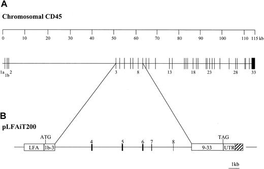
The 20-kb CD45 expression fragment of pLFAiT200 (Figure1) was injected into fertilized mouse oocytes for the production of transgenic mice. Three founder mice were initially identified by PCR amplification of minigene-specific sequence from genomic DNA isolated from tail and subsequently confirmed by Southern analysis. The 3 founders have similar properties, and here we report our findings with transgenic mice from founder F. The number of integrated copies of the transgene in transgenic F was determined to be approximately 15, with all copies transmitted to offspring as a single inheritable unit, indicating integration of all copies at one site. The transgene locus was placed on a CD45-negative background through matings with the exon 9–disrupted CD45-null mouse (CD45.2−/−).9 Unless specifically stated otherwise, reference to the transgenic mouse indicates the transgene is on the CD45.2−/− background.
Structure of the murine chromosomal CD45 gene and the minigene, pLFAiT200.
(A) The map of the 115-kb murine CD45 gene30 shows the exon and intron organization. The 12-kb genomic sequence contained in the minigene is indicated. (B) The structure of the CD45 minigene, pLFAiT200, is illustrated. LFA is the human LFA-1 (CD11a) promoter, and the UTR is the 3′ CD45 untranslated region. The CD45 5′ cDNA region containing exons 1b through 3 and the 3′ cDNA sequence region with exons 9 through 33 are designated 1b-3 and 9-33, respectively. The hatched box indicates the genomic sequence contiguous with the UTR 3′ of the CD45 poly(A) site.
CD45.1 expression
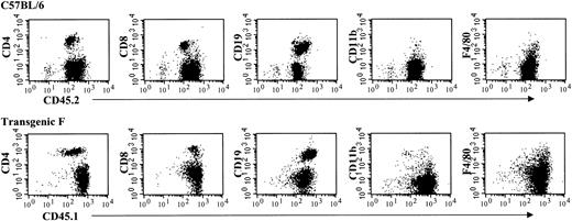
The expression of the transgene was investigated by flow cytometry of spleen cells labeled with monoclonal antibodies specific for CD45.1 and CD45.2 (Figure 2). As expected for the controls, high levels of CD45.2 protein are detected on C57BL/6 spleen cells, but no CD45.1 protein is found. Spleen cells from SJL/J mice express high amounts of CD45.1 but not CD45.2. No CD45.2 or CD45.1 protein is observed on cells isolated from CD45-null mice. Lymphocytes from transgenic mice show CD45.1 protein levels that approach those of CD45.1 expression in SJL/J mice. Both T (CD4+ and CD8+) and B lymphocytes (CD19+) and myeloid lineages (F4/80+ and CD11b+) express significant levels of CD45.1 protein that are comparable to those observed in C57BL/6 mice (Figure 3).
Comparison of CD45.1 transgene expression with endogenous CD45 gene expression in splenocytes from C57BL/6, SJL/J, CD45-null, and transgenic mice.
The source of spleen cells is indicated above each overlay. For each overlay the staining antibodies were CD45.1 (solid line) and CD45.2 (dotted line).
Comparison of CD45.1 transgene expression with endogenous CD45 gene expression in splenocytes from C57BL/6, SJL/J, CD45-null, and transgenic mice.
The source of spleen cells is indicated above each overlay. For each overlay the staining antibodies were CD45.1 (solid line) and CD45.2 (dotted line).
Analysis of cell surface expression of the CD45.1 transgene in lymphoid and myeloid lineages using flow cytometry.
The top row shows staining of C57BL/6 spleen cells and the bottom row staining of transgenic spleen cells as indicated. The antibodies used for staining are shown on the vertical (CD4, CD8, CD19, CD11b, and F4/80) and the horizontal (CD45.1 and CD45.2) axes.
Since the human LFA-1 promoter drives CD45.1 transcription in the transgene, the expression characteristics of CD45.1 were compared with those of endogenous LFA-1 and CD45.2. Spleen cells were labeled with anti-CD45.1, anti-CD45.2, or anti-CD11a (LFA-1). In C57BL/6 mice, the CD45.2-positive cells are also LFA-1 positive with variable levels of LFA-1 expression (Figure 4A). The brightest third of the LFA+CD45.2+ cells consists primarily of CD4+ and CD8+ T cells (data not shown). CD19+ B cells are in the less brightly labeled population. In the transgenic mouse the 2 populations of LFA+ CD45.1+ cells are more clearly defined. The labeling properties of the LFA+ population containing B cells closely resemble those of wild-type cells by exhibiting lower LFA and higher CD45 expression than the T-cell components (Figure 4A). However, the more brightly labeled LFA+ cells of the T-cell lineages are more heterogeneous with respect to CD45 expression and generally express less CD45 than their wild-type counterparts (Figures3-4A). Importantly, in transgenic mice hemizygous for CD45.2, all cells positive for CD45.1 are also positive for CD45.2 and vice versa (Figure4B).
Comparison of the LFA-1 promoter-driven CD45.1 expression to wild-type CD45.2.
The staining antibodies are given on the vertical and horizontal axes (CD45.1, CD45.2, and LFA-1 [CD11a]). (A) All transgene-positive cells were isolated from mice on a CD45-null background. (B) Cells from transgenic mice were on a CD45.2+/− background.
The CD45 isoform expression pattern from the transgene was evaluated in spleen cells from C57BL/6 and transgenic mice using monoclonal antibodies specific for exon 4 (CD45RA), exon 5 (CD45RB), or exon 6 (CD45RC) and flow cytometry. The percentage of positive cells with each of these antibodies is very similar for the transgenic mice and mice expressing the endogenous CD45 gene (CD45RA, 56% ± 6%; CD45RB, 80% ± 5%; CD45RC, 60% ± 4%).
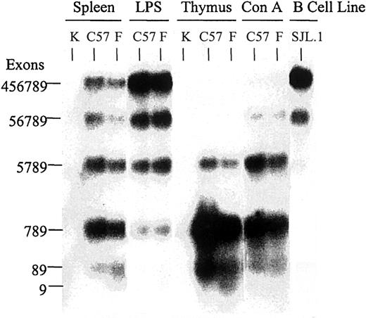
The similarity of isoform expression characteristics of transgenic and normal mice was confirmed through RT-PCR analysis. Lymphocytes from the spleen and thymus of C57BL/6, CD45-null, and transgenic mice were isolated. Total RNA was purified from untreated spleen and thymus cells and from spleen cells treated with ConA or LPS for 3 days. The CD45 isoforms were identified by RT-PCR, using primers that flank the region of alternatively spliced exons, allowing all isoforms to be detected (Figure 5). No CD45 mRNA was detected in cells from the CD45 knockout mouse with the conditions used. Multiple isoforms were observed in C57BL/6 spleen cells, as expected of a mixture of leukocyte populations. The same expression pattern is found in the spleen cells of the transgenic mouse. ConA-induced T-cell blasts show a reduction of the high molecular weight isoforms and an increase in the low molecular weight forms in both the C57BL/6 and transgenic mice. Enrichment of the B-cell population by activation of B-cell proliferation with LPS results in an increase in the proportion of high molecular weight isoforms. Primarily, the 789 and 89 isoforms are expressed in the thymus of C57BL/6 and transgenic mice.
RT-PCR analysis of RNA expression from the CD45 transgene.
The RT-PCR products were amplified from RNA isolated from splenocytes and thymocytes and visualized by Southern analysis. Cells were isolated from C57BL/6 (C57), transgenic F (F), or CD45-null (K) mice. RNA from the SJL pre–B-cell line, SJL.1, was used as a positive control. Spleen cells were untreated (Spleen), exposed for 3 days to ConA, or treated with LPS for 3 days. Thymocytes were untreated (Thymus). The amplification primer sequences are located in exon 2 and exon 9, flanking the region of alternative exon splicing. The oligonucleotide probe also is located in exon 2, downstream of the amplification primer. The exons present in each fragment are indicated.
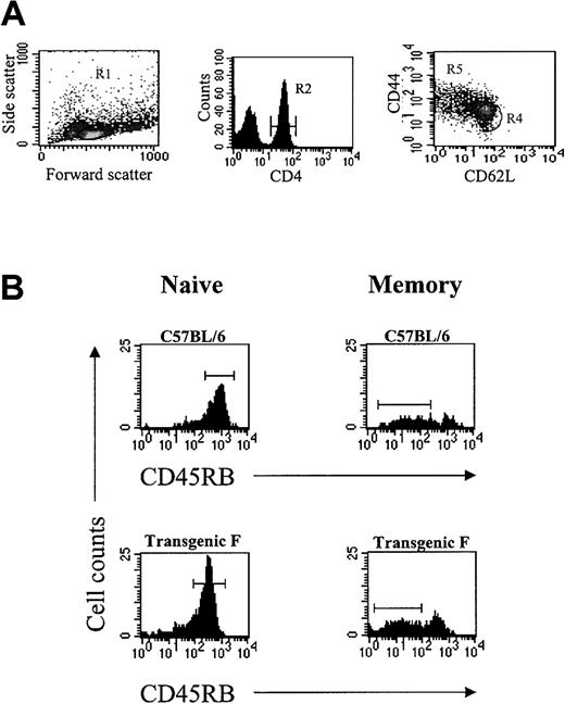
CD45 is commonly used to distinguish naive (CD45RBhi) and memory (CD45RBlo) T cells.31 To determine whether naive and memory T cells retain the same characteristic shift in isoform expression in the transgenic mice, we immunized transgenic and wild-type mice with DNP-KLH and analyzed the CD4+T-cell population. Draining lymph nodes were isolated 5 days after immunization, and the cells were stained with antibodies specific for CD44, CD62L, CD4, and CD45RB. In both wild-type and transgenic mice the CD4+, CD44lo, CD62Lhi cells are CD45RBhi as expected for naive T cells,31 and the percentage of CD45RBhi cells is essentially identical, 89% in wild-type and 88% in transgenic mice (Figure6). The CD4+ cells expressing the CD44hi, CD62Llo memory phenotype were primarily CD45RBlo for both wild-type and transgenic mice; 73% in C57BL/6 and 64% in transgenic mice (Figure 6).
CD45RB phenotype of CD4+ cells from draining lymph nodes of immunized mice.
(A) The gating parameters are shown for selection of naive and memory T cells. Naive CD4+ T cells were selected for CD4+, CD44lo, and CD62Lhi (R1 and R2 and R4), and for memory cells the CD4+, CD44hi, CD62Llo (R1 and R2 and R5) cells were selected.31 (B) The horizontal bars in the naive T-cell panels indicates the high CD45RB-expressing cells, and the memory T-cell panels illustrate the low CD45RB cell population.
Thymocyte development and T-cell function
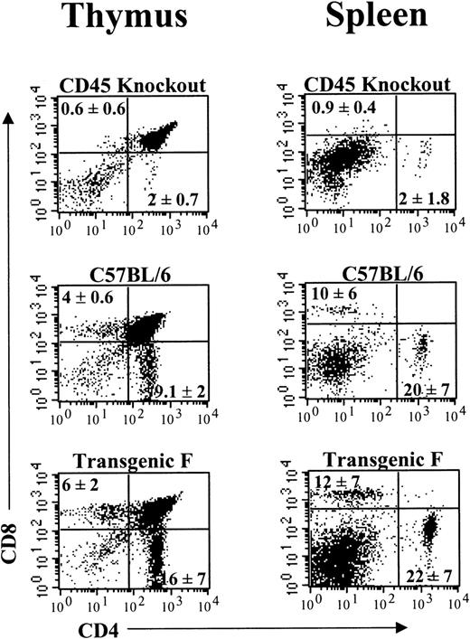
The CD45-null mutation causes a block in T-cell development at the CD4+CD8+ stage, resulting in reduced numbers of mature, single positive thymocytes.8-10 The presence of the CD45.1 transgene restores CD4+ and CD8+single positive thymic T cells in the transgenic mice (Figure7; Table1). The restoration of thymocyte development results in increased numbers of mature T cells in the spleens of these mice (Figure 7; Table 1).
Immunofluorescence of CD4+ and CD8+ T-cell subsets in thymus and spleen of CD45-null, C57BL/6, and transgenic mice.
The source of thymocytes and splenocytes is given above each plot. The staining antibodies (CD4 and CD8) are on the horizontal and vertical axes, respectively. The percentage of CD4 and CD8 single-positive cells are given in the corresponding quadrant and represent the average of 3 thymuses and 5 spleens.
T-cell restoration in transgenic mice
| Mouse . | Thymus . | Spleen . | ||
|---|---|---|---|---|
| CD4 . | CD8 . | CD4 . | CD8 . | |
| Knockout | 0.19 ± 0.06 × 10⁶ | 0.10 ± 0.02 × 10⁶ | 2.3 ± 1.8 × 10⁶ | 1.2 ± 0.8 × 10⁶ |
| C57BL/6 | 5.1 ± 2.0 × 10⁶ | 2.9 ± 1.5 × 10⁶ | 18 ± 5 × 10⁶ | 8.9 ± 2.7 × 10⁶ |
| Transgenic F | 4.0 ± 0.9 × 10⁶ | 3.3 ± 1.0 × 10⁶ | 16 ± 5 × 10⁶ | 7.7 ± 1.9 × 10⁶ |
The values are the total number of CD4 and CD8 single positive cells in the thymus and spleen of the mouse. Averages with standard deviations were derived from the separate analyses of 3 thymuses and 5 spleens.
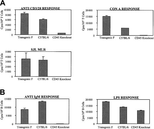
The functional capabilities of the peripheral T cells were determined by proliferation assays of spleen cells activated with ConA, a mixture of anti-CD3 and anti-CD28, or MHC-allogeneic stimulator cells. As expected, proliferative responses to both ConA and anti-CD3/CD28 are completely abrogated in the CD45-null mice (Figure8A). Expression of the CD45.1 transgene restores these proliferative responses to normal levels. Furthermore, in contrast to CD45-null cells, transgenic spleen cells are capable of normal levels of proliferation in reaction to allogeneic stimulator cells (Figure 8A).
Functional capability of T and B cells from spleen.
The data represent the means of triplicate wells with standard deviation from a single experiment and are representative of the results observed in 10 separate experiments. The source of the spleen cells is given below each bar. (A) The T-cell activators are shown above each graph. (B) The B-cell stimulators are indicated above the graphs. Error bars indicate standard deviation.
B-cell function
B-cell development is not impaired in CD45-deficient mice.9 Although the B cells of these mice respond to mitogenic stimulation by LPS, they do not proliferate in the presence of anti-IgM and do not produce an antibody response to antigen.9 Spleen cells isolated from transgenic, CD45-null, and C57BL/6 mice were activated with anti-IgM or LPS. The expression of the CD45.1 transgene in the knockout mice restores a substantial proliferative response to anti-IgM (Figure 8B). Responses to LPS are normal (Figure 8B).
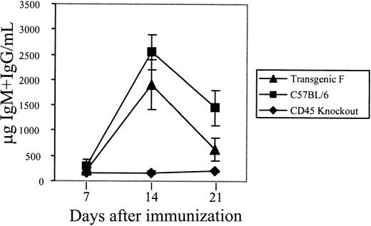
The ability of the CD45.1 transgenic mice to mount a primary immune response was examined. C57BL/6, transgenic, and CD45-null mice were immunized with DNP-KLH. Mice were bled 7, 14, and 21 days after immunization, and sera from each mouse were analyzed separately by ELISA for the presence of anti–DNP IgM and IgG antibodies. The CD45-null mice showed little to no antibody production, as expected (Figure 9). The titers of anti–DNP IgM and IgG in the sera from the transgenic mice were comparable to those in the C57BL/6 mice (Figure 9), with the peak response occurring 14 days after immunization for both.
Primary immune response of mice immunized with DNP-KLH.
The titer of anti–DNP IgM and IgG antibodies is shown. The mice were bled 7, 14, and 21 days after immunization as indicated. Each data point is the average of duplicate wells per dilution for 5 separate mice. The standard errors for the data points are indicated.
Discussion
The data presented demonstrate that the LFA-driven CD45.1 minigene in a CD45-null background restores normal splicing characteristics in the hematopoietic cells that express CD45. Importantly, the developmental characteristics of T cells and the functional properties of both T and B cells also are restored.
Multiple lines of evidence demonstrate that the LFA-1 controlled CD45.1 transgene is expressed in a pattern characteristic of the endogenous CD45 gene. Flow cytometry data indicate that in transgenic mice hemizygous for CD45.2, all cells positive for CD45.1 were positive also for CD45.2, strongly suggesting that the transgene is expressed in the same cells that express the endogenous gene. Furthermore the major hematopoietic lineages, lymphoid and myeloid, express the CD45.1 transgene. Both RT-PCR and flow cytometry results confirm that alternative splicing of transcripts from the CD45.1 transgene are accurately produced in vivo, since the correct cell type–specific isoform expression patterns are generated. Also, naive and memory CD4+ T cells exhibit the expected CD45RB isoform expression characteristics.
Comparison of endogenous LFA-1 and CD45.2 expression with that of the LFA-1–controlled CD45.1 transgene showed that the transgenic B-cell population has characteristics closely resembling the wild-type cells, whereas the transgenic T-cell lineages express less CD45 compared to wild type. The reason is not known but may be due to the particular properties of the transgene itself, such as the expression characteristics of the LFA-1 promoter. Another possibility is that the integration site has an effect on transgene expression in T cells.
The most important issue, however, is whether the presence of the CD45.1 transgene can overcome the developmental and functional defects resulting from the CD45-null mutation. The presence of the CD45.1 transgene fully restores the number of CD4+ and CD8+ single positive T cells in the thymus and, as a result, wild-type numbers of mature T cells are present in the spleen. Proliferation assays in response to antigen demonstrated that these T cells regained normal signal transduction characteristics through the antigen receptor complex. Therefore, these data confirm that the presence of the CD45.1 minigene restores both T-cell development and function.
With respect to B-cell function in the transgenic mice, the proliferative response to activation by anti-IgM is restored and suggests that these mice are capable of a humoral immune response to antigen. To test this parameter, the serum anti–DNP IgM and IgG levels of DNP-KLH immunized mice were assayed by ELISA and were found to be similar in both the transgenic and C57BL/6 mice. Thus, the presence of the CD45.1 transgene restores the ability to produce a significant antibody response in vivo.
Since the SCID phenotype exhibited by the CD45-null mice is overcome as a result of the expression of the CD45.1 minigene, a new opportunity can be considered now for treating the CD45-null form of human SCID. A similar human CD45 minigene, together with a gene transfer vehicle that can efficiently transfer the 20-kb construct into human hematopoietic cells, offers the potential for gene therapy for this otherwise life-threatening genetic disease.
The authors wish to acknowledge Drs Phyllis Linton, Joy Phillips, and Marilyn Thoman for the donation of their time for in-depth discussions.
Prepublished online as Blood First Edition Paper, September 19, 2002; DOI 10.1182/blood-2002-07-1969.
Supported by a grant from the National Institutes of Health (AI40259).
The publication costs of this article were defrayed in part by page charge payment. Therefore, and solely to indicate this fact, this article is hereby marked “advertisement” in accordance with 18 U.S.C. section 1734.
References
Author notes
William C. Raschke, 10835 Altman Row, San Diego, CA 92121; e-mail: braschke@skcc.org.


![Fig. 4. Comparison of the LFA-1 promoter-driven CD45.1 expression to wild-type CD45.2. / The staining antibodies are given on the vertical and horizontal axes (CD45.1, CD45.2, and LFA-1 [CD11a]). (A) All transgene-positive cells were isolated from mice on a CD45-null background. (B) Cells from transgenic mice were on a CD45.2+/− background.](https://ash.silverchair-cdn.com/ash/content_public/journal/blood/101/3/10.1182_blood-2002-07-1969/4/m_h80333788004.jpeg?Expires=1768493141&Signature=VlNz9EPoxrjP2v5JSuDTsme6C-ZfsvYZNxBPhGLzF1MjWDSJZH7U~Ot2z9GutnhcsIvLmAR42xptKpy9waQf6aMWoBdenrBGDIS2RA37ViPrsqlLDopRLe~wxSwf1kWqe0fLT~DtiJfMLemiY-nRo~h7iX1VmmCMTSApU16~c1mU6e8qTFyMonyttRDvyXinlwsClb3--dAtAIulW1nydtuMNWT7PfJsGqGkAZEjWObwSL5y6Lu3lBirIYj16wpbCozWMyWnm~SyDrVV7YEaewaE2pcfe4LsQQR5j6Y0esrTb4X8HUlCvPBdDZ3zharF0QNGPyEdwuim6vhJ96nxqw__&Key-Pair-Id=APKAIE5G5CRDK6RD3PGA)
This feature is available to Subscribers Only
Sign In or Create an Account Close Modal