Abstract
Sphingosine-1-phosphate (S1P) stimulates signaling pathways via G-protein-coupled receptors and triggers diverse cellular processes, including growth, survival, and migration. In S1P1 receptor-deficient embryos, blood vessels were incompletely covered by vascular smooth muscle cells (VSMCs), indicating the S1P1 receptor regulates vascular maturation. Because S1P1 receptor expression is not restricted to a particular cell type, it was not known whether the S1P1 receptor controlled VSMC coverage of vessels in a cell-autonomous fashion by functioning directly in VSMCs or indirectly through its activity in endothelial cells (ECs). By using the Cre/loxP system, we disrupted the S1P1 gene solely in ECs. The phenotype of the conditional mutant embryos mimicked the one obtained in the embryos globally deficient in S1P1. Thus, vessel coverage by VSMCs is directed by the activity of the S1P1 receptor in ECs. (Blood. 2003;102:3665-3667)
Introduction
Embryonic blood vessel development occurs via vasculogenesis, angiogenesis, and maturation.1-3 In vasculogenesis, endothelial cells (ECs) differentiate de novo and form the primary vascular plexus. In angiogenesis, the pre-existing network is remodeled by splitting and sprouting, producing a complex vascular tree of variably sized vessels. Finally, during maturation vascular smooth muscle cells (VSMCs) differentiate and are recruited to the channel wall to stabilize the new vessel, to protect it against rupture, and to provide hemostatic control. This final maturation process requires cell-to-cell communication and interactions between ECs and VSMCs.4,5
Although many of the signaling pathways that become engaged during blood vessel formation have been defined,1-3,6-14 the pathways regulating the interactions between ECs and VSMCs during vascular maturation, especially in larger vessels, remain unclear. The sphingosine-1-phosphate receptor (S1P1) (formally known as Edg1) was shown to be essential for vascular maturation during embryonic development.15 S1P1 is a widely distributed G-protein-coupled receptor for sphingosine-1-phosphate, a blood-borne bioactive lipid. Stimulation of the S1P1 receptor triggers a Gi-linked pathway, leading to growth, survival, migration, and morphogenesis.16-18 Disruption of the S1P1 gene in mice caused embryonic lethality because of massive hemorrhage at embryonic day (E) 12.5 to E14.5.15 Although in the S1P1-/- embryos, VSMCs were in the vicinity of aortae and other vessels, they failed to completely and productively surround the nascent endothelial tubes. Because the S1P1 receptor is expressed in both ECs and VSMCs, the cell type in which the receptor regulated vascular maturation was uncertain. It was not known whether the S1P1 receptor controlled VSMC coverage of vessels in a cell-autonomous fashion by functioning directly in VSMCs or indirectly through its activity in ECs.
To address this issue we have established mice with the S1P1 gene deleted specifically in ECs. Our results demonstrate that vessel coverage by VSMCs is directed by the activity of the S1P1 receptor in ECs.
Study design
Generation of S1P1loxP mice
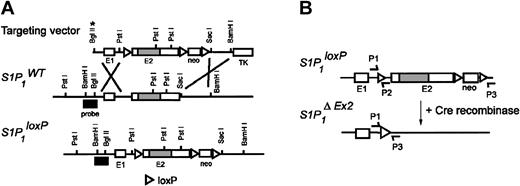
The structure of the targeting vector is shown in Figure 1. Gene targeting in TC1 ES cells and generation of chimeric and heterozygous mice were done as described.15 Mice were genotyped by PCR analyses using DNA from yolk sacs or tail biopsies.
Generation and characterization of EC-specificS1P1knock-out mice. (A) Targeting strategy to introduce the loxP sites into S1P1 gene. The S1P1 gene consists of 2 exons and 1 intron; the second exon (E2) contains the entire coding region. A 5.2-kilobase (kb) BamHI-SacI fragment, including exons 1 and 2 of the S1P1 gene, was subcloned into the XhoI site of the pLoxpneo vector19 upstream of the neo cassette to place one loxP site (▵) within the intron. A 3.8-kb SacI-BamHI fragment containing the 3′ untranslated region was subcloned downstream of the neo cassette which has associated 2 other loxP sites. *BglII restriction site was inactivated during construction of the targeting vector. (B) The loxed S1P1 gene (top; S1P1loxP) and the structure of the gene after Cre-mediated recombination (bottom; S1P1ΔEx2). For detecting the wild-type (S1P1WT), knock-out (S1P1Ko), and conditional alleles (S1P1loxP) by polymerase chain reaction (PCR), the following primers were used: P1, 5′GAGCGGAGGAAGTTAAAAGTG; P2, 5′CCTCCTAAGAGATTGCAGCAA. P1 and P2 amplify an approximately 250-bp fragment for the S1P1loxP allele and a 200-bp fragment for the S1P1WT and S1P1Ko alleles. To detect the S1P1ΔEx2 allele, P1 and P3 (5′GATCCTAAGGCAATGTCCTAGAATGGGACA) were used. P1 and P3 amplify a 200-bp fragment. When primers P1, P2, and P3 were used in the same PCR reaction, the PCR products were digested with SacI prior to the electrophoresis, which converted the S1P1ΔEx2 fragment to 180 bp.
Generation and characterization of EC-specificS1P1knock-out mice. (A) Targeting strategy to introduce the loxP sites into S1P1 gene. The S1P1 gene consists of 2 exons and 1 intron; the second exon (E2) contains the entire coding region. A 5.2-kilobase (kb) BamHI-SacI fragment, including exons 1 and 2 of the S1P1 gene, was subcloned into the XhoI site of the pLoxpneo vector19 upstream of the neo cassette to place one loxP site (▵) within the intron. A 3.8-kb SacI-BamHI fragment containing the 3′ untranslated region was subcloned downstream of the neo cassette which has associated 2 other loxP sites. *BglII restriction site was inactivated during construction of the targeting vector. (B) The loxed S1P1 gene (top; S1P1loxP) and the structure of the gene after Cre-mediated recombination (bottom; S1P1ΔEx2). For detecting the wild-type (S1P1WT), knock-out (S1P1Ko), and conditional alleles (S1P1loxP) by polymerase chain reaction (PCR), the following primers were used: P1, 5′GAGCGGAGGAAGTTAAAAGTG; P2, 5′CCTCCTAAGAGATTGCAGCAA. P1 and P2 amplify an approximately 250-bp fragment for the S1P1loxP allele and a 200-bp fragment for the S1P1WT and S1P1Ko alleles. To detect the S1P1ΔEx2 allele, P1 and P3 (5′GATCCTAAGGCAATGTCCTAGAATGGGACA) were used. P1 and P3 amplify a 200-bp fragment. When primers P1, P2, and P3 were used in the same PCR reaction, the PCR products were digested with SacI prior to the electrophoresis, which converted the S1P1ΔEx2 fragment to 180 bp.
Generation and analysis of S1P1-conditional mutant mice
S1P1loxP/loxP mice were bred with S1P1WT/Ko mice expressing Cre recombinase under the control of the EC-specific promoter Tie2 to obtain S1P1-conditional knock-out (S1P1loxP/KoTie2-Cre) and control littermates (S1P1loxP/Ko, S1P1loxP/WTTie2-Cre, or S1P1loxP/WT). To detect the Cre allele, primers Cre1 (5′GCCTGCATTACCGGTCGATGC) and Cre2 (5′CAGGGTGTTATAAGCAATCCC) were used. To detect the S1P1Ko allele, primers P4, 5′CCATCCTCTACTGCAGGATCT; P5, 5′TGCTGCGGCTAAATTCCATG; and P6, 5′TCGCCTTCTTGACGAGTTCTTCTGAG were used, giving a 250-bp fragment for the S1P1Ko allele and a 500-bp band for the WT band. Tie2-Cre mice were bred with ROSA26 Cre reporter mice20 to determine Cre-expression specificity. To detect the LacZ gene, primers LacZ-F (5′GATCCGCGCTGGCTACCGGC) and LacZ-R (5′GGATACTGACGAAACGCCTGCC) were used. Embryos were obtained from timed-pregnant females with the morning of vaginal plug considered E0.5. Immunohistochemistry and LacZ expression analysis were done as described previously.15 The S1P1 antibody was obtained from Suzanne Mandala (Merck, Whitehouse Station, NJ).
Results and discussion
Generation and characterization of mice with EC-specific S1P1 deletion
The S1P1 gene consists of 2 exons and 1 intron; the second exon contains the entire coding region21 (Figure 1A). To produce an S1P1 allele that could be conditionally disrupted, a gene-targeting vector was constructed that contained loxP sequences flanking exon 2. One loxP site was placed within the intron, and 2 others, associated with the neomycin selection cassette, were placed 3′ to the end of the gene (Figure 1A). This targeting vector was used to introduce these changes into the endogenous S1P1 gene in embryonic stem cells by homologous recombination. A clone that contained the S1P1loxP allele was injected into blastocytes to establish chimeric and, subsequently, heterozygous mice. Interbreeding the heterozygous mice yielded viable, homozygous S1P1loxP/loxP mice at the expected Mendelian frequency. The homozygous S1P1loxP/loxP mice appeared to be without abnormalities, indicating that the changes introduced into the S1P1 gene did not disrupt the locus. A null S1P1 allele (S1P1Ko) produced embryonic lethality when in homozygous form.15
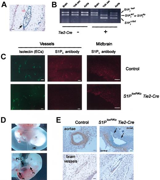
To delete the S1P1 gene specifically in ECs we used a previously characterized transgenic mouse line that expresses the Cre recombinase under the control of the promoter and enhancer regions of the mouse Tie2 gene.22 The specificity of Cre expression in ECs was corroborated by using the ROSA26 reporter line, which contains a LacZ gene that is activated by Cre-mediated recombination.20 Embryos containing the Tie2-Cre and ROSA26 reporter genes showed EC-specific Cre expression. Sections of X-gal-stained embryos revealed that LacZ expression was confined to ECs in vessels in brain (Figure 2A) and in the dorsal aorta (not shown).
Characterization of mice with EC-specific S1P1deletion. (A) Section of X-gal stained Tie2-Cre/ROSA26 E10.5 brain vessel showing lacZ staining specifically in ECs. (B) Tissue specificity of Tie2-Cre-mediated recombination of S1P1loxP using E12.5 embryo tissue DNA samples and PCR primers as shown in Figure 1B. In embryos carrying the Tie2-Cre gene, substantially more recombination of the S1P1loxP allele to produce the S1P1ΔEx2 allele was found in yolk sac and aorta as compared with embryonic neural tissue. No evidence of recombination was found in embryos without the Tie2-Cre gene. S1P1 loxP, S1P1 allele containing loxP sites; S1P1ΔEx2, S1P1 allele after Cre-mediated recombination; S1P1WT, wild-type S1P1 allele; S1P1Ko, null S1P1 allele.15 (C) S1P1 expression in ECs in control and S1P1loxP/KoTie2-Cre embryonic brains at E12.5. Sections were stained with both isolectin B4-fluorescein isothiocyanate (FITC; Vector Laboratories, Burlingame, CA), a specific marker for mouse ECs, and the anti-S1P1 antibody. Note that in control section essentially all ECs that are stained with isolectin B4 also stain with the antibody to S1P1. In the section from the S1P1loxP/KoTie2-Cre conditional mutant, only a few ECs stain strongly with the S1P1 antibody. Midbrain neuroepithelia from control and S1P1-conditional E12.5 embryos shown as positive control for S1P1 expression; both show about equal staining. (D) Phenotype of S1P1-conditional mutant embryos. At E13.5, the yolk sacs of S1P1loxP/KoTie2-Cre conditional mutant embryos show normal vasculature but less blood compared with control embryos (top, arrowhead). The mutant embryos display an enlarged pericardial cavity (PCC; right), undeveloped limbs, and intraembryonic hemorrhages (bottom; FL, front limb; HL, hind limb). (E) Vascular maturation in S1P1-conditional mutant embryos. Transverse sections of aortae (top) from an S1P1loxP/KOTie2-Cre embryo and a control embryo stained with anti-SM α-actin (DAKO, Carpinteria, CA). Note VSMCs are clustered to the ventral side of the aorta and fail to surround the vessel completely in the mutant. Also, ECs on the dorsal side of the aorta appear discontinuous (arrows). Cranial arteries (bottom) from S1P1loxP/KoTie2-Cre and control embryos stained with anti-SM α-actin. Note that VSMCs cluster on one side of the vessel of the S1P1-conditional mutant embryo. Scale bars = 50 μm.
Characterization of mice with EC-specific S1P1deletion. (A) Section of X-gal stained Tie2-Cre/ROSA26 E10.5 brain vessel showing lacZ staining specifically in ECs. (B) Tissue specificity of Tie2-Cre-mediated recombination of S1P1loxP using E12.5 embryo tissue DNA samples and PCR primers as shown in Figure 1B. In embryos carrying the Tie2-Cre gene, substantially more recombination of the S1P1loxP allele to produce the S1P1ΔEx2 allele was found in yolk sac and aorta as compared with embryonic neural tissue. No evidence of recombination was found in embryos without the Tie2-Cre gene. S1P1 loxP, S1P1 allele containing loxP sites; S1P1ΔEx2, S1P1 allele after Cre-mediated recombination; S1P1WT, wild-type S1P1 allele; S1P1Ko, null S1P1 allele.15 (C) S1P1 expression in ECs in control and S1P1loxP/KoTie2-Cre embryonic brains at E12.5. Sections were stained with both isolectin B4-fluorescein isothiocyanate (FITC; Vector Laboratories, Burlingame, CA), a specific marker for mouse ECs, and the anti-S1P1 antibody. Note that in control section essentially all ECs that are stained with isolectin B4 also stain with the antibody to S1P1. In the section from the S1P1loxP/KoTie2-Cre conditional mutant, only a few ECs stain strongly with the S1P1 antibody. Midbrain neuroepithelia from control and S1P1-conditional E12.5 embryos shown as positive control for S1P1 expression; both show about equal staining. (D) Phenotype of S1P1-conditional mutant embryos. At E13.5, the yolk sacs of S1P1loxP/KoTie2-Cre conditional mutant embryos show normal vasculature but less blood compared with control embryos (top, arrowhead). The mutant embryos display an enlarged pericardial cavity (PCC; right), undeveloped limbs, and intraembryonic hemorrhages (bottom; FL, front limb; HL, hind limb). (E) Vascular maturation in S1P1-conditional mutant embryos. Transverse sections of aortae (top) from an S1P1loxP/KOTie2-Cre embryo and a control embryo stained with anti-SM α-actin (DAKO, Carpinteria, CA). Note VSMCs are clustered to the ventral side of the aorta and fail to surround the vessel completely in the mutant. Also, ECs on the dorsal side of the aorta appear discontinuous (arrows). Cranial arteries (bottom) from S1P1loxP/KoTie2-Cre and control embryos stained with anti-SM α-actin. Note that VSMCs cluster on one side of the vessel of the S1P1-conditional mutant embryo. Scale bars = 50 μm.
The specificity of the Cre-mediated recombination of the S1P1loxP gene was demonstrated by examining tissues from embryos. In E12.5 embryos carrying the Tie2-Cre gene, substantially more recombination of the S1P1loxP allele to produce the S1P1ΔEx2 allele (Figure 1B) was found in yolk sac and aorta (tissues highly enriched in ECs) as compared with embryonic neural tissue (Figure 2B). Tie2-Cre mediated deletion of S1P1 receptor was confirmed by immunohistochemistry, using an S1P1-specific antibody. On sections of E12.5 embryos, S1P1 expression in ECs was substantially reduced in S1P1loxP/loxP embryos carrying Tie2-Cre compared with control embryos. By contrast, in brain, epithelium S1P1 expression was similar in S1P1loxP/loxPTie2-Cre embryos and in control embryos (Figure 2C).
EC-specific deletion of S1P1 impairs vascular maturation
When S1P1loxP/loxP mice were bred with S1P1WT/Ko mice carrying the Tie2-Cre transgene, no S1P1loxP/KoTie2-Cre mice were found among 74 offspring, suggesting embryonic lethality. To determine the time of lethality in utero, embryos were analyzed at different days after coitum. At E12.5, the S1P1-conditional mutant embryos (S1P1loxP/KoTie2-Cre) (n = 34) appeared phenotypically similar to the global S1P1 knock-out embryos (S1P1Ko/Ko).15 The S1P1-conditional mutant embryos showed an enlarged pericardial cavity, undeveloped and rounded limbs, and spots of bleeding along the body. Yolk sacs from S1P1-conditional mutant embryos were edematous and with less blood in vessels than in control embryos. At E13.5, the appearance of the S1P1-conditional embryos worsened with massive bleeding (n = 9) (Figure 2D). All S1P1-conditional mutant embryos examined were without a heartbeat at E14.5 (n = 13). Thus, these conditional mutant embryos died by E14.5 similar to the S1P1Ko/Ko embryos,15 which died between E12.5 and E14.5.
Maturation of vessels in S1P1-conditional mutant embryos was studied using smooth muscle (SM) α-actin as a marker for VSMCs15 (Figure 2E). In transverse sections of aortae from control embryos, VSMCs were found to completely surround the vessels. In contrast, the aortae in S1P1-conditional knock-out embryos were incompletely covered by VSMCs. Cells positive for SM α-actin were found clustered on the ventral side of the vessel. The endothelial cell layer on the uncovered dorsal side of aortae appeared discontinuous, possibly because of the lack of VSMC support.23 Similarly, vessels in the brains of the S1P1-conditional mutant embryos were incompletely surrounded by VSMCs. These vessel abnormalities in the S1P1-conditional mutants resembled those described for the global S1P1 knock-out embryos.15
Because the S1P1 receptor is expressed in both ECs and VSMCs, as well as in other cells types, it had been uncertain whether S1P1 functioned in ECs during vascular maturation. The results shown here indicate that vessel coverage by VSMCs is indeed directed by the activity of the S1P1 receptor in ECs. In addition, we have found that S1P1loxP/loxP mice carrying a Cre transgene under the control of the smooth muscle myosin heavy chain (SM-MHC) promoter24 were viable as adults with an S1P1 gene deletion in smooth muscle. These smooth muscle-specific S1P1 KO mice did not show any evidence of hemorrhage or vascular maturation defects (M.L.A., G.K. Owens, R.L.P., unpublished results, 2003). These data suggest that S1P1 receptor expression in VSMCs may not be essential for the process of vessel coverage during development. However, because the expression of Cre recombinase driven by the SM-MHC promoter begins at about the time that VSMCs start to migrate around the vessels, we cannot be certain that the S1P1 deletion was timely enough to affect significantly the vascular maturation process. In any case, the evidence indicates that the expression of S1P1 receptor within ECs has an essential function to regulate the coverage of vessels by VSMCs. At this time an additional function of S1P1 within VSMCs during this process cannot be totally ruled out.
The promotion of functional EC-VSMC interactions by the S1P1 receptor may be incorporated into strategies for therapeutic angiogenesis: the formation of new blood vessels into ischemic tissues.25,26 Endothelial growth factors such as vascular endothelial growth factor (VEGF) promote nascent vessel formation; however, these vessels are leaky and will regress unless stabilized by VSMC interactions.27 Highly effective angiogeneic therapies may ultimately require stimulation of multiple signaling pathways, including the S1P1 receptor to produce mature, stabile vessels.
Prepublished online as Blood First Edition Paper, July 17, 2003; DOI 10.1182/blood-2003-02-0460.
The publication costs of this article were defrayed in part by page charge payment. Therefore, and solely to indicate this fact, this article is hereby marked “advertisement” in accordance with 18 U.S.C. section 1734.
We thank Cuiling Li and Chu-Xia Deng for establishing the chimeric mice, Masashi Yanagisawa for the Tie2-Cre mice, and Suzanne Mandala for the antibody to S1P1.


This feature is available to Subscribers Only
Sign In or Create an Account Close Modal