Abstract
IRTA1 (immunoglobulin superfamily receptor translocation-associated 1) is a novel surface B-cell receptor related to Fc receptors, inhibitory receptor superfamily (IRS), and cell adhesion molecule (CAM) family members and we mapped for the first time its distribution in human lymphoid tissues, using newly generated specific antibodies. IRTA1 was selectively and consistently expressed by a B-cell population located underneath and within the tonsil epithelium and dome epithelium of Peyer patches (regarded as the anatomic equivalents of marginal zone). Similarly, in mucosa-associated lymphoid tissue (MALT) lymphomas IRTA1 was mainly expressed by tumor cells involved in lympho-epithelial lesions. In contrast, no or a low number of IRTA1+ cells was usually observed in the marginal zone of mesenteric lymph nodes and spleen. Interestingly, monocytoid B cells in reactive lymph nodes were strongly IRTA1+. Tonsil IRTA1+ cells expressed the memory B-cell marker CD27 but not mantle cell-, germinal center-, and plasma cell-associated molecules. Polymerase chain reaction (PCR) analysis of single tonsil IRTA1+ cells showed they represent a mixed B-cell population carrying mostly mutated, but also unmutated, IgV genes. The immunohistochemical finding in the tonsil epithelial areas of aggregates of IRTA1+ B cells closely adjacent to plasma cells surrounding small vessels suggests antigen-triggered in situ proliferation/differentiation of memory IRTA1+ cells into plasma cells. Collectively, these results suggest a role of IRTA1 in the immune function of B cells within epithelia. (Blood. 2003;102: 3684-3692)
Introduction
The IRTA1 and IRTA2 (immunoglobulin superfamily receptor translocation-associated 1 and 2) genes encode new members of the immunoglobulin receptor superfamily, which have been recently identified through their involvement in chromosomal translocations affecting band 1q21 in various types of B-cell tumors.1,2 IRTA2 expression was found to be deregulated in Burkitt lymphoma with 1q21 abnormalities, whereas IRTA1 was involved in a rare chromosomal translocation that fuses it to the immunoglobulin (Ig) C-alpha domain to generate a chimeric IRTA1/C-alpha fusion protein.1 Subsequently, it was discovered that IRTA1 and 2 belong to a family of 5 new genes named IRTA 1-52 or FcRH 1-5 (Fc receptor homologs 1 to 5),3 which are continuously located at band 1q21 and encode cell surface receptors related to the Fc receptor,4 inhibitory receptor superfamily (IRS),5 and cell adhesion molecule (CAM) families.6 All IRTA receptors have multiple Ig-related extracellular domains, a transmembrane domain, and an intracellular domain containing multiple ITIM (immunoreceptor tyrosine-based inhibitory motif) and ITAM (immunoreceptor tyrosine-based activation motif)-like motifs.2 These features, together with preliminary RNA-based data showing expression in mature B-cell compartments,1,2 suggest that IRTA molecules may be involved in B-cell-mediated immune responses (like Fc receptors), intercellular communication (like IRS and CAM family members), and cell migration (like CAM family members).
The IRTA1 gene encodes a receptor characterized by 4 extracellular Ig-type domains and 3 potential ITIM motifs in its intracellular domain.1 In situ hybridization results indicated that the IRTA1 mRNA is located in cells within the tonsil epithelium.1 However, no information is available on the distribution and function of the IRTA1 protein in lymphoid tissues. To address this issue, we have generated monoclonal and polyclonal antibodies against recombinant proteins corresponding to the intracytoplasmic and extracellular portions of the IRTA1 molecule, using previously established strategies.7 Immunohistochemical analysis of normal and reactive lymphoid tissues with these antibodies showed that IRTA1 is selectively expressed by a population of B cells located underneath and within the tonsil epithelium and the dome epithelium of Peyer patches (regarded as the anatomic equivalents of marginal zone). Differently from mucosa-associated lymphoid tissues (MALT), no or a low number of IRTA1+ cells was usually observed in the marginal zone of mesenteric lymph nodes and spleen. Interestingly, monocytoid B cells in reactive lymph nodes were consistently IRTA1+. Further characterization of IRTA1+ tonsil B cells by flow cytometry and single-cell polymerase chain reaction (PCR) analysis revealed that they represent memory B cells carrying mostly mutated, but also unmutated, IgV genes. These results suggest a role of IRTA1 in the immune function of B cells in the epithelia.
Materials and methods
Generation of recombinant GST-IRTA1 protein and IRTA1 peptides
A cDNA fragment encoding amino acids (AAs) 102-373 of the extracellular portion (N-terminus) of the human IRTA1 protein was subcloned into the BamH1 and EcoR1 cloning sites of the pGEX 3X (Pharmacia Biotech, Piscataway, NJ) bacterial expression vector downstream and in frame with glutathione S-transferase (GST) coding sequences. The GST-IRTA1 fusion protein was then expressed in Escherichia coli strain BL21-competent bacteria and purified by affinity chromatography following the manufacturer's instructions.
A 15-amino acid peptide corresponding to the cytoplasmic portion of the IRTA1 protein (CDNSAGKISSKDEES) was synthesized and conjugated to keyhole limpet hemocyanin (KLH). The cysteine at position 1 was added to facilitate KLH conjugation. All procedures were performed by Research Genetics (Huntsville, AL).
Production of antibodies specific for the IRTA1 protein
Monoclonal antibodies. BALB/c mice were injected intraperitoneally (3 times, at a 10-day interval) with 150 μg of the GST-IRTA1 fusion protein plus Freund adjuvant. A booster was performed by intraperitoneal injection of 150 μg of the recombinant GST-IRTA1 protein and the fusion was carried out 3 days later, as described previously.8 Hybridoma supernatants were screened by the immunoalkaline phosphatase (APAAP) technique9 on formalin-fixed paraffin sections of normal human tonsils. One of approximately 1000 hybridomas (named monoclonal IRTA1 [M-IRTA1]) reacted strongly with a small population of lymphoid-looking cells located beneath and within the tonsil epithelium; this hybridoma was cloned by a limiting-dilution technique and selected for further studies.
Polyclonal antibodies. Polyclonal antibodies were obtained by injecting rabbits with a 15-AA synthetic peptide corresponding to the C-terminus of the IRTA1 protein (CDNSAGKISSKDEES). The antiserum was then affinity purified against his cognate synthetic peptide (Research Genetics). One aliquot of the antibody was conjugated to biotin. Antibodies were used at 0.5 μg/mL concentration for immunohistochemistry or flow cytometry. Blocking of specific staining was obtained by preincubation with 100 × concentrated immunizing peptide.
Other antibodies
Other antibodies used for double immunoenzymatic or immunofluorescence studies were as follows: mouse monoclonal antibodies (mAbs) directed against BCL2, CD3ϵ, CD20, CD30, CD79a, Ki-67, and IgD (Dako A/S, Glostrup, Denmark); PAX5 (paired box-5; Transduction Laboratories, San Diego, CA); CD138 (Serotec, Raleigh, NC); and CD27 (Novocastra, Newcastle upon Tyne, United Kingdom). CD2, CD5, CD68 were from the Second and Third International Workshop on Human Leukocyte Differentiation Antigens. Ki-67 (MIB1) antibody was generously donated by J. Gerdes (Research Center Borstel, Borstel, Germany); the anti-MUM1/IRF48 (anti-multiple myeloma oncogene 1/interferon regulatory factor 4) and anti-BCL610,11 mAbs were generated in one author's laboratory (B.F.). CD5-phycoerythrin (CD5-Pe), CD23-Pe, CD27-Pe, CD38-Pe, CD95-Pe, and CD3-fluorescein isothiocyanate (CD3-FITC) were purchased from Pharmingen/BD Biosciences (San Diego, CA). Anti-isotype specific, TRITC-(tetramethylrhodamine-5(and 6)-isothiocyanate)-, FITC-, or biotin-conjugated antibodies were from Southern Biotechnology Associates (Birmingham, AL) and Dako A/S. FITC- and 9-amino-6-chloro-2-methoxyacridine (AMCA)-conjugated avidin, alkaline phosphatase (AP)-conjugated goat anti-FITC, and Vectashield mounting medium were purchased from Vector (Burlingame, CA).
Cell suspensions
Tonsils were processed on ice immediately after removal, finely minced in phosphate-buffered saline (PBS), washed, and resuspended in azide and phenol-free medium with 1% calf serum. T cells were removed by sheep red blood cell rosetting (Cocalico Biologicals, Reamstown, PA) and separation on a Ficoll-Hypaque gradient. This procedure reproducibly yields B cells purer than 95% with more than 98% viability.
Cell transfection
A pMT2T-IRTA1-hemagglutinin (HA) expression vector was constructed by generating a PCR product spanning the entire IRTA1 coding region and in which the natural stop codon of IRTA1 was replaced with an NcoI site (in the 3′ end primer). The PCR product was cloned in-frame into an HA-encoding pBluescript vector (Stratagene, Cedar Creek, TX). After reamplification and subcloning into pGEM-T (plasmid gemcitabine transfector), the IRTA1-HA open-reading frame (ORF) was excised with NotI and cloned into pMT2T. The construct was sequenced to verify lack of mutations. The pMT2T-IRTA1-HA and pMT2T (as a control) plasmids were used for transient transfection of COS cells (monkey's kidney) by the diethylamino ethanol (DEAE)-dextran method or 293T cells (American Type Culture Collection, Manassas, VA) by the calcium phosphate precipitation method. IRTA1-transfected and control cells were grown in DMEM (Dulbecco modified essential medium) containing 10% bovine calf serum, penicillin (100 IU/mL), and streptomycin (100 μg/mL). Cells were then lysed and analyzed by Western blotting (“Western blotting”). Cells were also grown on slides, air-dried overnight, fixed in acetone for 10 minutes, and immunostained by APAAP and immunofluorescence techniques.
Western Blotting
M-IRTA1 monoclonal antibody. Western blotting was performed on cell lysates from the IRTA1-transfected and control COS cells. Cells were lysed with sodium dodecyl sulfate (SDS)-loading buffer and an aliquot of each lysate was loaded onto an 8% sodium dodecyl sulfate acrylamide gel and electrotransferred to nitrocellulose sheets, as previously described.8 The membrane was subsequently incubated with nonfat dry milk (Biorad, Hercules, CA) for 1 hour at room temperature (RT), followed by the M-IRTA1 mAb supernatant (net) overnight at 4°C. Following washing in TBS-T (0.05 M Tris-buffered saline, pH 7.5, plus 0.1% Tween-20), the secondary goat antimouse horseradish peroxidase (HRP)-conjugated antibody (Biorad) was applied at 1:10 000 dilution in TBS-T at RT for 40 minutes. Results were visualized by enhanced chemiluminescence (ECL) detection using ECL Plus reagents (Amersham Pharmacia Biotech, Little Chalfont, England).
Polyclonal anti-IRTA1 antibodies. Lysates from IRTA1 transiently transfected 293T cells were solubilized in Triton X-100 lysis buffer (150 mM NaCl; 10 mM Tris-HCl, pH 7.4; 1% Tx-100; 0.1% bovine serum albumin [BSA]) in the presence of a protease inhibitor cocktail (Roche Biochemicals, Indianapolis, IN). Lysates (1.2 × 10⁶ cell equivalents) were electrophoresed on 7% Tris glycine-SDS gels and transferred to polyvinylidene fluoride (PVDF) membranes (Amersham Pharmacia Biotech) for immunoblotting. The purified anti-IRTA1 polyclonal antibodies were diluted to a final concentration of 2 μg/mL in 0.05 M TBS, pH 7.5; 0.05% Tween-20; and 3% BSA and incubated with the membrane overnight at 4°C. The membrane was subsequently incubated with a secondary goat antirabbit HRP-conjugated antibody (Amersham Pharmacia Biotech) at 1:30 000 dilution at RT for 1 hour. Results were visualized by enhanced chemiluminescence detection using ECL Plus reagents (Amersham Pharmacia Biotech).
Immunoprecipitation
IRTA1-transfected and control COS cells were lysed with a lysis buffer (1.5 mM MgCl2; 50 mM Tris-HCl, pH 8.0; 150 mM NaCl; 1% Triton X-100; 5 mM EGTA [ethyleneglycotetraacetic acid]; 50 mM NaF, pH 8.0; and 10% glycerol) containing a protease inhibitor cocktail (leupeptin, aprotinin, pepstatin A, and phenhylmethylsulfonyl fluoride) and immunoprecipitated with the M-IRTA1 mAb coupled to Protein A Sepharose beads (Amersham Pharmacia Biotech). The immunoprecipitates were run onto 10% SDS polyacrylamide gel, blotted, and then incubated overnight at 4°C with an anti-HA mAb (BAbCO, Richmond, CA). Finally, the blots were incubated for 40 minutes at RT with a secondary goat antimouse HRP-conjugated antibody (Biorad) diluted 1:10.000 in TBS-T and stained by the ECL system (Amersham Pharmacia Biotech).
Tissue specimens
Expression of the IRTA1 protein was studied in formalin-fixed paraffin sections from the following samples: 15 palatine tonsils, 5 spleens, 3 thymi, 5 Peyer patches, 10 lymph nodes with follicular hyperplasia, 15 reactive lymph nodes rich in monocytoid B cells (10 toxoplasmic, 2 HIV-associated, and 3 infectious mononucleosis-associated lymphadenitis), 7 samples with acquired MALT (3 Helicobacter pylori-associated gastritis, 3 Hashimoto thyroiditis, 1 Sjogren syalodenitis), and 3 representative examples of MALT lymphomas12,13 (2 gastric, 1 lung). Tonsil specimens were also snap-frozen in liquid nitrogen and used to isolate single cells for PCR studies.
Immunohistochemical analysis
M-IRTA1 monoclonal antibody. Formalin-fixed paraffin sections (3- to 5-μm thick) were subjected to antigen retrieval (750 W × 3 cycles of 5 minutes each) in 1 M ethylenediaminetetraacetic acid (EDTA) buffer, pH 8.0.8 Tonsil frozen sections (8- to 9- μm thick) for micromanipulation (“Micromanipulation of single cells”) were air-dried for 2 hours and fixed in acetone for 10 minutes. Both paraffin and frozen sections were immunostained by the APAAP procedure.9 Slides were then counterstained for 5 minutes in hematoxylin and mounted in Kaiser glycerol gelatin.
Polyclonal anti-IRTA1 antibody. Following EDTA-antigen retrieval, slides were cooled and sequentially incubated in the following: (a) TBS 0.05 M, pH 7.5, + 0.01% Tween 20 for 5 minutes; (b) filtered egg white 1:10 in PBS14 for 15 minutes; (c) TBS-T for 5 minutes; and (d) 5% skim milk in TBS-T14 for 30 minutes. After a final wash, sections were incubated overnight with the rabbit anti-IRTA1 diluted in TBS + 1% BSA + 1% sodium azide (TBS-BSA) followed by AP-conjugated species-specific secondary antibodies (Jackson ImmunoResearch Laboratories, West Grove, PA) diluted 1:2.000 in TBS-BSA + 1% skim milk. Slides were developed with NBT-BCIP (p-nitro blue tetrazolium chloride-5-bromo-4-chloro-3-indolyl phosphate; Roche Molecular Biochemicals) for 4 hours to 18 hours at RT.
Double immuno-enzymatic labeling. Antigen-retrieved tonsil paraffin sections were double stained for IRTA1 and the following nuclear-associated molecules: IRTA1/PAX5, IRTA1/BCL6, IRTA1/MUM1-IRF4, and IRTA1/MIB1. Antigens other than IRTA1 were usually detected by a biotin-avidin peroxidase technique using diaminobenzidine/hydrogen peroxide as substrate (diaminobenzidine was purchased from Sigma-Aldrich, Milan, Italy).15 The IRTA1 molecule was then revealed by the APAAP procedure9 using naphthol AS-MX plus Fast red TR or Fast Blue BB salt (all purchased from Sigma-Aldrich) as substrates. In some experiments, a double alkaline phosphatase immunostaining procedure using contrasting chromogens was employed to detect the 2 antigens. Slides were mounted in Kaiser gelatin following counterstain for 30 seconds in hematoxylin or without counterstain.
Double immunofluorescence labeling. This was performed by using the tyramide amplification method.16 Briefly, the first primary antibody was amplified with biotin tyramide or biotin fluorochrome (Perkin Elmer, Boston, MA) followed by non-cross-reactive indirect immunofluorescence with a second and a third antibody and counterstaining with contrasting fluorochrome-labeled secondary antibodies (Southern Biotechnology Associates) and with avidin-FITC or avidin-AMCA (Vector Labs).
Flow cytometry
Since the M-IRTA1 mAb is weakly reactive with fresh cells, studies on cell suspensions were performed with the anti-IRTA1 polyclonal antibody. Validation of the specific staining was obtained by analysis of cell lines positive or negative for IRTA1 RNA or by use of a biotin-conjugated negative control antibody. Total tonsil or purified B-cell suspensions were fixed in fluorescence-activated cell sorter (FACS) lysing solution (BD Immunocytometry Systems, San Jose, CA) for 10 minutes, washed in avidin- and biotin-free phenol-free medium plus serum, blocked with 1 mg/mL purified rabbit immunoglobulins (Sigma, St Louis), and incubated overnight with either biotin-conjugated anti-IRTA1 polyclonal antibody or a biotin-conjugated rabbit antihen egg lysozyme as a negative control (Research Diagnostics, Flanders NJ), both at 0.5 μg/mL in 1 mg/mL unconjugated rabbit Ig in TBS-BSA. The cells were then washed, stained with lineage-specific fluorochrome-labeled mAbs, and counterstained with avidin-allophycocyanin (APC) 1:400 (Pharmingen). Fifty thousand events were then acquired on a 6-parameter FACSCalibur instrument (BD Immunocytometry Systems). The data were elaborated with FlowJo (TreeStar, San Carlos, CA).
PCR analysis of single tonsil IRTA1+ cells
Micromanipulation of single cells. Single IRTA1+ cells were isolated from 8- to 9- μm-thick tonsil frozen sections of 3 healthy donors using a hydraulic micromanipulator. Isolated cells were then transferred for PCR reaction into tubes containing 20 μL of 1 × Expand high-fidelity PCR buffer (Roche) with 1 ng/μL of 5s rRNA (Roche) and stored at -20°C, as previously described.17 Before and after isolating each cell, a photograph was taken of its localization in the histologic section. As negative controls for the micromanipulation procedure, aliquots of buffer covering the section were taken. For one of the tonsils, single CD3+ cells from CD3-stained adjacent sections were isolated and used as additional negative controls.
Single-cell PCR. Immunoglobulin heavy-chain variable (IgVH) gene rearrangements of the 3 most frequently used VH families (VH1, VH3, and VH4) were amplified using a seminested PCR approach, as previously described.18 In the present study, for amplification of VH1 gene rearrangements a different forward primer was used (5′-CAG-TCT-GGG-GCT-GAG-GTG-AAG-A-3′) and the second round of amplification was performed in the presence of 2.5 mM MgCl2. Five or 10 μL of each reaction were analyzed on 2% agarose gels.
DNA sequence analysis. PCR products were purified either directly or through 2% agarose gel electrophoresis using the QIAquick purification kit or the QIAEX II gel extraction kit (Qiagen, Hilden, Germany) and directly sequenced using the same primers as in the second round of amplification.
A fraction of the amplificates was sequenced from both sides. The procedure was performed by the dye-deoxy-terminator method on an automated DNA sequencer (ABI377A; Perkin-Elmer, Applied Biosystem Division, Foster City, CA) and sequences were compared with the most homologous human germ line VH genes using the IMGT (ImMunoGeneTics) database (http://www.dnaplot.de) and the GenBank data library. A rearrangement was regarded as mutated when its mutation frequency was greater than 1%. As an indication of antigen selection, the ratio between replacement and silent mutations (R/S) in the framework regions (FWRs) 2 and 3 was calculated and a sequence was regarded as being selected when this ratio was no more than 1.5.19 To rule out contamination, the rearranged sequences were compared with each other and with our own and published databank sequences. The VH gene sequences have been deposited in the European Molecular Biology Laboratory (EMBL) data library under accession numbers AJ557032-AJ557071.
Results
The monoclonal (M-IRTA1) and polyclonal antibodies react specifically with the IRTA1 protein
The M-IRTA1 mAb identified a strong band of 50 kDa on Western blot lysates from COS cells transfected with an expression vector encoding the extracellular portion of the IRTA1 protein (amino acids 102-373; Figure 1A). A single 50-kDa band was also detected on lysates from COS cells transfected with the extracellular portion of IRTA1, which were immunoprecipitated with the M-IRTA1 mAb and revealed by Western blotting using an anti-HA mAb (Figure 1B). No bands were observed in COS cells transfected with the empty vector (Figure 1A-B) or with cDNAs encoding for the other members of the IRTA family (IRTA2 to 5; not shown).
Identification of the IRTA1 protein with specific antibodies. (A-B) The M-IRTA1 mAb. (A) A strong band of 51 kDa (arrow) of the expected molecular size for the external part of the IRTA1 protein is seen in the lane corresponding to lysates of IRTA1-HA-transfected COS cells but not in the control cells lane. (B) Identical results were observed following immunoprecipitation of the protein with M-IRTA1 and detection of Western blotted immunoprecipitates with anti-HA mAb; the arrow points to the IRTA1 band. (C-D) The anti-IRTA1 polyclonal antibody. (C) Northern blot (top) analysis of IRTA1 expression in transfectants and cell lines. Two specific RNA species (*) are shown in the ER-EB cell line 24 and 48 hours after induction2 and in the CA46 cell line; the lower molecular weight bands represent the GAPDH (glyceraldehyde phosphate dehydrogenase) mRNA obtained by hybridization with the cognate probe in the same reaction (loading control). The bottom panel shows the Western blot analysis with the anti-IRTA1 purified polyclonal antibody on total cell extracts from vector- or full-length IRTA1-HA-transfected 293T cells and cell lines. Western blot with β-actin is shown as a control for protein loading. IRTA1 appears as 2 closely migrating bands of 70 to 75 kDa (black bars), which represent glycosylation isoforms (G.C., unpublished results, December 2002). (D) Colocalization of anti-IRTA1 polyclonal rabbit antibody (FITC, green) and anti-HA tag (TRITC, red) on transfected 293T cells. The bottom small panel shows DAPI staining on nuclei. Colocalization is seen as yellow signals in the superimposed image (large panel). Original magnifications: × 400 (large panel); × 4000 (small panels).
Identification of the IRTA1 protein with specific antibodies. (A-B) The M-IRTA1 mAb. (A) A strong band of 51 kDa (arrow) of the expected molecular size for the external part of the IRTA1 protein is seen in the lane corresponding to lysates of IRTA1-HA-transfected COS cells but not in the control cells lane. (B) Identical results were observed following immunoprecipitation of the protein with M-IRTA1 and detection of Western blotted immunoprecipitates with anti-HA mAb; the arrow points to the IRTA1 band. (C-D) The anti-IRTA1 polyclonal antibody. (C) Northern blot (top) analysis of IRTA1 expression in transfectants and cell lines. Two specific RNA species (*) are shown in the ER-EB cell line 24 and 48 hours after induction2 and in the CA46 cell line; the lower molecular weight bands represent the GAPDH (glyceraldehyde phosphate dehydrogenase) mRNA obtained by hybridization with the cognate probe in the same reaction (loading control). The bottom panel shows the Western blot analysis with the anti-IRTA1 purified polyclonal antibody on total cell extracts from vector- or full-length IRTA1-HA-transfected 293T cells and cell lines. Western blot with β-actin is shown as a control for protein loading. IRTA1 appears as 2 closely migrating bands of 70 to 75 kDa (black bars), which represent glycosylation isoforms (G.C., unpublished results, December 2002). (D) Colocalization of anti-IRTA1 polyclonal rabbit antibody (FITC, green) and anti-HA tag (TRITC, red) on transfected 293T cells. The bottom small panel shows DAPI staining on nuclei. Colocalization is seen as yellow signals in the superimposed image (large panel). Original magnifications: × 400 (large panel); × 4000 (small panels).
A single band corresponding to the expected molecular weight of the full-length IRTA1 protein (70-75 kDa; Figure 1C, bottom panel) was detected with the anti-IRTA1 polyclonal antibody on Western blot lysates from 293T cells transfected with a vector expressing the IRTA1 protein fused to the HA tag. No bands were observed with 293T cells transfected with the empty vector. The same size protein band was also detectable in 2 cell lines in which the expression of the IRTA1 mRNA was either induced (ER-EB)2 or was constitutively present (CA46) but not in other B-cell lines lacking IRTA1 (Figure 1C, top panel, and not shown). The polyclonal antibody reacted in immunofluorescence with permeabilized IRTA1 transfectants, but not with controls (Figure 1D), and colocalized with the HA-tagged IRTA1 protein. Identical results were obtained with the M-IRTA1 mAb (not shown).
These findings demonstrate that both the mouse monoclonal (M-IRTA1) and the rabbit polyclonal antiserum react specifically with the IRTA1 protein.
IRTA1 is expressed by intraepithelial and subepithelial MALT marginal zone B cells and by monocytoid B cells
Optimal labeling of the IRTA1 protein was observed in formalin-fixed tissue sections but not in B5-fixed tissue sections. Reactivities of the polyclonal and monoclonal antibody were superimposable.
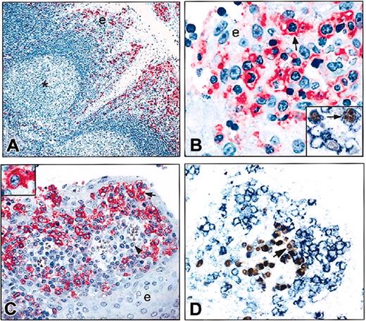
As shown in Figure 2A, tonsil IRTA1+ cells were mainly distributed in the subepithelial and intraepithelial regions; occasional IRTA1+ cells were also seen at the border of mantle zones and in the interfollicular area. In contrast, mantle and germinal center B cells, as well as T cells in the interfollicular areas, were consistently IRTA1-. Thus, the topographical location of IRTA1+ cells by immunohistochemical analysis closely matched the distribution of the IRTA1 mRNA transcripts detectable by in situ hybridization.1 IRTA1+ cells were small to medium in size with a slightly irregular nuclear outline and wide cytoplasm and showed a strong IRTA1 surface positivity that characteristically highlighted villous projections (Figure 2B).
IRTA1 expression in paraffin sections from normal tonsil immunostained with the mAb M-IRTA1. (A) IRTA1+ cells are selectively distributed within the tonsil epithelium (e) and at the border of mantle zones. * indicates a IRTA1- germinal center. (B) IRTA1+ cells often show a villous cell surface (arrow). (Inset) Blue indicates staining for IRTA1; brown, the proliferation antigen MIB1. Most IRTA1+ intraepithelial B cells are not proliferating. The arrow points to a large IRTA1+/MIB1+ cell. (C) IRTA1+ B cells within the epithelium (e) often form a rim (arrow) around clusters of subepithelial plasma cells (arrowhead) arranged around small vessels. Inset shows higher magnification of an IRTA+ cell. (D) Double staining for IRTA1 (blue) and MUM1/IRF4 (brown) shows a rim of IRTA1+/MUM1- negative intraepithelial B cells surrounding a cluster of IRTA1-/MUM1+ subepithelial plasma cells (arrow). The APAAP technique was used in panels A-C; double immunoperoxidase/APAAP staining, panels D and B inset. Original magnifications × 100 (A); × 1000 (B, larger panel and inset); × 400 (C-D).
IRTA1 expression in paraffin sections from normal tonsil immunostained with the mAb M-IRTA1. (A) IRTA1+ cells are selectively distributed within the tonsil epithelium (e) and at the border of mantle zones. * indicates a IRTA1- germinal center. (B) IRTA1+ cells often show a villous cell surface (arrow). (Inset) Blue indicates staining for IRTA1; brown, the proliferation antigen MIB1. Most IRTA1+ intraepithelial B cells are not proliferating. The arrow points to a large IRTA1+/MIB1+ cell. (C) IRTA1+ B cells within the epithelium (e) often form a rim (arrow) around clusters of subepithelial plasma cells (arrowhead) arranged around small vessels. Inset shows higher magnification of an IRTA+ cell. (D) Double staining for IRTA1 (blue) and MUM1/IRF4 (brown) shows a rim of IRTA1+/MUM1- negative intraepithelial B cells surrounding a cluster of IRTA1-/MUM1+ subepithelial plasma cells (arrow). The APAAP technique was used in panels A-C; double immunoperoxidase/APAAP staining, panels D and B inset. Original magnifications × 100 (A); × 1000 (B, larger panel and inset); × 400 (C-D).
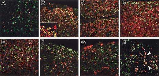
To further define the phenotype of the tonsil IRTA1+ cells, double immunoenzymatic and immunofluorescence stainings were performed on tonsil sections. Double staining for IRTA1 and PAX5, a B-cell-restricted nuclear antigen, revealed that all IRTA1+ cells were of B lineage (not shown). IRTA1+ cells were also CD20+ (Figure 3B); CD79a partially positive (Figure 3C); weakly positive or negative for BCL2 (Figure 3D); and negative for CD3 (Figure 3E), CD68 (Figure 3F), and BCL6 (not shown). IRTA1+ cells did not express the plasma cell-associated markers CD138 (Figure 3G) and MUM1/IRF4 (Figure 2D). Occasional interfollicular large cells were IRTA1+/CD30+ (Figure 3H). Between 5% and 10% of intraepithelial IRTA1+ cells (Figure 2B inset) coexpressed the proliferation antigen Ki-67.
The phenotype of IRTA1+ tonsil cells. (A) Double immunofluorescence staining of tonsil sections. IRTA1 is green. Negative control is red. No double-positive cells are identified (yellow-green). (B) IRTA1+ cells are CD20+ B cells; (inset) higher power of the same. (C) CD79a is weakly coexpressed on IRTA1+ B cells. (D) BCL2 is weak or negative on IRTA1+ extrafollicular B cells. The epithelium surface is at the top edge of the image. (E) CD3 and IRTA1 do not costain. (F) CD68 labels macrophages but not IRTA1+ B cells. (G) IRTA1+ cells and CD138 are mutually exclusive. (H) Rare interfollicular large cells (arrowheads) are IRTA1+ (green)/CD30+ (red) (green + red = yellow). All immunostaining for IRTA1 was performed with the specific polyclonal antibody. Original magnifications: × 400 (A-H); × 1000 (B inset).
The phenotype of IRTA1+ tonsil cells. (A) Double immunofluorescence staining of tonsil sections. IRTA1 is green. Negative control is red. No double-positive cells are identified (yellow-green). (B) IRTA1+ cells are CD20+ B cells; (inset) higher power of the same. (C) CD79a is weakly coexpressed on IRTA1+ B cells. (D) BCL2 is weak or negative on IRTA1+ extrafollicular B cells. The epithelium surface is at the top edge of the image. (E) CD3 and IRTA1 do not costain. (F) CD68 labels macrophages but not IRTA1+ B cells. (G) IRTA1+ cells and CD138 are mutually exclusive. (H) Rare interfollicular large cells (arrowheads) are IRTA1+ (green)/CD30+ (red) (green + red = yellow). All immunostaining for IRTA1 was performed with the specific polyclonal antibody. Original magnifications: × 400 (A-H); × 1000 (B inset).
We then tested whether the polyclonal antibody could detect IRTA1 in cell suspensions. Three-color flow cytometric analysis of cell suspensions from tonsils showed that IRTA1+ cells were CD20+, IgD+ or IgD-, CD27+, CD95+, CD11b+, CD5-, CD23-, CD38dim or CD38- (Figure 4). Attempts to define the Ig isotype of IRTA1+ cells, besides IgD, were unsuccessful due to the large quantity of blocking rabbit Ig required to block nonspecific binding of the polyclonal antibody to the low-abundance antigen.
Flow cytometric detection and phenotype of IRTA1+ cells. A tonsil total cell suspension was labeled with a control antibody (gray histogram) and IRTA1 or lineage-specific markers (black lines). (Left) IRTA1+ cells (5.5% in this sample) are gated. (Right) Top row shows total and purified B-cell phenotype; bottom row, IRTA1+ cell gating. All staining was performed with the anti-IRTA1 polyclonal antibody.
Flow cytometric detection and phenotype of IRTA1+ cells. A tonsil total cell suspension was labeled with a control antibody (gray histogram) and IRTA1 or lineage-specific markers (black lines). (Left) IRTA1+ cells (5.5% in this sample) are gated. (Right) Top row shows total and purified B-cell phenotype; bottom row, IRTA1+ cell gating. All staining was performed with the anti-IRTA1 polyclonal antibody.
The topographical relationships between the IRTA1+ cells and plasma cells was investigated on tonsil tissue sections double stained for IRTA1 and the plasma cell marker MUM1/IRF4. Expression of IRTA1 and MUM1/IRF4 was mutually exclusive. We also noticed the presence of aggregates of IRTA1+/MUM1- cells that were mostly proliferating (Ki67+) and characteristically arranged in close proximity to subepithelial MUM1+/IRTA1- plasma cells usually distributed around small vessels (Figure 2C-D).
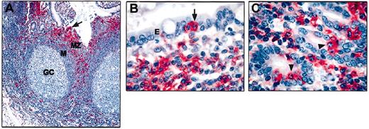
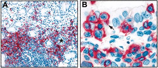
In order to assess whether the epitheliotropism of IRTA1+ B cells observed in the tonsil is also found in normal, reactive, and neoplastic MALT, we investigated 5 samples of Peyer patches (normal MALT of intestine), 7 samples with reactive MALT, and 3 representative examples of MALT lymphomas (2 gastric, 1 lung). Notably, in Peyer patches IRTA1 was selectively expressed by marginal zone B cells (Figure 5A) including intraepithelial B cells (IRTA1+/PAX5+/MUM1-) in the dome epithelium (Figure 5B-C). Similar findings were observed in acquired MALT of the stomach, thyroid, and salivary glands (not shown). In the 3 cases of MALT lymphomas, IRTA1 was predominantly expressed by the tumor cells involved in the formation of lympho-epithelial lesions (Figure 6A-B), whereas the neoplastic cells distant from the epithelium displayed weak or no expression.
IRTA1 expression in Peyer patches. (A) Expression of IRTA1 is topographically restricted to the marginal zone (MZ) including isolated or small clusters of intraepithelial B cells (arrows) in the dome epithelium; cells in the germinal center (GC) and mantle (M) of B-cell follicles are IRTA1-. (B-C) Other areas of Peyer patches showing, at higher magnification, that IRTA1 is selectively expressed by marginal zone (MZ) B cells including intraepithelial B cells. The arrow in panel B and the arrowheads in panel C point to IRTA1+ intraepithelial B cells. APAAP technique with mAb M-IRTA1; hematoxylin counterstain. Original magnifications × 100 (A); × 1000 (B-C).
IRTA1 expression in Peyer patches. (A) Expression of IRTA1 is topographically restricted to the marginal zone (MZ) including isolated or small clusters of intraepithelial B cells (arrows) in the dome epithelium; cells in the germinal center (GC) and mantle (M) of B-cell follicles are IRTA1-. (B-C) Other areas of Peyer patches showing, at higher magnification, that IRTA1 is selectively expressed by marginal zone (MZ) B cells including intraepithelial B cells. The arrow in panel B and the arrowheads in panel C point to IRTA1+ intraepithelial B cells. APAAP technique with mAb M-IRTA1; hematoxylin counterstain. Original magnifications × 100 (A); × 1000 (B-C).
IRTA1 expression in MALT lymphomas. (A) Gastric MALT lymphoma. Tumor cells forming lympho-epithelial lesions are IRTA1+. * indicates a gland lumen. Tumor cells located far from the glandular epithelium are IRTA1-. (B) Lympho-epithelial lesion in a MZ lymphoma of the lung. The arrow points to a IRTA1+ lymphoma cells invading the ciliated bronchial epithelium. The APAAP technique with mAb M-IRTA1 was used with a hematoxylin counterstain. Original magnifications × 200 (A); × 1000 (B).
IRTA1 expression in MALT lymphomas. (A) Gastric MALT lymphoma. Tumor cells forming lympho-epithelial lesions are IRTA1+. * indicates a gland lumen. Tumor cells located far from the glandular epithelium are IRTA1-. (B) Lympho-epithelial lesion in a MZ lymphoma of the lung. The arrow points to a IRTA1+ lymphoma cells invading the ciliated bronchial epithelium. The APAAP technique with mAb M-IRTA1 was used with a hematoxylin counterstain. Original magnifications × 200 (A); × 1000 (B).
Rare IRTA1+ cells were found in 3 thymi, mainly around Hassal corpuscles. Expanded marginal zone in the 3 mesenteric lymph nodes studied showed no or rare IRTA1+ cells in 2 of them and a “corona” of 2 to 4 layers of IRTA1+ cells marginally to the lymphoid follicles in the third one. Ten cases of reactive follicular hyperplasia were investigated. In 8 of 10 cases, only rare IRTA1+ cells were seen at the border of mantle zones, whereas in 2 samples IRTA1 was expressed by a thin rim of loosely arranged, medium-sized lymphoid cells located at the external part of the mantle zones of the secondary follicles (Figure 7A) and by cells filling the subcapsular sinus (Figure 7B). IRTA1 was strongly expressed by the so-called monocytoid B cells in all cases of toxoplasmic (n = 10; Figure 7C), infectious mononucleosis (n = 3), and HIV-associated (n = 2) lymphadenitis that were tested (not shown). In 4 of 5 spleens investigated, only occasional scattered IRTA1+ cells were seen in the areas corresponding to the splenic marginal zone (MZ); in one case, several IRTA1+ cells were seen in the marginal zone (Figure 7D).
IRTA1 expression in reactive lymph nodes. (A) Double APAAP staining for IRTA1 (mAb M-IRTA1) and the proliferating antigen MIB1 in a reactive lymph node. IRTA1 strongly labels in red B cells (arrow) at the border of the mantle zone (m) while MIB1 (blue) is mainly expressed by germinal center B cells. A small percentage of double-stained IRTA1+/MIB1+ cells is present (arrowhead and inset). (B) Double APAAP staining for IRTA1 (mAb M-IRTA1) and BCL6 in a reactive lymph node. IRTA1 strongly labels in red B cells of the subcapsular sinus (arrow), whereas BCL6 (blue) is strongly expressed by germinal center B cells. (C) Monocytoid B cells of toxoplasmic lymphadenitis strongly express IRTA1 (APAAP technique with mAb M-IRTA1). (D-F) IRTA1 expression in human spleen. (D) Spleen stained with polyclonal anti-IRTA1 (H&E stain). The GC is marked by *; the marginal zone is bracketed in white. (E) Adjacent section stained for IRTA1 (black) and the B-cell antigen PAX5 (red nuclear staining). (F) Serial sections stained for IgD (black) and PAX5 (red). IRTA1+ B cells are distributed in the IgD-dim marginal portion of the marginal zone. Other areas are negative. Original magnifications × 100 (A); × 1000 (A inset); × 200 (B); × 100 (C); × 100 (D-F, top); × 400 (D-F bottom).
IRTA1 expression in reactive lymph nodes. (A) Double APAAP staining for IRTA1 (mAb M-IRTA1) and the proliferating antigen MIB1 in a reactive lymph node. IRTA1 strongly labels in red B cells (arrow) at the border of the mantle zone (m) while MIB1 (blue) is mainly expressed by germinal center B cells. A small percentage of double-stained IRTA1+/MIB1+ cells is present (arrowhead and inset). (B) Double APAAP staining for IRTA1 (mAb M-IRTA1) and BCL6 in a reactive lymph node. IRTA1 strongly labels in red B cells of the subcapsular sinus (arrow), whereas BCL6 (blue) is strongly expressed by germinal center B cells. (C) Monocytoid B cells of toxoplasmic lymphadenitis strongly express IRTA1 (APAAP technique with mAb M-IRTA1). (D-F) IRTA1 expression in human spleen. (D) Spleen stained with polyclonal anti-IRTA1 (H&E stain). The GC is marked by *; the marginal zone is bracketed in white. (E) Adjacent section stained for IRTA1 (black) and the B-cell antigen PAX5 (red nuclear staining). (F) Serial sections stained for IgD (black) and PAX5 (red). IRTA1+ B cells are distributed in the IgD-dim marginal portion of the marginal zone. Other areas are negative. Original magnifications × 100 (A); × 1000 (A inset); × 200 (B); × 100 (C); × 100 (D-F, top); × 400 (D-F bottom).
These findings provide evidence that tonsil IRTA1+ cells represent B cells with a memory phenotype (as defined by CD27 expression) homing to those epithelial areas that are regarded as the putative equivalents of the spleen MZ.
IRTA1+ tonsil B cells contain mutated or unmutated IgV genes
To investigate whether IRTA1+ cells harbor mutated IgVH genes and therefore had transited through the germinal center (GC), 100 and 120 single IRTA1+ cells were isolated from tonsillar sections of donors 1 and 2, respectively, and analyzed by PCR amplification and sequence analysis of rearranged VH1, VH3, and VH4 family genes. Seventeen VH genes were amplified from 15 of the cells from donor 1 and 23 VH genes were obtained from 21 cells of donor 2 (Tables 1-2). Conversely, no PCR product was obtained from 20 buffer samples and 25 CD3+ single cells analyzed as negative controls from donor 1; all 40 buffer controls from case 2 were also negative (Table 1). Sequencing analysis of the PCR products (Table 2) revealed that all VH gene sequences were unique. In donor 1, 14 of 17 rearrangements were productive, showing a preferential usage of VH3 family genes (overall, 11 of 14 productive rearrangements). Likewise, in donor 2, 20 of 23 VH gene rearrangements were productive (the reading frame could not be determined for one VH gene) and 16 of those used genes of VH3 family. In donor 1, somatically mutated VH genes were observed in 8 of 15 cells (53%), with an average mutation frequency of 5.6% (range, 2.6%-9.4%) in the mutated cells (Tables 1-2). In donor 2, the fraction of mutated cells was higher (19 of 21 cells with mutated IgH genes) with an average mutation frequency of 7.5% (range, 0.5-15.9%) in the mutated cells (Tables 1-2). The average R/S value in the FWRs 2 and 3 of the productive VH gene rearrangements (1.3 and 1.2 for donors 1 and 2, respectively) showed strong counterselection of replacement mutations and is thus consistent with antigen-driven selection for the expression of an antigen receptor. Twenty-four VH genes amplified from 100 IRTA1+ cells of a third donor confirmed the heterogeneous mutational status of the V genes in the IRTA1+ population, although the occurrence of a contamination prevented further evaluation of the results (data not shown). Interestingly, 3 mutated IRTA1+ cells from this donor were clonally related and showed intraclonal diversity, suggesting that this clone had expanded in a germinal center.
Single-cell PCR analysis of human tonsillar IRTA1+ cells
Tonsil . | Cell phenotype . | No. of cells positive/cells analyzed (%) . | No. of cells mutated/cells positive for IRTA1 (%) . | Average mutation frequency, % (range)* . |
|---|---|---|---|---|
| 1 | IRTA1+ | 15/100 (15) | 8/15 (53.3) | 5.6 (2.6−9.4) |
| 2 | IRTA1+ | 21/120 (18) | 19/21 (90.5) | 7.5 (0.5−15.9) |
| Controls | ||||
| 1 | CD3+ | 0/25 | — | — |
| 1 | Buffer controls† | 0/20 | — | — |
| 2 | Buffer controls† | 0/40 | — | — |
— indicates not applicable.
Considering the mutated V gene rearrangements only.
See “Materials and methods.”
Analysis of VH gene rearrangements from IRTA1+ cells
IRTA1+ cell . | V gene family . | VH gene . | JH gene . | Potentially functional . | Mutation frequency (%) . | R/S mutations in FWRs 2 and 3 . |
|---|---|---|---|---|---|---|
| Donor 1 | ||||||
| 5.24 | VH3 | V3-19P | 6 | − | 0 | NA |
| 5.24 | VH4 | V4-39 (DP79) | 3b | + | 0 | NA |
| 5.32 | VH3 | V3-30.5 (DP49) | 6 | + | 4.8 | 3/3 |
| 5.39 | VH3 | V3-8 | 4 | − | 0.5 | 0/1 |
| 5.43 | VH3 | V3-33 | 3 | + | 0 | NA |
| 5.43 | VH3 | V3-22P | 3/6 | − | 0 | NA |
| 5.45 | VH3 | V3-9 | 4 | + | 0.9 | 1/1 |
| 5.63 | VH3 | V3-33 (DP50) | 4 | + | 3 | 1/2 |
| 5.71 | VH3 | V3-48 (DP51) | 6 | + | 9.4 | 4/4 |
| 5.77 | VH3 | V3-33 | 6 | + | 2.6 | 1/1 |
| 5.100 | VH3 | V3-30 | 4 | + | 0 | NA |
| 5.104 | VH3 | V3-7 | 6 | + | 8.8 | 7/5 |
| 5.114 | VH3 | V3-49 (Rm) | ni | + | 4.0 | 2/0 |
| 5.120 | VH3 | V3-33 (DP50) | 6 | + | 8.5 | 5/3 |
| 5.122 | VH4 | V4-39 | 2 | + | 3.5 | 3/2 |
| 5.130 | VH3 | V3-15 | 6 | + | 0 | NA |
| 5.133 | VH1 | V1-2 | 5 | + | 0 | NA |
| Donor 2 | ||||||
| 3.6 | VH3 | V3-53 | 6 | + | 1.0 | 1/0 |
| 3.9 | VH1 | DP-7 | 3 | + | 7.6 | 3/3 |
| 3.11 | VH3 | V3-23 | 5 | + | 10.5 | 8/1 |
| 3.13 | VH3 | V3-30 | 1 | + | 9.4 | 7/5 |
| 3.18 | VH3 | DP-46 | 6 | + | 1.0 | 10/0 |
| 3.23 | VH3 | V3-30 | 4 | − | 0.5 | NA |
| 3.23 | VH4 | DP-79 | 5b | + | 12.2 | 10/6 |
| 3.29 | VH3 | DP-50 | 6 | + | 5.8 | 2/3 |
| 3.30 | VH4 | DP-64 | 6 | + | 4.2 | 2/2 |
| 3.37 | VH3 | DP-53 | 3b | + | 11.6 | 8/7 |
| 3.42 | VH3 | V3-9P | 6 | + | 12.7 | 11/5 |
| 3.43 | VH3 | V3-7 | 6 | + | 0 | NA |
| 3.45 | VH3 | DP-50 | 1 | + | 3.8 | 2/2 |
| 3.46 | VH4 | DP-64 | 6 | + | 15.9 | 9/10 |
| 3.50 | VH1 | DP-7 | 5b | + | 0 | NA |
| 3.50 | VH3 | V3-30 | 4 | − | 0 | NA |
| 3.60 | VH4 | DP-71/66 | 2 | + | 2.8 | 4/0 |
| 3.72 | VH3 | V3-9P | 3b | + | 3.0 | 1/2 |
| 3.86 | VH4 | V4-34 | ni | ni | 13.0 | NA |
| 3.98 | VH3 | 8-1B | 6 | + | 10.9 | 7/4 |
| 3.100 | VH3 | LSG4.1 | ni | + | 11.7 | 5/8 |
| 3.108 | VH3 | V3-9P | 6 | + | 2.1 | 1/2 |
| 3.112 | VH3 | V3-9P | 4 | + | 10.4 | 7/7 |
NA indicates not applicable; ni, not identifiable; +, productive sequence; −, nonproductive sequence (V3-22P and V3-19P are pseudogenes, the V3-8 gene of cell 5.39, the V3-30 genes of cell 3.23 and 3.50 are rearranged out of frame).
These results indicate that tonsillar IRTA1+ cells represent a mixed population of B cells carrying either mutated or unmutated IgV genes in variable proportion in different individuals.
Discussion
In this paper, specific anti-IRTA1 antibodies were employed to study the distribution in lymphoid tissues, as well as the phenotypic and genotypic profile, of the cells expressing the product of the human IRTA1 gene.1
The most consistent expression of the IRTA1 molecule in lymphoid tissues was found in a B-cell population located underneath and within the tonsil epithelium and the dome epithelium of Peyer patches. The topographical distribution of IRTA1+ cells, their cytologic appearance (medium-sized lymphoid cells with abundant cytoplasm, centrocytic-like nuclei, and villous surface), and their memory B-cell phenotype (coexpression of B-cell molecules and CD27 in the absence of germinal center-, mantle cell-, and plasma cell-associated markers) represent distinctive features of the MALT20-33 and support the view that these cells belong to the MZ. The tonsil IRTA1+ cells are likely to correspond topographically, cytologically, and phenotypically to the subset of intraepithelial and subepithelial tonsil B cells22 that were originally identified by using complex physical separation procedures23 and regarded as the equivalent of the splenic MZ B cells in the human tonsil. Antibodies against IRTA1 for the first time provide a tool for directly identifying this subpopulation of tonsil B cells.
The tropism for the epithelia, which is the most distinctive feature of IRTA1+ B cells, was observed not only in the tonsil but also in normal and reactive MALT of different sites and in a few cases of extranodal MALT lymphomas that were tested for comparison. This trait may reflect a specific function of IRTA1 as an Fc receptor homolog functioning in B cells within epithelia. Consistent with this hypothesis is the preliminary observation that cells expressing IRTA1 (but not other IRTA receptors) can bind heat-aggregated IgA in vitro.1 The homology between IRTA1 and adhesion receptors of the cell adhesion molecule (CAM) family1,2 also suggests that IRTA1 may be involved in determining the tropism of B cells for epithelial cells.
In order to molecularly characterize tonsil IRTA1+ cells we performed PCR analysis on single cells isolated by a mechanical micromanipulator. These studies provided evidence that tonsil IRTA1+ cells carry either mutated or unmutated IgV genes with significant interindividual variations. This finding is consistent with previous observations in animals34-36 and in humans33,37-40 that marginal zone B cells are heterogeneous for IgV gene status.
Tonsil IRTA1+ cells bearing mutated IgV genes display a mature B-cell phenotype, lack germinal center markers (eg, BCL-6), and display memory B-cell markers (eg, CD27), suggesting that they represent cells that, following stimulation by T-cell-dependent antigens, have transited through the germinal centers where they accumulate point mutations in their IgV genes. Like typical memory B cells, these cells may then move from the germinal centers to the subepithelial and intraepithelial areas or migrate to other MZ sites or to the peripheral blood.
The nature of tonsil IRTA1+ cells showing unmutated IgV genes is more difficult to determine. Since they express the CD27 marker and are otherwise indistinguishable from IRTA1+ cells with mutated IgV genes, the unmutated IRTA1+ cells may also represent memory B cells. This hypothesis is consistent with previous observations that a fraction of the normal memory B-cell pool in the peripheral blood29 and the majority of CD27+/IgM-only tonsil subepithelial B cells37 carry unmutated IgV genes. Thus, unmutated IRTA1+ cells might have entered the memory cell pool without acquiring IgV mutations, possibly because of an already high affinity for the antigen or via encounter with a T-cell-independent (and thus GC-independent and IgV mutation-independent) pathway.41-43 One possible scenario is that memory IRTA1+ B cells seeded beneath and within the epithelium proliferate and differentiate into plasma cells in situ, following exposure to T-dependent (for mutated IRTA1+ cells) or T-independent (for unmutated IRTA1+ cells) antigens. Notably, immunohistochemical analysis of tonsil epithelial/subepithelial area (Figure 2C-D) highlights aggregates of IRTA1+/MUM1-/Ki-67+ cells (suggestive of local proliferation and activation) closely adjacent to IRTA1-/MUM1+ plasma cells, usually located around small vessels (suggestive of local differentiation and direct release of the produced antibodies into the vessels). This model requires further validation including the analysis of the clonal relationship between the cellular components of this putative “morpho-functional” unit.
Unlike tonsil and Peyer patches, in both the spleen and mesenteric nodes, IRTA1 expressing cells were overall remarkably rare. Nonetheless, in the spleen the rare IRTA1+ cells are located in the MZ. A thin rim of IRTA1+ cells was occasionally observed at the external border of the mantle zone in reactive lymph nodes. These cells are likely to correspond to the population of sIgM+, sIgD-, alkaline phosphatase-positive B cells that had been previously identified as the MZ equivalent in the lymph node.27 Taken together, these results indicate that IRTA1 is differentially expressed in marginal zone B cells depending on their anatomic location and further suggest that the MZ of spleen and lymph nodes may be different from other organs. The significance of the consistent and strong IRTA1 expression by monocytoid B cells, whose relationship to marginal zone B cells yet remains a matter of controversy,38,44,45 is presently unclear and further investigations are required to clarify this issue.
Based on our preliminary finding of IRTA1 association to lympho-epithelial lesions in extranodal MZ lymphomas, studies are also in progress on a large number of lymphoid tumors to assess whether IRTA1 may serve as diagnostic marker for this distinct subtype of lymphoma.
Prepublished online as Blood First Edition Paper, July 24, 2003; DOI 10.1182/blood-2003-03-0750.
Supported by AIRC (Associazione Italiana per la Ricerca sul Cancro). E.T. was supported by “Livia Benedetti” grant.A.P. was supported by FIRC (Federazione Italiana per la Ricerca sul Cancro). G.C. is an Esther Aboodi Associate Professor in Pathology. L.P. is a Special Fellow of the Leukemia-Lymphoma Society of America.
B.F. and E.T. contributed equally to the work.
The publication costs of this article were defrayed in part by page charge payment. Therefore, and solely to indicate this fact, this article is hereby marked “advertisement” in accordance with 18 U.S.C. section 1734.
We would like to thank Mrs Roberta Pacini, Alessia Tabarrini, Federica Frenguelli, and Celestine Ashlyn for their excellent technical assistance, and Mrs Claudia Tibidò for the secreterial assistance. We are also most grateful to Prof Stefano Pileri for kindly providing some of the tissue samples investigated in this study.


This feature is available to Subscribers Only
Sign In or Create an Account Close Modal