Abstract
We report the generation and use of pseudotyped adeno-associated viral (AAV) vectors for the liver-specific expression of human blood coagulation factor IX (hFIX). Therefore, an AAV-2 genome encoding the hfIX gene was cross-packaged into capsids of AAV types 1 to 6 using efficient, large-scale technology for particle production and purification. In immunocompetent mice, the resultant vector particles expressed high hFIX levels ranging from 36% (AAV-4) to more than 2000% of normal (AAV-1, -2, and -6), which would exceed curative levels in patients with hemophilia. Expression was dose- and time-dependent, with AAV-6 directing the fastest and strongest onset of hFIX expression at all doses. Interestingly, systemic administration of 2 × 10¹² vector particles of AAV-1, -4, or -6 resulted in hFIX levels similar to those achieved by portal vein delivery. For all other serotypes and particle doses, hepatic vector administration yielded up to 84-fold more hFIX protein than tail vein delivery, corroborated by similarly increased vector DNA copy numbers in the liver, and elicited a reduced immune response against the viral capsids. Finally, neutralization assays showed variable immunologic cross-reactions between most of the AAV serotypes. Our technology and findings should facilitate the development of AAV pseudotype-based gene therapies for hemophilia B and other liver-related diseases. (Blood. 2003;102:2412-2419)
Introduction
Hemophilia B is the clinical manifestation of an inherited deficiency or defect in human blood coagulation factor IX (hFIX), resulting in spontaneous soft tissue and joint bleeding episodes, most severely pronounced in patients with circulating hFIX levels less than 1% of normal (5 μg/mL). The clinical end points of hemophilia treatment are well defined: an increase in hFIX level up to 5% of normal (therapeutic range) improves the disease phenotype from severe to moderate, and higher levels (curative range) result in a mild phenotype. These low thresholds make the disease one of the most promising candidates for eventual cure through somatic gene therapy,1 whose aim is to introduce a functional hfIX gene into the hemophilia patient to provide sustained expression of clotting factor in the circulation. Developing a gene therapy for hemophilia B has been the focus of intense efforts, and a wide variety of strategies have been evaluated in murine and canine models over the last decade. Generally, they differed in the promoters (ubiquitous or tissue-specific) driving expression of the hfIX gene, the tissues specifically targeted (liver, muscle, or lung), and the vector (nonviral or viral) used to deliver the hfIX expression cassette.2-4
Of the combinations tried so far, those involving vectors derived from adeno-associated virus type 2 (AAV-2), a single-stranded human DNA parvovirus, hold particular promise. This vector system provides numerous advantages over other viral vector candidates,5 including the ability to direct long-term gene expression from integrated or episomal forms of the viral genome, a lack of toxicity or pathogenicity associated with the wild-type virus and vectors derived thereof, and the feasibility to produce AAV-2 vector particles at high titer and purity.6 Preclinical analyses of AAV-2 vectors in hemophilia B have thus far yielded encouraging results, with the most remarkable reports demonstrating expression of curative hFIX levels in hemophilic mice and dogs for more than 17 months.2,7 Moreover, ongoing phase 1 clinical trials of muscle- or liver-directed gene transfer with hFIX-expressing AAV-2 vectors so far show that the recombinant particles are well tolerated.8,9 Despite these promising results, 2 major drawbacks warrant further development of this system: first, the efficiency of in vivo liver transduction with AAV-2 is low because of a limited subpopulation of hepatocytes susceptible to AAV-2 infection10 ; second, the high prevalence of neutralizing antibodies against AAV-2 in the population11 is likely to hamper AAV-2 vector-mediated gene transfer in a large number of patients.
Alternative serotypes of AAV represent vectors that could circumvent these drawbacks. Seven other serotypes have thus far been described, denoted AAV-1 to AAV-8, with AAV-6 likely representing a hybrid between AAV-1 and AAV-2.11-17 Unique cell-type specificities for the different AAV variants and their ability to escape anti-AAV-2 immune responses have been demonstrated in a variety of tissues (for a review, see Grimm18 ), but reports of AAV serotype transduction of the liver have so far been limited and inconsistent.11,19,20 In view of these limitations, we aimed to provide a thorough side-by-side comparison of AAV serotype vectors for liver-directed gene transfer, including all serotypes that were available at the time of our study (ie, AAV-1 to AAV-6). We developed tools and technology that allowed us to cross-package an AAV-2 vector genome, containing our most robust hfIX expression cassette, into capsids derived from AAV-1 to -6. These pseudotyped AAV vectors were then evaluated in immunocompetent mice for their ability to express hFIX protein. Expression depended on vector serotype, virus particle dose, time after vector injection, and route of particle administration. We also show that the different AAV pseudotypes varied in tissue distribution of the vector genomes and in their ability to generate (cross-) neutralizing antibodies. The large-scale production technology, together with the results and conclusions reported here, should further improve the prospects of AAV-mediated gene therapy for liver diseases such as hemophilia B.
Materials and methods
Plasmids
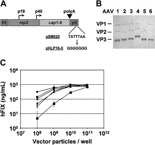
The AAV-2 vector plasmid phF.IX-16 has been described.3 Briefly, this construct contains the apolipoprotein E hepatic locus control region (ApoEHCR)-human α1-antitrypsin promoter (hAAT)-driven hfIX minigene (including the 1.4-kb intron A) and a bovine growth hormone polyadenylation signal, flanked by inverted terminal repeats of AAV-2. The adenoviral helper plasmid pladeno5, used for helpervirus-free AAV vector production (next paragraph), has also been described.21 A second helper expressing AAV-2 Rep and capsid (VP) proteins, pHLP19-2, was constructed by subcloning AAV-2 nucleotides 315 to 4534 from pSM62022 into pBluescript s/k+ (Stratagene, La Jolla, CA). The AAV-2 p5 promoter was moved to a position 3′ of the AAV polyadenylation site and was replaced by a 5′ untranslated region primarily composed of an Flp recombinase protein (FLP) recombinase recognition sequence (frt). Additionally, the 7-bp TATA box of the p5 promoter was destroyed by mutation to GGGGGGG (Figure 1A). Initially part of efforts to generate an inducible AAV-2 expression construct, these modifications were found to result in enhanced AAV vector yields and to suppress the generation of replication-competent pseudo-wild-type virus.
Characterization of AAV vector types 1 to 6 for expression of hfIX. (A) Structure of AAV pseudotyping helper plasmids. All helper constructs contain the AAV-2 rep gene, together with the cap gene of one of the 6 AAV serotypes. Other elements shown are the AAV-2 promoters (p5, p19, and p40), the AAV polyA signal, and the frt sequence upstream of the rep gene. Depicted also is the mutation of the p5 TATA box, from the genuine sequence as present in wild-type AAV-2 plasmid pSM620 (top) to a modified sequence (bottom). As a result, the constructs express lower amounts of large Rep but greater amounts of small Rep proteins than a nonmodified helper plasmid (data not shown). (B) Analysis of vector particle purity. A recombinant AAV-2 genome expressing the hfIX gene was cross-packaged into capsids of AAV serotypes 1 to 6. Then 5 × 10¹⁰ genome-containing particles were subjected to SDS-PAGE silver stain. This revealed 3 protein bands (VP1, VP2, and VP3) in each sample, corresponding to the proteins of the particular AAV capsid (as a reference, capsid proteins of AAV-2 are marked on the left side). Notable is the absence of other protein bands demonstrating the high purity of the preparations. The fact that ratios of capsid proteins to genome copy numbers were constant for all AAV types was also evident. Note that the capsid proteins of the 6 AAVs differ slightly in their gel migration pattern, though the corresponding cap genes have nearly identical lengths. (C) Analysis of hFIX expression in cell culture. HepG2 cells were infected with the 6 different vector preparations, and hFIX protein levels in the media were quantified as detailed in “Materials and methods.” ▪ indicates AAV-1; ▪, AAV-2; ▴, AAV-3; ×, AAV-4; ♦, AAV-5; and •, AAV-6. Values shown are mean ± SD (n = 3).
Characterization of AAV vector types 1 to 6 for expression of hfIX. (A) Structure of AAV pseudotyping helper plasmids. All helper constructs contain the AAV-2 rep gene, together with the cap gene of one of the 6 AAV serotypes. Other elements shown are the AAV-2 promoters (p5, p19, and p40), the AAV polyA signal, and the frt sequence upstream of the rep gene. Depicted also is the mutation of the p5 TATA box, from the genuine sequence as present in wild-type AAV-2 plasmid pSM620 (top) to a modified sequence (bottom). As a result, the constructs express lower amounts of large Rep but greater amounts of small Rep proteins than a nonmodified helper plasmid (data not shown). (B) Analysis of vector particle purity. A recombinant AAV-2 genome expressing the hfIX gene was cross-packaged into capsids of AAV serotypes 1 to 6. Then 5 × 10¹⁰ genome-containing particles were subjected to SDS-PAGE silver stain. This revealed 3 protein bands (VP1, VP2, and VP3) in each sample, corresponding to the proteins of the particular AAV capsid (as a reference, capsid proteins of AAV-2 are marked on the left side). Notable is the absence of other protein bands demonstrating the high purity of the preparations. The fact that ratios of capsid proteins to genome copy numbers were constant for all AAV types was also evident. Note that the capsid proteins of the 6 AAVs differ slightly in their gel migration pattern, though the corresponding cap genes have nearly identical lengths. (C) Analysis of hFIX expression in cell culture. HepG2 cells were infected with the 6 different vector preparations, and hFIX protein levels in the media were quantified as detailed in “Materials and methods.” ▪ indicates AAV-1; ▪, AAV-2; ▴, AAV-3; ×, AAV-4; ♦, AAV-5; and •, AAV-6. Values shown are mean ± SD (n = 3).
Chimeric helper plasmids encoding the AAV-2 rep gene and capsid genes from AAV types 1 or 3B to 6 were derived from pHLP19-2 and named pHLP19-1 and pHLP19-3 through -6, respectively. The first chimeric construct, pHLP19-6, was obtained by polymerase chain reaction (PCR) amplification of the AAV-6 capsid gene from an AAV-6 plasmid (provided by David Russell, University of Washington, Seattle) and its subsequent cloning into the rep-containing fragment of pHLP19-2 cut with SwaI and SnaBI. PCR primers used in the construction of all chimeric plasmids are provided at the end of this paragraph. Other helpers were derived from pHLP19-6 by cloning PCR-amplified cap genes of AAV-1 (using wild-type AAV-1 DNA as template; ATCC VR-645), or of AAV-3B, -4, or -5 (using provided plasmids as templates) into the rep-containing fragment of pHLP19-6 cut with SwaI and AgeI. This cloning strategy, by which AAV-2 rep and the various capsid genes were joined in the central AAV intron, ensured that critical features of the introns of AAV-2 and the cap-providing serotype were maintained. PCR primer pairs used were as follows (5′ to 3′ direction): AAV-1, CACACATTTAAATCAGGTATGGCTGC and TTTATCGCGAAGCGCAACCAAGCAG; AAV-3B, AAATGACTTAAACCAGGTATGGCTGC and GAATTAACCGGTTTATTGATTAACCAGG; AAV-4, AAATCAGATATGACTGACGGTTACC and GGACACGGAGACCAAAGTTC; AAV-5, GTAAATAAATTTAGTAGTCATGTCTTTTGTTGATCACC and GAATAACCGGTTTATTGAGGGTATGCG; AAV-6, AAATCAGGTATGGCTGCCGAT and GGGGATCTGGTGACCAGATAAG.
Before ligation, the PCR fragments were digested with AgeI, and the AAV-1 and -5 fragments were also digested with SwaI. All PCR reactions were performed using Pfu DNA polymerase (Stratagene). The integrity of all 5 chimeric helpers was verified by sequencing the cap regions.
Vector production
Vented cap roller bottles (850 cm2; Corning, New York, NY) were seeded with 1.8 × 10⁷ HEK293 cells in 140 mL Dulbecco modified Eagle medium (DMEM) containing 10% fetal calf serum (FCS). The cultures were incubated at 37°C with 5% CO2 and rotated at 0.2 rounds per minute for 3 days before transfection. For each roller bottle, 10 mL of 300 mM CaCl2 containing 100 μg each of the phF.IX-16 vector plasmid and the 2 helpers (pladeno5 and one of the pHLP19 plasmids) was rapidly mixed with 10 mL of 2 × HBS (HEPES-buffered saline; 50 mM HEPES [N-2-hydroxyethylpiperazine-N′-2-ethanesulfonic acid], 280 mM NaCl, 1.5 mM NaH2PO4, pH 7.1) and was added to the roller bottle within 10 minutes. The media were changed after 6-hour transfection and replaced with 100 mL serum-free DMEM. Cultures were then incubated for 72 hours. Vector-containing cells were dislodged from the sides of the roller bottles by gentle swirling and collected by centrifugation at 1000g for 15 minutes 4°C.
Titration of vector particles (“Vector titration”) at this point showed that the serotypes differed in the amount of vector released into the production media from the cells. Of the total vector produced (cells + media), AAV-2 cultures released approximately 10% into the media. In contrast, AAV-1 cultures released approximately 25%, and AAV-3 to -6 released approximately 40% to 60%, of the total vector into the media. Consequently, cultures involving AAV-3 to -6 helpers produce roughly twice as much total vector particles as AAV-2.
Vector purification
The collected HEK293 cells were dispersed in TBSM buffer (50 mM Tris-Cl, 150 mM NaCl, 2 mM MgCl2, pH 8.0, 15 mL per roller bottle) before disruption by microfluidization (3 passes, model HC 2000; Microfluidics Corporation, Newton, MA). Tissue debris was removed by centrifugation (2000g, 30 minutes, 4°C), and the supernatant was adjusted to 25 mM CaCl2 by adding the appropriate amount of a 1 M stock solution. After 1-hour incubation at 4°C, the resultant precipitate was removed by centrifugation (2000g, 30 minutes, 4°C), and the supernatant was digested with Benzonase (100 U/mL) for 1 hour at 37°C. Vector particles were then precipitated for 2 hours at 0°C with 8% polyethylene glycol 8000 (in 650 mM NaCl) and collected by centrifugation (2000g, 30 minutes, 0°C). The pellet was completely dissolved in 50 mM HEPES, 150 mM NaCl, 20 mM EDTA (ethylenediaminetetraacetic acid), 1% Sarcosyl, pH 8.0 containing 10 μg/mL RNase A (2 mL per roller bottle) and was applied to a CsCl step gradient consisting of 5 mL of 1.5 g CsCl/mL for the bottom layer, 10 mL of 1.3 g CsCl/mL for the middle layer, then a 22-mL sample (in 25 × 89 mm polyallomer tubes, spun in an SW28 rotor at 28 000 RPM for 17 hours at 20°C). The lower full capsid band was identified visually and collected with an 18-gauge hypodermic needle and a 10-mL syringe through the side of the tube. The vector-containing solution was again centrifuged on a linear CsCl gradient (14 × 95-mm polyallomer tubes, spun in an SW40 rotor at 38K RPM for 24 hours at 20°C). The full capsid band was collected with a hypodermic needle, diafiltered against phosphate-buffered saline (PBS) containing 5% sorbitol and 0.1% Tween-80, pH 7.4, and stored frozen at -80°C.
Vector titration
Vector genome titers were established by taking the average of 3 quantitative real-time PCR (Q-PCR) determinations. All samples were DNaseI treated before measurement and were subjected to 45 cycles of amplification (2 steps per cycle: 62°C for 60 seconds, 95°C for 15 seconds) in a 7700 Q-PCR machine (Applied Biosystems, Foster City, CA) using the following primer and probe sets: forward, ACC AGC AGT GCC ATT TCC A; reverse, GAA TTG ACC TGG TTT GGC ATC T; probe, 6FAM-TTG GAT AAC ATC ACT CAA AGC ACC CAA TCA-TAMRA.
Analysis of vector transduction in vitro and in vivo
In vitro transduction activities of the vectors were assessed by infecting HepG2 cells for 8 hours in serum-free media with serial dilutions of each vector (1 × 10⁸ to 1 × 10¹¹ genome-containing particles per well, in 12-well plates containing 3 × 10⁵ cells per well). Media were then replaced with fresh media containing 10% FCS and 20 μM etoposide (to speed up the transduction process). Human FIX concentrations in the media were measured 24 hours after transduction by hFIX enzyme-linked immunosorbent assay (ELISA), as reported.23
For assessment of in vivo transduction, vectors were infused as previously described24 into the portal or tail vein of 6-week-old C57Bl/6 mice (Jackson Laboratories, Bar Harbor, ME) at the particle doses specified in the text. At various points in time after vector infusion, blood was collected from the animals by retro-orbital plexus bleed, and hFIX levels in the plasma were determined by hFIX ELISA.23
Analyses of vector integrity
Sodium dodecyl sulfate-polyacrylamide gel electrophoresis (SDS-PAGE) analysis was performed according to the manufacturer's instructions (Invitrogen, Carlsbad, CA), using 8 × 6 × 0.1-cm 10% acrylamide gels, and proteins were visualized by silver staining (Daiichi, Tokyo, Japan). Dynamic light-scattering analysis was performed using a DynaPro model according to the manufacturer's recommendations (Protein Solutions, Charlottesville, VA). Analysis of 1 × 10¹⁰ to 1 × 10¹¹ particles in a volume of 12 μL showed hydrodynamic radii of 12 nm for 100% of the vectors in each preparation, indicating that the particles were monomeric and not aggregated.
Vector DNA tissue distribution analyses
Mice were killed 6 weeks after portal or tail vein injection of 2 × 10¹¹ AAV vector particles (n = 3 per group), and the following tissues were extracted, minced with scissors, and immediately frozen in liquid nitrogen: liver, heart, kidney, spleen, brain and lung. Genomic DNA was extracted from approximately 10 to 25 mg of each tissue using the DNeasy Kit (Qiagen, Valencia, CA) according to the manufacturer's instructions and was eluted from the supplied columns in 100 μL provided elution buffer. Total DNA concentrations were quantified by spectrophotometry and confirmed by Q-PCR using rodent glyceraldehyde-3-phosphate dehydrogenase (GAPDH) control reagents (Applied Biosystems) and one of our samples (DNA from an AAV-1 portal vein-injected mouse) as an internal standard. The number of AAV vector genomes in each sample (10 ng genomic DNA) was determined by Q-PCR, with the following primers and probe: forward, AAG ATG CCA AAC CAG GTC AAT T; reverse, CGA TAG AGC CTC CAC AGA ATG C; probe, 6FAM-CCT TGG CAG GTT GTT TTG AAT GGT AAA GTT GA-TAMRA. For the standard curve, 8 serial 4-fold dilutions of the phF.IX-16 plasmid were used, corresponding to 1 × 10⁶ down to 61 DNA molecules. However, because neither this standard nor any other would accurately correlate with vector genome forms in a complex tissue DNA background, absolute numbers were not used. Instead, all Q-PCR values were standardized, using as internal reference maximum genome copy numbers, which were obtained from animals injected with the AAV-6 vector through the portal vein (see “Results”). Accordingly, those numbers were set to 1, and all other data were then calculated as relative genome copy numbers. The Q-PCR reactions were performed on a GeneAmp 5700 Q-PCR machine (Applied Biosystems), and the reaction conditions were 50°C for 2 minutes and 95°C for 10 minutes, followed by 40 cycles of 95°C for 15 seconds and 60°C for 1 minute.
AAV vector genome copy numbers in the livers of the mice were additionally quantified by Southern blot analysis using previously reported protocols for extraction and Southern blot analyses of liver genomic DNA.24
Virus neutralization assay
Murine sera were assessed for anti-AAV neutralizing antibodies by testing their ability to inhibit transduction of Huh-7 cells by the hFIX-expressing AAV vectors of the 6 different types. Therefore, 2 × 10⁹ genome-containing particles of each type were incubated for 1 hour at 37°C with heat-inactivated (56°C, 1 hour) sera that had been serially 4-fold diluted (from 1:25 to 1:6400) or with PBS or sera from naive mice as negative controls. The vectors were then added to Huh-7 cells in 24-well plates (2 × 10⁴ cells per well) at a multiplicity of infection of 2 × 10⁵. After 16 hours of incubation at 37°C with 5% CO2, the medium was replaced with 400 μL fresh DMEM, and the cells were further incubated for 5 days before the amount of hFIX protein in the media was determined by hFIX ELISA.23 For each sera and AAV type, a titer of (cross-) neutralizing antibodies was calculated as the reciprocal of the highest dilution of the sera that inhibited transduction of the particles by at least 50% of that from the corresponding negative controls.
Results
Large-scale production of hfIX-expressing AAV serotype vectors
For generation of vectors based on serotypes 1 to 6 of AAV, a set of novel helper plasmids, called pHLP19-1 through -6, was developed, consisting of the AAV-2 rep gene positioned upstream of the cap genes from AAV-1 to -6 (Figure 1A; see “Materials and methods” for details). We used these helpers to cross-package an AAV-2 vector genome, containing AAV-2 inverted terminal repeats flanking an hfIX minigene under the control of a strong liver-specific promoter, into capsids of AAV-1 to -6. Large-scale vector production was performed in 293 cells in roller bottles, as described in “Materials and methods.” Vector particles were extracted from the cells 72 hours after transfection and quantified by Q-PCR. Vector yields from the crude extracts were similar for all 6 serotypes, ranging from 5.7 × 10¹² vector-genome (vg)-containing particles (AAV-1) to 1.44 × 10¹³ vg (AAV-4) per roller bottle. No particular serotype has proven superior in subsequent vector production, suggesting this range is attributed entirely to experimental variation.
AAV vector preparations were purified with a CsCl-based process that resulted in good recoveries of 30% to 58% relative to the crude extract, with final yields of 2.04 × 10¹² to 7 × 10¹² vg per roller bottle. Again, subsequent purification experience indicated that the yield variation did not correlate with AAV serotype. Purity and integrity of the vectors were analyzed by subjecting equal numbers of particles to SDS-PAGE with silver staining (Figure 1B). The 3 AAV capsid bands were predominant in each lane and displayed equal relative staining intensities among the 6 preparations, indicating that encapsidation of the AAV-2 vector DNA into the different capsids had occurred equally efficiently.
To assess functionality of the particles in vitro, we infected HepG2 cells with increasing multiplicities of infection from 333 to 3.3 × 10⁶ under conditions described in “Materials and methods.” Human FIX levels in media harvested 24 hours after infection were measured by ELISA (Figure 1C). All 6 vectors expressed hFIX, and vectors derived from AAV-2 to -6 responded similarly to increasing dose, reaching a plateau of hFIX expression at the second highest dose. Interestingly, the AAV-6 vector produced greater responses at the lower doses, and overall the AAV-1 vector transduced the cells less efficiently than the other 5 serotypes.
In vivo expression of human factor IX from AAV serotype vectors
To determine the efficiency of pseudotyped AAV vector-mediated transduction of hepatocytes in vivo, we injected C57Bl/6 mice with low (2 × 10¹⁰ vg), middle (2 × 10¹¹), or high (2 × 10¹²) doses of each serotype, through the portal or the tail vein. Plasma hFIX levels were monitored for 12 weeks.
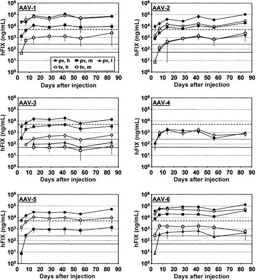
At all doses, hFIX expression from all 6 different AAV vectors gradually increased in 14 days and thereafter stabilized for the duration of the experiment (84 days) (Figure 2). Maximal protein levels varied up to 75-fold between the serotypes. Strongest expression was observed with the AAV-6 vector, with maximal hFIX expression at 134.9 μg/mL hFIX (corresponding to 2700% of normal hFIX levels in humans), seen 84 days after portal vein delivery of the highest dose. This is the strongest expression of hFIX protein thus far reported from an AAV vector. Similarly high hFIX levels were achieved with the AAV-1 and -2 vectors injected at high doses into the hepatic circulation, with maximal levels of 114.4 μg/mL (day 42) and 103.9 μg/mL (day 84), respectively, both representing more than 2000% of normal levels. For the other vectors, maximal hFIX levels after hepatic infusion of the highest dose were at least 2-fold lower, with 53.4 μg/mL for AAV-5 (day 84), 17.8 μg/mL for AAV-3 (day 42), and 1.8 μg/mL for AAV-4 (day 14). Importantly, these lower hFIX levels were still approximately 36% (AAV-4) of normal or greater, which would be in excess of curative levels in humans.
In vivo expression of hFIX from AAV serotype vectors. C57Bl/6 mice (n = 5 or 8) were injected with AAV serotype 1 to 6 vectors expressing the hfIX gene at 3 different particle doses—2 × 10¹⁰ (low dose, l), 2 × 10¹¹ (middle dose, m), or 2 × 10¹² (high dose, h) genome-containing particles. Vectors were injected through the portal vein (pv) or the tail vein (tv). Shown are plasma levels (mean ± SD) of FIX protein measured by ELISA between day 3 after particle injection up to day 84. The lines in each graph indicate 1% of normal FIX levels (solid line), 5% (dotted line), or 100% (dashed line). An hFIX plasma level of 5 ng/mL was considered the detection limit of the assay; protein levels below this limit are thus not shown.
In vivo expression of hFIX from AAV serotype vectors. C57Bl/6 mice (n = 5 or 8) were injected with AAV serotype 1 to 6 vectors expressing the hfIX gene at 3 different particle doses—2 × 10¹⁰ (low dose, l), 2 × 10¹¹ (middle dose, m), or 2 × 10¹² (high dose, h) genome-containing particles. Vectors were injected through the portal vein (pv) or the tail vein (tv). Shown are plasma levels (mean ± SD) of FIX protein measured by ELISA between day 3 after particle injection up to day 84. The lines in each graph indicate 1% of normal FIX levels (solid line), 5% (dotted line), or 100% (dashed line). An hFIX plasma level of 5 ng/mL was considered the detection limit of the assay; protein levels below this limit are thus not shown.
Human FIX levels above the curative threshold were also found after portal vein delivery of the middle particle dose for all vectors, with the exception of AAV-4, which expressed only at the highest dose. Portal vein administration of the low dose resulted in expression from 3 of the 6 serotypes, with hFIX levels above the therapeutic (AAV-3) or curative (AAV-2 and -6) threshold.
Tail vein delivery of the high dose gave hFIX levels in the curative range from all 6 serotypes, whereas at the middle dose, only the 3 overall strongest expressing AAV types (1, 2, and 6) resulted in curative hFIX levels. AAV-3 expressed close to the therapeutic threshold, and expression from AAV-4 and -5 was undetectable. Human FIX levels were also undetectable from all vectors after tail vein delivery of the low dose.
In most cases, portal vein particle infusion resulted in stronger hFIX expression than tail vein delivery of the same vector dose, as was most obvious from the 84-fold difference seen after injection of the middle dose of AAV-3. Surprisingly, however, at the high dose, AAV-1, -4, and -6 expressed almost equally efficiently from both routes of administration.
Dose response and kinetics of AAV serotype vector-mediated gene expression
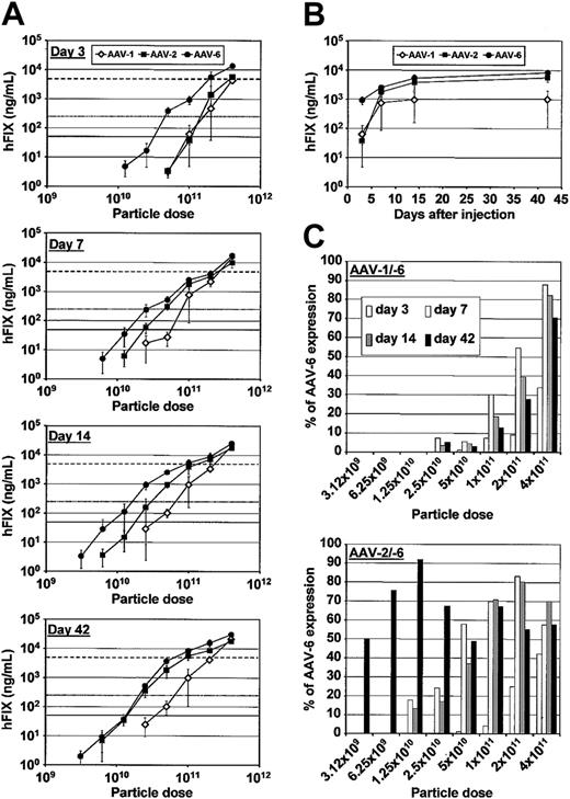
Despite the more than 99% sequence homology between AAV-1 and AAV-6,12 we found that AAV-6 mediated consistently higher levels of hFIX expression than AAV-1 and expressed the transgene with faster kinetics (Figure 2). To confirm this finding, we injected new mice through the portal vein with the 3 most strongly expressing AAV pseudotypes (1, 2, and 6) at 9 different doses, which increased in 2-fold increments from 1.56 × 10⁹ to 4 × 10¹¹ particles per mouse. Expression of hFIX protein was monitored starting 3 days after particle injection and then at days 7, 14, and 42, when hFIX levels were expected to be stable (Figure 2).
As evident from the expression kinetics (Figure 3A-B) and the quantitative comparison of hFIX expression (Figure 3C), there were indeed significant differences in dose response and kinetics in all 3 AAV vectors. Although all serotypes exhibited an approximately linear dose response, the AAV-6 vector gave strongest expression at all doses tested (Figure 3A) and reached a plateau of maximal expression at earlier points in time (Figure 3B). The faster onset of expression was striking at day 3 after injection, when AAV-6 expressed at 2 lower doses than AAV-1 and -2 and yielded up to greater than 100-fold more hFIX protein at higher doses (Figure 3A, top). At this early time, 5 × 10¹⁰ particles of AAV-6 were already sufficient to reach potentially curative hFIX levels, whereas 4-fold higher doses of AAV-1 or -2 were required to achieve similar levels. At later times, AAV-6 consistently expressed hFIX from lower doses than AAV-1 or -2 (Figure 3A).
Dose-dependence and kinetics of hFIX expression from AAV serotype vectors. (A) hFIX expression as a function of vector dose and time after injection. hFIX-expressing AAV vectors of type 1, 2, or 6 were injected into the portal veins of C57Bl/6 mice (n = 4 or 5) at 9 different doses, in 2-fold increments ranging from 1.56 × 10⁹ to 4 × 10¹¹ genome-containing particles. hFIX levels in the plasma were measured using ELISA, from day 3 after injection until day 42, and were plotted as mean ± SD against the vector dose. The lines in each graph indicate 1% (solid line), 5% (dotted line), or 100% (dashed line) of normal hFIX levels. (B) Kinetics of hFIX expression. As an example to highlight the different kinetics of AAV-1, -2, and -6 vectors, the graph shows hFIX levels obtained between 3 and 42 days after portal vein injection of 1 × 10¹¹ particles. (C) Ratio of hFIX expression from AAV vector types 1 or 2, respectively, to the AAV-6 vector. For each vector dose and point in time after injection, plasma hFIX levels from the AAV-1 (left) or -2 (right) vectors were plotted as a percentage of expression from the AAV-6 vector, which was always set at 100%.
Dose-dependence and kinetics of hFIX expression from AAV serotype vectors. (A) hFIX expression as a function of vector dose and time after injection. hFIX-expressing AAV vectors of type 1, 2, or 6 were injected into the portal veins of C57Bl/6 mice (n = 4 or 5) at 9 different doses, in 2-fold increments ranging from 1.56 × 10⁹ to 4 × 10¹¹ genome-containing particles. hFIX levels in the plasma were measured using ELISA, from day 3 after injection until day 42, and were plotted as mean ± SD against the vector dose. The lines in each graph indicate 1% (solid line), 5% (dotted line), or 100% (dashed line) of normal hFIX levels. (B) Kinetics of hFIX expression. As an example to highlight the different kinetics of AAV-1, -2, and -6 vectors, the graph shows hFIX levels obtained between 3 and 42 days after portal vein injection of 1 × 10¹¹ particles. (C) Ratio of hFIX expression from AAV vector types 1 or 2, respectively, to the AAV-6 vector. For each vector dose and point in time after injection, plasma hFIX levels from the AAV-1 (left) or -2 (right) vectors were plotted as a percentage of expression from the AAV-6 vector, which was always set at 100%.
Although both AAV-1 and -2 expressed hFIX less efficiently than AAV-6 at earlier points in time, the expression profile of AAV-2 shifted over time from being identical to AAV-1 to more closely matching the profile of AAV-6 (Figure 3A, C). AAV-1 remained less efficient than AAV types 2 and 6, except at the highest dose (4 × 10¹¹ vg), at which all 3 vectors expressed with similar efficiency. These data demonstrate that all 3 AAV serotypes have distinct expression kinetics and further support that AAV-1 and -6 are distinct members of the AAV family, despite their extensive homology.
Tissue distribution of AAV serotype vector DNA
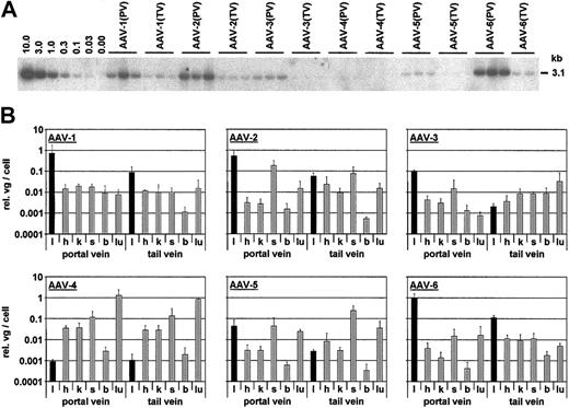
The promoter used to drive expression of the hfIX gene in all 6 AAV vectors was liver specific, implying that the plasma hFIX protein detected originated in the livers of the mice. To confirm that AAV vector copy numbers in the livers corroborated protein levels and to analyze whether vector DNA was also present in other tissues, we extracted genomic DNA from liver, heart, kidney, spleen, brain, and lung from mice killed 6 weeks after portal or tail vein injection with middle vector doses of each serotype. Human fIX vector genomes were quantified in identical amounts of genomic DNA using Southern blotting (livers only; a representative blot is shown in Figure 4A) and Q-PCR (all tissues; Figure 4B).
Distribution of AAV vector DNA in murine tissues. (A) Southern blot analysis for determination of double-stranded vector genome copy numbers per diploid genomic equivalent. Total DNA was extracted from livers of C57Bl/6 mice 6 weeks after portal vein (pv) or tail vein (tv) injection with 2 × 10¹¹ hFIX-expressing AAV vector particles of types 1 to 6. Twenty micrograms DNA was digested with a combination of BamHI and XhoI (leaving a 3.1-kb fragment), separated on a 0.8% agarose gel, and hybridized with a vector sequence-specific probe. Lanes labeled 10.0 to 0.00 are vector genome copy number standards, and all other lanes represent samples from individual mice. (B) Genomic DNA was isolated from 6 different tissues of the same mice described in (A), and AAV genomes were detected and quantified by Q-PCR. Tissues were liver (l), heart (h), kidney (k), spleen (s), brain (b), and lung (lu). Shown are relative genome copy numbers (liver samples from mice injected with AAV-6 through the portal vein served as internal standard and were set to 1; see Table 1) per diploid cell (mean ± SD; n = 3), with liver samples highlighted by dark bars.
Distribution of AAV vector DNA in murine tissues. (A) Southern blot analysis for determination of double-stranded vector genome copy numbers per diploid genomic equivalent. Total DNA was extracted from livers of C57Bl/6 mice 6 weeks after portal vein (pv) or tail vein (tv) injection with 2 × 10¹¹ hFIX-expressing AAV vector particles of types 1 to 6. Twenty micrograms DNA was digested with a combination of BamHI and XhoI (leaving a 3.1-kb fragment), separated on a 0.8% agarose gel, and hybridized with a vector sequence-specific probe. Lanes labeled 10.0 to 0.00 are vector genome copy number standards, and all other lanes represent samples from individual mice. (B) Genomic DNA was isolated from 6 different tissues of the same mice described in (A), and AAV genomes were detected and quantified by Q-PCR. Tissues were liver (l), heart (h), kidney (k), spleen (s), brain (b), and lung (lu). Shown are relative genome copy numbers (liver samples from mice injected with AAV-6 through the portal vein served as internal standard and were set to 1; see Table 1) per diploid cell (mean ± SD; n = 3), with liver samples highlighted by dark bars.
AAV vector DNA was detected in all tissues examined, but copy numbers of recombinant genomes per cell varied significantly between the samples. For most AAV types and routes of administration, most vector genomes were detected in the liver. Additional Southern blot analyses showed the predominance of supercoiled circular molecules in head-to-tail configuration for all pseudotyped vectors in this tissue (data not shown). Interestingly, AAV-2 and -5 gave equal or higher genome numbers in the spleen, than in the liver, and hfIX DNA copy numbers in the liver were lower than in all other tissues in mice in which the AAV-3 vector was injected through the tail vein. Overall, genome numbers were lowest in livers of mice injected with AAV-4, which, after hepatic or systemic particle delivery, directed most vector DNA to the lungs of the animals, suggesting an unknown inherent tropism of AAV-4 for this tissue.
In all samples, the liver vector genome numbers correlated with the plasma levels of hFIX protein (Table 1). The ratios of plasma hFIX levels to liver genome copy numbers were consistent among the groups; only the AAV-3 vector gave slightly higher ratios, indicating that for unknown reasons, relative protein expression per genome was stronger for AAV-3. When comparing the ratios of liver DNA copy numbers between mice injected through the portal or tail vein, we noted a good correlation to the ratios of the plasma hFIX protein levels for AAV serotypes 1 to 3 and 6. For AAV-4 and -5, this correlation was not evident because of the low hFIX expression, though the equal numbers of vector genomes found after portal or tail vein delivery of AAV-4 were reminiscent of the similar protein levels seen at the high dose of this vector (Figure 2).
AAV vector genome copy numbers per diploid liver cell genome and hFIX levels
. | Southern blot . | . | Q-PCR . | . | hFIX, μg/mL . | . | . | |||
|---|---|---|---|---|---|---|---|---|---|---|
| Vector . | Mean ± SD . | pv/tv . | Mean ± SD . | pv/tv . | Mean ± SD . | pv/tv . | hFIX/genome . | |||
| AAV-1 pv | 1.25 ± 0.6 | 11.36 | 0.725 ± 1.042 | 8.15 | 12.25 ± 6.16 | 9.49 | 16.9 | |||
| AAV-1 tv | 0.11 ± 0.06 | — | 0.089 ± 0.078 | — | 1.29 ± 0.78 | — | 14.49 | |||
| AAV-2 pv | 1.96 ± 1.02 | 28 | 0.52 ± 0.503 | 9.12 | 10.76 ± 3.33 | 7.63 | 20.69 | |||
| AAV-2 tv | 0.07 ± 0.01 | — | 0.057 ± 0.023 | — | 1.41 ± 0.75 | — | 24.74 | |||
| AAV-3 pv | 0.38 ± 0.15 | — | 0.097 ± 0.019 | 48.5 | 4.68 ± 1.27 | 78 | 48.25 | |||
| AAV-3 tv | < 0.03* | — | 0.002 ± 0.001 | — | 0.06 ± 0.03 | — | 30 | |||
| AAV-4 pv | < 0.03* | — | 0.001 | 1 | ND | — | — | |||
| AAV-4 tv | < 0.03* | — | 0.001 ± 0.001 | — | ND | — | — | |||
| AAV-5 pv | 0.03 ± 0.01 | — | 0.044 ± 0.041 | 14.67 | 0.93 ± 0.59 | — | 21.14 | |||
| AAV-5 tv | < 0.03* | — | 0.003 ± 0.001 | — | ND | — | — | |||
| AAV-6 pv | 1.61 ± 0.12 | 11.5 | 1 ± 0.587 | 8.93 | 18.7 ± 3.4 | 10.11 | 18.7 | |||
| AAV-6 tv | 0.14 ± 0.06 | — | 0.112 ± 0.036 | — | 1.85 ± 1.33 | — | 16.52 | |||
AAV genomes were detected in livers from vector-treated animals and were quantified using Southern blot analysis or Q-PCR, as described in “Materials and methods.” Shown are absolute (Southern blot) or relative (Q-PCR) genome copy numbers per cell, together with hFIX levels expressed (mean ± SD; n = 3 per group). Within the Q-PCR data, values obtained from AAV-6 portal-vein-injected animals served as an internal reference and were set to 1 because of the lack of an appropriate absolute standard (see “Materials and methods”). Also shown are ratios of copy numbers of hFIX levels after portal or tail vein delivery of each vector (pv/tv). The rightmost column lists ratios of hFIX levels to relative genome copy numbers.
pv indicates portal vein; tv, tail vein;—, not applicable; and ND, not detected.
Below detection limit of the method.
Emergence of cross-neutralizing antibodies from AAV serotype vector delivery
To determine whether the mice had generated neutralizing antibodies against the different vectors and whether these would cross-react among the AAV types, we performed a virus neutralization assay (see “Materials and methods”) using sera from the same animals analyzed in the tissue distribution study.
As summarized in Table 2, delivery of each AAV vector yielded the expected neutralizing antibodies against the same AAV type (boldface data). This was most pronounced for AAV-4, which after portal or tail vein injection elicited high titers of neutralizing antibodies above the detection limit of our assay. AAV-4 was also the only serotype that was not neutralized by antibodies against other serotypes. The opposite was observed for AAV-5, which generated only low levels of self-neutralizing antibodies but was inhibited by antibodies against all other types, with the exception of AAV-4.
Generation of (cross-) neutralizing antibodies in response to infection with AAV serotype vectors
Initial vector . | AAV-1 . | AAV-2 . | AAV-3 . | AAV-4 . | AAV-5 . | AAV-6 . |
|---|---|---|---|---|---|---|
| AAV-1pv | 1600 | ND | ND | ND | ND to 400 | 1600 |
| AAV-1 tv | 1600-6400 | ND | ND | ND | ND to 400 | 1600 |
| AAV-2pv | ND | 400-1600 | ND | ND | ND to 25 | ND |
| AAV-2 tv | ND | 1600 | 25 | ND | 100-400 | ND |
| AAV-3pv | ND | ND | 100-400 | ND | ND | ND |
| AAV-3 tv | ND | 25 | 400-1600 | ND | ND to 25 | ND |
| AAV-4pv | ND | ND | ND | 6400 | ND | ND |
| AAV-4 tv | ND | ND | ND | 6400 | ND | ND |
| AAV-5pv | ND to 25 | ND | ND | ND | 25-100 | 25-100 |
| AAV-5 tv | 1600-6400 | ND | ND | ND | 100 | 100 |
| AAV-6pv | 100-400 | ND to 25 | ND | ND | 400 | 1600 |
| AAV-6 tv | 400-6400 | 100 | ND to 25 | ND | 1600 | 1600 |
Values represent the titers of neutralizing antibodies against the vectors and subtypes indicated in each column. Four-fold serial dilutions of sera from mice injected with recombinant AAV serotypes 1 to 6 were incubated with the same viruses in all possible combinations, and residual virus infectivity was determined (see “Materials and methods” for details). Three independent samples were analyzed per group. Antibody titers are given as reciprocals of the highest sera dilutions that caused a decrease in vector infectivity of at least 50%. Boldface values indicate titers of neutralizing antibodies against the same serotype with which the mice were initially injected. Abbreviations as in Table 1.
Cross-reactivity among AAV serotypes was noted in 12 cases and usually resulted in moderate titers of neutralizing antibodies. Interesting exceptions were the AAV-1 and -6 vectors, each of which yielded antibodies that efficiently cross-neutralized the other AAV type. This supports the notion that AAV-1 and -6 are closely related serotypes.
Noteworthy was that in 14 of the 18 total cases of antibody emergence, systemic particle injection led to stronger immune response against the same or other AAV types than direct hepatic delivery. This generally suggested that the hepatic route of particle administration is favorable for liver-directed gene transfer strategies that rely on the readministration of AAV serotype vectors.
Discussion
The present work was inspired by recent reports demonstrating that vectors based on AAV serotypes differ in cell tropism and serology from the commonly used AAV vector type 2 (for review, see Grimm18 ). This suggests they can be used to target tissues that are resistant to efficient infection with AAV-2 or to circumvent neutralizing anti-AAV-2 antibodies, which are highly prevalent in the human population. Here, we evaluated vectors based on AAV serotypes 1 to 6 for the purpose of improving hepatic gene transfer. Our experiments were designed to answer the following questions: How would these vectors compare for in vivo transduction of hepatocytes? In particular, how efficiently would they transduce after systemic or hepatic infusion, and would they differ in dose response and kinetics of gene expression? Would they result in different tissue distribution of the transgene, and would each vector yield immune responses limited to itself or extending to other serotypes?
To address these issues accurately, we aimed to ensure that differences between the vectors were limited to the particle capsids that originated from the different AAV types. This was achieved by pseudotyping the identical hfIX-expressing AAV-2 vector DNA with the 6 different AAV capsids, using the same large-scale plasmid transfection protocol. Resultant particles were purified and concentrated by an identical CsCl-based procedure, which further ensured that the 6 preparations were comparable. Importantly, the high degree of particle purity achieved, together with the high particle yields obtainable from transfected cells in roller bottles, make the technology presented here appealing for clinical-grade vector production.
We found that all 6 AAV vectors were able to direct high-level hFIX expression from the liver, yielding maximum levels ranging from 36% to more than 2700% of normal, which is in great excess of the curative threshold for patients with hemophilia. With a peak of 134.9 μg/mL (AAV-6), our hFIX levels were also in excess of previously reported levels from any viral or nonviral vector systems. This was likely because of the conditions for efficient hepatocyte transduction used here—direct hepatic infusion of high vector doses and a strong liver-specific enhancer/promoter driving hfIX expression—but it also clearly highlights the great potential of AAV serotypes for liver-directed gene delivery.
Our finding is clinically relevant considering that anti-AAV-2 antibodies prevalent in the population might prevent a substantial number of patients from efficient treatment with the AAV-2 vector system.11,12 Hepatic gene delivery could be achieved by simply pseudotyping a therapeutic gene with an alternative AAV shell to which the patient is not already immune. In this respect, it is interesting to note that Gao et al11 recently described successful hepatic gene transfer with 2 further serotypes, AAV-7 and -8, thus extending the number of AAV serotypes to target the human liver to 8. Importantly, antibodies against most AAV serotypes other than 2 appear to be rare in the human population.11,12,25
In our hands, none of the alternative serotypes expressed significantly more strongly in the liver than the AAV-2 vector. Mingozzi et al20 reported up to 25-fold higher efficiency from AAV-5 in the murine liver, and Rabinowitz et al19 similarly claimed that serotypes 1, 3, and 5 worked better in liver than AAV-2. In contrast, we found that maximal expression from AAV-1 was comparable to that of the AAV-2 vector, and our most efficient vector, AAV-6, gave only 1.3-fold higher hFIX levels than AAV-2. Only approximately 50% of the maximum hFIX protein level from AAV-2 was obtained from the AAV-5 vector. Although this is difficult to reconcile with published results, the discrepancies may be attributed to a number of issues. For instance, in one recent study, the AAV-2 vector was purified using a different method from the other vector pseudotypes.11 These authors reported that pseudotypes 1, 5, and 8 outperformed serotype 2, with AAV-8 transducing up to 110-fold more efficiently, but the inconsistencies between vector preparation, coupled with the finding of dramatically lower AAV-2 vector genomes in the liver than reported by others,10 raise doubts as to the validity of such comparisons. Moreover, as exemplified by the faster kinetics of expression from AAV-6, we showed that the transduction profile of different AAV serotypes is dependent on virus dose and on when examination takes place. Differences in expression kinetics and doses administered could further explain some of the disagreements with previous reports, in which only one vector dose and point in time after virus administration were investigated.19
The findings of unique dose responses and expression kinetics for different AAV serotypes are of interest, not only because they provide novel insight into virus biology but because they imply that lower particle doses could be used to achieve therapeutic protein levels or that these levels could be reached earlier after vector administration. From a clinical standpoint, both would be highly desirable because lower vector doses provoke a reduced antibody response and save cost and time for vector preparation, and faster transgene expression translates into a shortened lag phase to reach therapeutic protein levels.
Our findings that the AAV-6 vector expressed more strongly and with faster kinetics than comparable doses of the other vector serotypes are consistent with our previous observations of faster expression kinetics for AAV-6 vectors in various cultured cells26 and were found here in a human liver cell line. The fact that the vector DNA was in all cases identical among the serotypes suggests that AAV-6 is faster in the early steps of cell entry, such as receptor binding and intracellular events after virus uptake. Because this requires further exploration, it is noteworthy that these kinetics were not observed for AAV-1, though the capsid proteins of AAV-1 and -6 differ in only 6 amino acids,12 providing a good basis for future investigation.
Our comparison of the hepatic and systemic routes for particle administration indicated another potential benefit of AAV serotype vectors. For AAV-2, hFIX levels after direct hepatic delivery were at least 3-fold higher than those from systemic delivery of the same particle dose, corroborated by increased numbers of liver vector genomes. This is consistent with previous reports from our group and others,20,24 and similar results were also found here for all other serotypes after injection of low or moderate doses. Interestingly however, at the high dose (2 × 10¹² particles) of AAV types 1, 4, and 6, hFIX protein levels were nearly identical by the hepatic and systemic routes, whereas protein levels were still significantly different for the other serotypes at this dose. The feasibility of expressing hFIX from intravenous injection at levels equal to those from intrahepatic infusion is attractive for the clinic because of the minimal invasiveness of the process compared with the surgical procedure needed to access the portal vein. A drawback may be that systemic application yielded increased titers of antibodies, neutralizing the viral capsids, which might prevent repeated vector delivery. Thus, additional studies are required to fully evaluate the advantages and disadvantages of either route of vector particle administration.
It will also be of interest to delineate the molecular basis for the differences in protein expression among the AAV serotypes after hepatic or systemic delivery. One explanation is that when delivered systemically, AAV-2 becomes sequestered in extracellular heparan sulfate proteoglycans (HSPGs), which serve as the primary receptor for AAV-2,27 resulting in reduced vector diffusion and transduction capabilities. This may not affect AAV-1 or -6; though the putative receptors remain unknown, our findings indicate that they are not extracellularly abundant. HSPG may also not affect AAV-4, which requires α2-3 O-linked sialic acid and possibly other membrane-bound determinants for efficient infection.28 In this respect, it was interesting to obtain evidence for an AAV-4 lung tropism in our vector tissue distribution analyses given that α2-3 O-linked sialic acid is present on human airway epithelia cells but that mucins prevent the virus from infecting the cells from the apical surface.29 Thus, systemic administration might provide an intriguing alternative means of AAV-4 vector-mediated gene delivery to the lung.
In summary, our study yielded strong evidence that vectors based on AAV serotypes might offer 3 important benefits for clinical liver-directed gene transfer: (1) the ability to express a transgene more quickly and from lower vector doses, (2) the ability to obtain therapeutic expression through a noninvasive systemic route of administration, and (3) the ability to circumvent preexisting immunity in patients with circulating anti-AAV neutralizing antibodies. The latter might also allow for vector readministration, which, particularly in the liver, might yield an additive effect because only a subpopulation of hepatocytes is susceptible to infection at a given time.10 In this respect, promising candidates worth testing are AAV types 1 to 3, which yielded no or low levels of cross-reacting antibodies, and in particular AAV-4, which did not cross-react with any other AAV type. Finally, coadministration of vector pseudotypes may increase the total number of hepatocytes transduced. Mingozzi et al20 recently investigated this approach with capsids of AAV-2 and -5 and observed widely overlapping populations of cells in the murine liver. Results may be different, however, with other combinations of serotypes or in human liver.
The development of vectors allowing for the efficient transfer of the hfIX gene to the liver remains a crucial goal for the eventual cure of hemophilia B. Our study has provided compelling evidence that serotypes of AAV could provide a solid basis for such vectors because they offer many advantages over the established AAV-2 vector system. It will be exciting to extend their analysis to larger animal models, which should help delineate the exact parameters required for optimum gene transfer to the liver in humans and which will be important for predicting the eventual success of the approach.
Prepublished online as Blood First Edition Paper, June 5, 2003; DOI 10.1182/blood-2003-02-0495.
Supported by National Institutes of Health grant HL66948.
D.G. and S.Z. contributed equally to this work.
S.Z., T.M., J.A., R.S., M.L., A.M., and P.C. are employed by Avigen Inc, whose potential product was studied in the present work.
The publication costs of this article were defrayed in part by page charge payment. Therefore, and solely to indicate this fact, this article is hereby marked “advertisement” in accordance with 18 U.S.C. section 1734.
We thank John Chiorini, Robert Kotin, and David Russell for the kind gifts of plasmids containing AAV-3B, -4, and -5 and AAV-6 rep and cap genes.


This feature is available to Subscribers Only
Sign In or Create an Account Close Modal