Abstract
Background: Controversy persists regarding diagnostic accuracy as well as added value for quantitative assessment of neutrophil PRV-1 expression in polycythemia vera (
Methods: The current prospective study involves 141 subjects, including 49 with PV, 40 with secondary or spurious polycythemia (SP), 23 with ET, 11 with AMM, 4 with PPMM, 3 with PTMM, and 11 normal controls. Peripheral blood neutrophil PRV-1 expression was quantified by real-time PCR according to published methods (Klippel et al. Blood 2003). The results were first compared among the different disease categories and controls. Subsequently, within the specific disease categories of both PV and ET, PRV-1 expression was correlated with several laboratory [leukocyte count, absolute neutrophil count (ANC), leukocyte alkaline phosphatase score (LAP) score, CD34 count, platelet count, hematocrit, serum EPO level, MCV, serum ferritin, reticulin fibrosis] and clinical (age, sex, disease duration, spleen size, thrombosis history, and presence of treatment with cytoreductive agent, aspirin, or phlebotomy) parameters.
Results: The incidence of increased neutrophil PRV-1 expression was the highest in PV and PPMM (p<0.0001) while the results in the other disease categories (ET, SP, AMM, PTMM) were similar among each other and controls ( p=0.89).
Table 1
| Parameters . | Cont. (n=11) . | SP (n=40) . | PV ( n=49) . | ET (n=23) . | AMM (n=11) . | PPMM (n=4) . | PTMM (n=3) . |
|---|---|---|---|---|---|---|---|
| Age in years median | NA | 51 | 58 | 46 | 60 | 63 | 47 |
| range | NA | 19–78 | 18–83 | 18–81 | 41–80 | 56–71 | 47–72 |
| Females | NA | 20% | 55% | 65% | 18% | 25% | 33% |
| PRV-1/GAPDH median | 1.25 | 1.25 | 1.01 | 1.24 | 1.26 | 1.04 | 1.25 |
| range | 1.14–1.43 | 0.99–1.41 | 0.83–1.28 | 0.9–1.44 | 1.08–1.43 | 0.93–1.11 | 1.23–1.27 |
| PRV-1 ↑ | 9% | 15% | 76% | 17% | 18% | 100% | 0% |
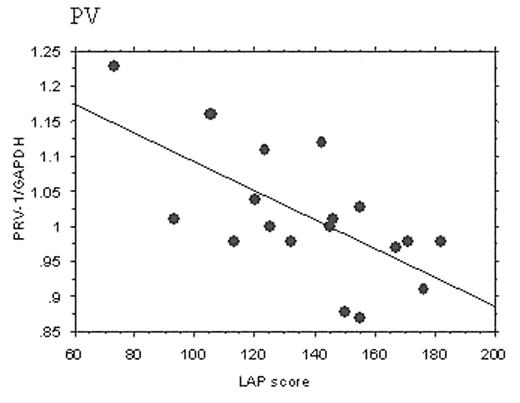
In both PV and ET, neutrophil PRV-1 expression strongly correlated with LAP score (p=0.003 and 0.04, respectively). In addition, in PV, univariate analysis revealed significant correlations between PRV-1 expression and leukocyte count, ANC, and reticulin fibrosis. On multivariate analysis, only LAP score retained its significance.
Conclusion:The current study confirms the previously recognized close association between increased neutrophil PRV-1 expression and PV as well as suggests a similarly strong association with PPMM but not with ET, MMM, or PTMM. The study also underscores the suboptimal diagnostic accuracy of the neutophil PRV-1 assay in distinguishing PV from SP. Most importantly, the demonstrated tight correlation between PRV-1 transcript level and LAP score raises the issue of added value to currently available diagnostic tests.
Author notes
Corresponding author


This feature is available to Subscribers Only
Sign In or Create an Account Close Modal