Abstract
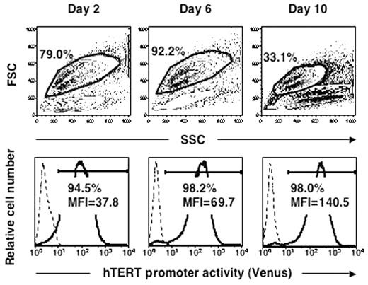
Acute myeloid leukemia (AML) is generally believed to originate from hematopoietic stem cells and to constitute a hierarchy of heterogeneous populations in terms of proliferation. To gain insight into the hierarchical organization of AML, a lentiviral reporter assay, based on the expression of novel yellow fluorescent protein variant (Venus) using flow cytometry, was applied to the promoter activity of the human telomerase reverse transcriptase (hTERT) gene in primary AML cells. hTERT promoter activity correlated well with endogenous telomerase activity in experiments inducing apoptosis and differentiation of leukemia cell lines. Next, AML cells or purified cord blood (CB) CD34+ cells transduced with hTERT-Venus (the lentiviral vector containing the expression cassette of the Venus gene driven by the hTERT promoter) were cultured in the presence or absence of colony-stimulating factors (CSF) mixtures (SCF, IL3, GM-CSF, and G-CSF) for 48 hrs before analysis. In primary AML cases, hTERT promoter activity revealed considerable patient-to-patient variation, regardless of stimulation by CSF, but its distribution appeared as a single peak or two narrowly separated peaks, suggesting that hTERT expression occurs along a continuous gradient in the leukemia cell population. The mean promoter activity of the 19 AML cells was 2-fold higher than that of CB CD34+ cells without CSF, but almost equal to that with CSF. In four cases, the MFI was not responsive to CSF stimulation, and in three cases, the basal and stimulated MFI were prominent. In contrast, three lots of CB CD34+ cells uniformly revealed very low activity (MFI) without stimulation (mean=1.2), but 5-fold up-regulation with stimulation (mean=5.8). Without CSF stimulation, hTERT promoter activity was slightly higher in CD34+CD38− cells than in CD34+CD38+ cells in most AML cases, and leukemia cells in S/G2/M phase had greater promoter activity than those in G0/G1 phase. Next, to monitor the changes in hTERT promoter activity during short-term culture, hTERT-Venus transduced leukemia cells were maintained with CSF mixtures, and Venus expression was analyzed using flow cytometry at Days 2, 6, and 10. A representative case is shown in the Figure. Both the viable cell population and its hTERT promoter activity increased at Day 6. The viable cell population then decreased, but the fluorescence intensity of this fraction still increased markedly at Day 10. This might be due to the gradual disappearance of the cell fraction with lower hTERT promoter activity, which suggests that leukemia cells with higher telomerase activity survive in a mass population. Furthermore, AML cells expected to have high telomerase activity could be identified under fluorescent microscopy, making it possible to track their behavior in culture. In conclusion, we presented here novel evidence for a hierarchy of telomerase activity, deduced from hTERT promoter activity, among individual leukemia cells in AML.
Author notes
Corresponding author


This feature is available to Subscribers Only
Sign In or Create an Account Close Modal