Abstract
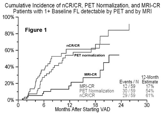
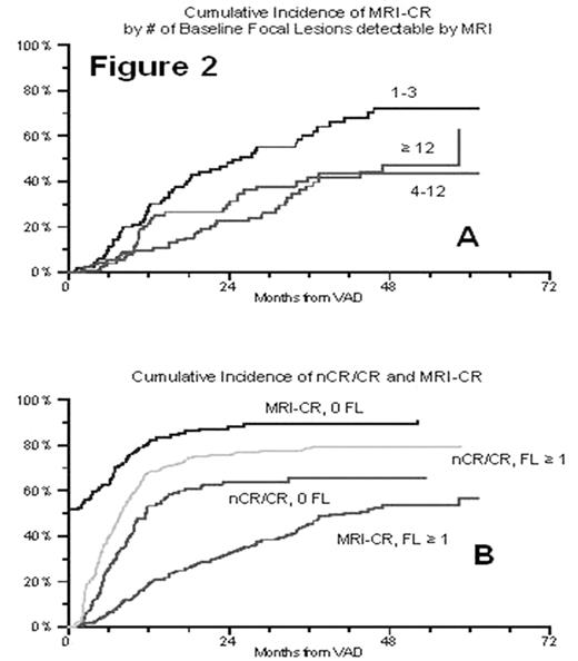
Multiple myeloma results in both diffuse infiltration and/or focal lesions (FL) of the marrow. MRI detects FL long before X-ray. We routinely perform MRI of skull, entire spine, pelvis, sternum and shoulders. MRI-defined number of FL (#FL) is an adverse prognostic variable for both EFS and OS for patients treated according to Total Therapy 2, 2nd in importance only to cytogenetic abnormalities (CA) (univariate and Cox multivariate models using standard prognostic factors, CA, and #FL). Of these patients who also had FDG PET (PET), baseline analysis revealed the same axial skeleton FL as on MRI (r=0.47, p<0.001). Another 268 FL in long bones and 11 sites of extramedullary tumor were seen with PET due to MRI’s restricted field of view. Serial MRI and PET in 59 patients with FL on MRI (Fig. 1) and 123 patients with hyperintense MRI signal on STIR (no FL) revealed that patients without FL normalize MRI and PET scans synchronously with clinical (M-protein and bone marrow) response (nCR/CR) (Fig. 2a). However, in patients with FL, PET normalization follows nCR/CR very closely, while regression of MRI-FL (MRI-CR) lags in proportion to MRI #FL (Fig. 2b). We conclude PET is superior to MRI for detection of extent of disease and monitoring treatment response. However, PET-negative MRI-FL usually contain viable myeloma cells on biopsy which eventually progress. Thus, long term serial MRI is superior to PET in assessing completeness of response.
Author notes
Corresponding author


This feature is available to Subscribers Only
Sign In or Create an Account Close Modal