Abstract
Cholesterol, in addition to providing rigidity to the fluid membrane, plays a critical role in receptor function, endocytosis, recycling, and signal transduction. In the present study, we examined the effect of membrane cholesterol on functional expression of tissue factor (TF), a cellular receptor for clotting factor VIIa. Depletion of cholesterol in human fibroblasts (WI-38) with methyl-β-cyclodextrin–reduced TF activity at the cell surface. Binding studies with radiolabeled VIIa and TF monoclonal antibody (mAB) revealed that reduced TF activity in cholesterol-depleted cells stems from the impairment of VIIa interaction with TF rather than the loss of TF receptors at the cell surface. Repletion of cholesterol-depleted cells with cholesterol restored TF function. Loss of caveolar structure on cholesterol removal is not responsible for reduced TF activity. Solubilization of cellular TF in different detergents indicated that a substantial portion of TF in fibroblasts is associated with noncaveolar lipid rafts. Cholesterol depletion studies showed that the TF association with these rafts is cholesterol dependent. Overall, the data presented herein suggest that membrane cholesterol functions as a positive regulator of TF function by maintaining TF receptors, probably in noncaveolar lipid rafts, in a high-affinity state for VIIa binding.
Introduction
Cholesterol is a lipid precursor for steroid hormones and bile salts and is present in cell membranes and circulation. Cholesterol in the membrane regulates flexibility and mechanical stability of the membrane.1 Further, cholesterol plays a critical role in differentiating and maintaining cell surface microdomains of differing lipid composition, particularly sphingolipid rafts. Lipid rafts are shown to contribute to the regulation of various cellular functions, including receptor function, endocytosis, intracellular trafficking of receptors, and signaling pathways.2-5
Tissue factor (TF) is the cellular receptor for clotting factor VIIa, and the formation of TF-VIIa complexes on cell surfaces triggers the coagulation cascade.6 Studies suggest that exposure of TF to circulating blood on rupture of atherosclerotic plaque plays an important role in the pathogenesis of thrombus formation at sites of plaque rupture, resulting in acute coronary events and myocardial infarction.7-10 Since cholesterol/oxidatively modified low-density lipoprotein (LDL) present in atherosclerotic plaques is thought to play an important role in the atherogenesis through its biologic effects, including TF expression, many earlier studies were focused on investigating the effect of cholesterol on TF expression. 3-hydroxy-3-methylglutaryl coenzyme A (HMG-CoA) reductase inhibitors, widely used to suppress plasma LDL cholesterol levels in patients with primary hypercholesterinemia, were shown to inhibit TF expression in both in vitro and in vivo.11,12 Consistent with this, dietary lipid lowering was found to reduce TF expression in rabbit atheroma.13 However, in vitro studies on effects of cholesterol on TF expression gave conflicting results. Cholesterol loading, by exposing monocytes/macrophages or endothelial cells to modified LDL or cholesterol, was shown to induce TF expression in some studies,14-19 whereas no effect was found in other studies.20-23 Most of these previous studies were focused primarily on investigating the role of LDL or cholesterol in modulating transcriptional or translational regulation of TF. At present, there is little information on how cholesterol regulates TF functional expression, independent of transcription/translational control.
Studies show that cholesterol, either through a direct molecular interaction or other mechanisms, can have a strong influence on the affinity state, binding capacity, and signal transduction property of membrane receptors.2,24-34 Cholesterol- and sphingolipid-rich rafts in association with a structural protein, caveolin, form caveolae, flask-shaped invaginations of 50- to 100-nm diameter in the plasma membrane.5 These structures are present in many cell types, including endothelial cells35,36 and smooth muscle cells.37 The structure of caveolae is dependent on cholesterol,4,5 as the removal of cholesterol disrupts caveolae.31,32 Studies suggest that TF in smooth muscle cells was associated with caveolae and speculated that caveolae-associated TF may function as a latent pool, which can become active when the vessel wall integrity is lost.37 Studies of Ruf and colleagues (Sevinsky et al35 ) demonstrated that TF redistributes into caveolae following a series of events, which include binding of VIIa to TF, generation of factor Xa, and subsequent formation of a transient ternary complex with tissue factor pathway inhibitor (TFPI) localized in glycosphingolipid-rich microdomains.
In the present study, we investigated the role of membrane cholesterol on the regulation of TF receptor function by depleting the membrane cholesterol of fibroblasts with methyl-β-cyclodextrin (mβCD) and evaluating TF functional activity for its ability to support VIIa binding and TF-VIIa activation of factor X. These data show that cholesterol depletion impairs the functional expression of TF by reducing its affinity to VIIa. Our data also suggest that the reduced cholesterol at the cell surface per se, and not the loss of caveolar structure, is responsible for reduced TF activity in cholesterol-depleted cells.
Materials and methods
Cell culture
A human fibroblast cell line (WI-38), derived from normal embryonic lung tissue, was obtained from ATCC (Rockville, MD) and was cultured as described earlier.38
Radiolabeling of proteins
VIIa and other proteins were labeled by using Iodo-Gen–coated tubes and Na125I according to the manufacturer's (Pierce Biotechnology, Rockford, IL) technical bulletin and as described previously.39 Our earlier studies40,41 established that the radiolabeled proteins were intact with no apparent degradation, and 125I-labeled VIIa retained 80% or more of the functional activity of the unlabeled material.
Cholesterol depletion and loading of cholesterol
To deplete cholesterol, unless specified otherwise, monolayers of fibroblasts were treated with mβCD (10 mM in buffer A, 10 mM N-2-hydroxyethylpiperazine-N′-2-ethanesulfonic acid [HEPES], 0.15 M NaCl, 4 mM KCl, 11 mM glucose, pH 7.5) for 30 to 45 minutes at 37°C. Then the cells were washed with buffer A and immediately used for functional activity or binding assays. Cholesterol (water-soluble) (Sigma Chemical, St Louis, MO) was loaded to cells by incubating control untreated cells or cholesterol-depleted cells with cholesterol (1 mM cholesterol:10 mM mβCD) for 30 minutes at 37°C. The unincorporated cholesterol was removed, and the cells were washed with buffer A before they were used in experiments. For loading of other steroids, first, steroid-mβCD complexes were prepared as described earlier.24,26 Briefly, the steroids were dissolved in 200 mM mβCD solution, preheated to 80°C, to make a 20 mM stock concentration of steroids. The complexes were protected from light, incubated at 80°C, and mixed by vortexing occasionally, until a clear solution was obtained (approximately 30 minutes). The complexes were then stored at –20°C. Immediately prior to their use, they were diluted 1 to 10 in buffer A.
Cholesterol determination
Cells were removed from culture dishes by scraping them in buffer A, and the cell suspension was centrifuged for 5 minutes at 3000 rpm in an Eppendorf 5415 C microcentrifuge (Eppendorf AG, Hamburg, Germany). The cell pellets were suspended in TBS (Tris-buffered saline; 50 mM tris(hydroxymethyl)aminomethane [Tris]–HCl, 0.15 M NaCl, pH 7.5) containing 0.1% Tween-20. Cholesterol was determined spectrophotometrically using Cholesterol CII kit (Wako Chemicals, Richmond, VA), following the manufacturer's instructions. We also determined cholesterol levels in cell membrane fractions by first isolating the cell membranes by ultracentrifugation as described earlier 42 and suspending the membrane pellet in TBS containing 0.1% Tween-20.
Binding studies
Cell surface binding of 125I-VIIa (TF-specific) or 125I-TF mAb (TF9H10) was performed essentially as described previously.38
Determination of cell-surface TF-VIIa activity
Monolayers of control cells, cholesterol-depleted or cholesterol-loaded cells were incubated with VIIa (10 nM) in buffer B (buffer A containing 5 mM CaCl2 and 1 mg/mL bovine serum albumin [BSA]) for 5 minutes at 37°C, followed by the addition of substrate factor X (175 nM). Unless otherwise specified, an aliquot was removed at a specific time point (usually at 5 minutes) into stopping buffer (TBS containing 1 mg/mL BSA and 10 mM ethylenediaminetetraacetic acid [EDTA]), and factor Xa in the sample was measured in a chromogenic assay as described earlier.43
Electron microscopy
Following control and experimental treatments, cells were fixed for 1 hour at 4°C in 4% paraformaldehyde and 1% glutaraldehyde in 0.1 M sodium cacodylate buffer, pH 7.2. Following the fixation, cells were washed thrice with the cacodylate buffer and rinsed once with Milli-Q water. Cells were first stained with anti-TF mAB (a mixture TF9H10, TF9-5B7, and TF9-11D12, 30 μg/mL) for 90 minutes at 4°C in phosphate-buffered saline (PBS) containing 0.2% BSA, followed by secondary antibody, gold (10 nm particle size)–conjugated goat anti–mouse immunoglobulin G (IgG; 25-fold dilution) for 90 minutes at 4°C in PBS containing 0.2% BSA. After quick washes in PBS, the cells were refixed in paraformaldehyde as described earlier in this section and exposed to 1% OsO4 for 1 hour at room temperature in the cacodylate buffer. The fixed cells were stained in 1% aqueous uranyl acetate for 30 minutes in the dark at 4°C, washed in deionized water, subsequently dehydrated in graded ethanol, and embedded in epoxy resin. Thin sections (0.5 μm) were cut perpendicular to the dish. The sections were mounted on copper grids (300 mesh size) and stained in 0.5% aqueous uranyl acetate for 10 minutes, followed by 2% lead citrate for 5 to 10 minutes. Grids were washed thoroughly in deionized water and dried. Sections were viewed and photographed with a JOEL 12 EX electron microscope fitted with a BIOTEM SCAN camera (JOEL USA, Peabody, MA) at 30 000× magnification under 60 kV acceleration. Micrographs shown in Figures 1 and 5 were reproduced from original photographs without any manipulation.
Ultrastructural localization of TF in fibroblasts and loss of caveolar structure on cholesterol depletion. Fibroblasts (WI-38 cells) were fixed and immunostained with TF mAb as described in “Materials and methods” (A-D). TF was localized on the cell membrane (A), on cellular processes (B), and in caveolae (C). The tip of a cellular process that was in contact with other cell/cellular process are stained densely for TF (D). Thin arrows point out gold particles, whereas arrowheads point out caveolae. Monolayers of WI-38 fibroblasts were treated with a control vehicle (E) or mβCD (10 mM) (F) for 45 minutes at 37°C. The cells were then fixed, sectioned, stained, and viewed under transmission electron microscope. Bar indicates 200 nm.
Ultrastructural localization of TF in fibroblasts and loss of caveolar structure on cholesterol depletion. Fibroblasts (WI-38 cells) were fixed and immunostained with TF mAb as described in “Materials and methods” (A-D). TF was localized on the cell membrane (A), on cellular processes (B), and in caveolae (C). The tip of a cellular process that was in contact with other cell/cellular process are stained densely for TF (D). Thin arrows point out gold particles, whereas arrowheads point out caveolae. Monolayers of WI-38 fibroblasts were treated with a control vehicle (E) or mβCD (10 mM) (F) for 45 minutes at 37°C. The cells were then fixed, sectioned, stained, and viewed under transmission electron microscope. Bar indicates 200 nm.
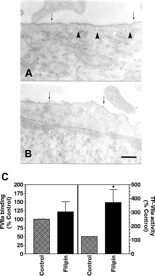
Effect of filipin on caveolar structure and TF expression. Monolayers of WI-38 fibroblasts were treated with a control vehicle (A) or filipin (5 μg/mL) (B) for 15 minutes at 37°C. Cells were fixed, sectioned, stained for TF by immunogold, and visualized under transmission electron microscope. Thin arrows point out gold particles whereas arrowheads point out caveolae. Bar indicates 200 nm. (C) Control or filipin-treated monolayers were incubated with either 125I-VIIa (10 nM) or unlabeled VIIa (10 nM) and factor X (175 nM) to determine VIIa binding and TF-VIIa activity (n = 4, mean ± SE). * denotes significantly differs (P < .05) from the control.
Effect of filipin on caveolar structure and TF expression. Monolayers of WI-38 fibroblasts were treated with a control vehicle (A) or filipin (5 μg/mL) (B) for 15 minutes at 37°C. Cells were fixed, sectioned, stained for TF by immunogold, and visualized under transmission electron microscope. Thin arrows point out gold particles whereas arrowheads point out caveolae. Bar indicates 200 nm. (C) Control or filipin-treated monolayers were incubated with either 125I-VIIa (10 nM) or unlabeled VIIa (10 nM) and factor X (175 nM) to determine VIIa binding and TF-VIIa activity (n = 4, mean ± SE). * denotes significantly differs (P < .05) from the control.
Separation of Triton X-100–insoluble complexes by sucrose gradient ultracentrifugation
Triton X-100–insoluble complexes were prepared by sucrose gradient ultracentrifugation fractionation essentially as described earlier.35,44 From each fraction, 30 μg protein was precipitated using 10% vol/vol trichloroacetic acid (TCA), and the pellets were suspended in 50 μL sodium dodecyl sulfate–polyacrylamide gel electrophoresis (SDS-PAGE) sample buffer. Aliquots (20 μL) were subjected to SDS-PAGE, followed by Western blot analysis.
Detergent lysis and fractionation
Cells were solubilized in various detergents and fractionated as described earlier.34 Briefly, control and cholesterol-depleted cells (2 T-75 flasks each) were harvested in ice-cold buffer A by detaching the cells from the bottom of the dish with a cell scraper. The cells were sedimented by centrifugation, resuspended in buffer A, and split into 3 equal aliquots. The cells in each aliquot were lysed in an equal volume of 1% ice-cold detergent, either Triton X-100, Brij 56 or Brij 58, by gentle mixing at 4°C for 30 minutes. The cell lysates were centrifuged at 800g for 10 minutes at 4°C to remove nuclei and cell debris. The postnuclear supernatants were centrifuged at 16 000g for 30 minutes at 4°C. Pellets, which contain insoluble membrane domains, were resuspended in buffer A containing 0.5% appropriate detergent. Both pellets and supernatants were subjected to SDS-PAGE on 12% polyacrylamide gels and processed for immunoblot analysis using standard approaches.
Results
Ultrastructural localization of TF
To determine the role of membrane cholesterol on cell surface TF expression, we first investigated the cellular distribution of TF in fibroblasts by immunogold electron microscopy. To avoid the possibility of nonspecific clustering due to secondary antibody cross-linking, we first fixed the cells before they were immunostained. Tissue factor was predominantly localized on the cell membrane and on cellular processes (Figure 1A-B). In general, cellular processes were stained heavily with anti-TF antibodies. It is interesting to note that when a cellular process from 1 cell comes in contact with another cell, the tip of the cellular process is decorated with TF (Figure 1D). Similar observations were also made with confocal microscopy using fibroblasts transfected with TF–green fluorescent protein (GFP; data not shown). In addition to localizing on the cell membrane and cellular processes, TF was also found in noncoated membrane invaginations, caveolae, mostly at the neck of caveolae (Figure 1C). Quantitation of the cellular distribution of TF from a total of 41 sections revealed that about 15% of gold particles were associated with caveolae.
Depletion of cholesterol and loss of caveolar structure
We have used mβCD, a membrane-impermeable agent that binds to cholesterol with high specificity, to deplete cholesterol.24,25 Incubation of fibroblasts with increasing concentrations of mβCD (1 to 10 mM) reduced the cholesterol content in a dose-dependent manner (Figure 2A). Treatment with 10 mM mβCD for 1 hour reduced the cholesterol content by about 60%. Examination of cells treated with mβCD (10 mM) for varying times (up to 4 hours) under light microscopy revealed no gross differences in morphology between control (untreated) and mβCD-treated cells. mβCD treatment neither reduced the cell viability (cell viability at the end of 1-hour treatment: control, 91% ± 2%; mβCD-treated [10 mM], 90% ± 1%) nor the number of cells attached to the plate (control, 157 500 ± 13 500 cells/well; mβCD-treated, 153 750 ± 15 190 cells/well).
Effect of varying doses of cyclodextrin treatment on cholesterol depletion and TF-VIIa activity in fibroblasts. Monolayers of WI-38 cells were treated with varying doses (1 to 10 mM) of mβCD (A-B) for 45 minutes. At the end of 45 minutes, cells were washed and used for determining cholesterol content (A) or cell surface TF-VIIa activity by adding factor VIIa (10 nM) and factor X (175 nM) to the monolayers (B). (n = 4 to 6, mean ± SE). * denotes significantly differs (P < .05) from the control (untreated cells). Panel C depicts the time course of factor X activation by TF-VIIa in control and cholesterol-depleted cells (10 mM mβCD treatment for 45 minutes). Two different concentrations of factor X were used, 175 nM (circles) and 1 μM (squares). Filled symbols represent control cells, and open symbols represent mβCD-treated cells.
Effect of varying doses of cyclodextrin treatment on cholesterol depletion and TF-VIIa activity in fibroblasts. Monolayers of WI-38 cells were treated with varying doses (1 to 10 mM) of mβCD (A-B) for 45 minutes. At the end of 45 minutes, cells were washed and used for determining cholesterol content (A) or cell surface TF-VIIa activity by adding factor VIIa (10 nM) and factor X (175 nM) to the monolayers (B). (n = 4 to 6, mean ± SE). * denotes significantly differs (P < .05) from the control (untreated cells). Panel C depicts the time course of factor X activation by TF-VIIa in control and cholesterol-depleted cells (10 mM mβCD treatment for 45 minutes). Two different concentrations of factor X were used, 175 nM (circles) and 1 μM (squares). Filled symbols represent control cells, and open symbols represent mβCD-treated cells.
Removal of cholesterol from the plasma membrane by mβCD treatment, as revealed by transmission electron microscopy (TEM), completely disrupted the structural integrity of caveolae. While caveolae invaginations are clearly visible in control cells (Figure 1E), there are very few morphologically recognizable caveolae in the cell membrane after cholesterol depletion (Figure 1F). TEM analysis of a total of 28 sections revealed that mβCD treatment disrupted more than 80% of the caveolar structures from the membrane (average number of caveolae per section: control, 17.5 ± 2.1; mβCD-treated, 2.1 ± 0.5). No noticeable differences in the membrane ultrastructure or integrity were observed between control cells and cells treated with mβCD. Immunogold staining of sections with TF mAB showed a similar number of gold particles associated with cell surfaces of control cells and cells treated with mβCD (number of gold particles/section: control, 9.5 ± 1.5; mβCD-treated, 10.5 ± 0.3, n = 25 to 28).
Cholesterol depletion inhibits functional expression of TF at the cell surface
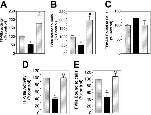
To determine the role of membrane cholesterol on TF functional expression, WI-38 cells were treated with varying concentrations of mβCD for 45 minutes to deplete membrane cholesterol. After removing mβCD and washing the cells, VIIa was added to the cells, and TF-VIIa proteolytic activity was measured by adding a plasma concentration of factor X (175 nM) to monolayers and determining factor Xa generation. As shown in Figure 2B, depletion of cholesterol from the plasma membrane reduced TF-VIIa activity, and the extent of decrease in cell surface TF-VIIa activity is correlated with the extent of cholesterol depletion from the plasma membrane. To assure that the reduced TF-VIIa activity seen in cholesterol-depleted cells was not due to limited availability of substrate factor X, we also measured TF-VIIa activity in control and cholesterol-depleted cells with a saturating concentration of factor X (1 μM). The data confirmed the finding that cholesterol depletion reduced cell surface TF-VIIa activity (Figure 2C). Consistent with the observation that cholesterol modulates TF functional activity, loading fibroblasts with cholesterol increased TF functional activity by about 2-fold (Figure 3A). Binding studies with 125I-labeled VIIa (10 nM) revealed that cholesterol depletion reduced VIIa binding to cell surface TF, whereas cholesterol loading increased VIIa binding to TF (Figure 3B). To investigate whether cholesterol depletion reduces the effective concentration of TF at the cell surface, we performed binding studies with TF mAB. These studies showed no significant differences in TF mAB binding among control, cholesterol-depleted, and cholesterol-loaded cells (Figure 3C). These data indicate that cholesterol modulates TF functional expression by impairing TF interaction with VIIa. To strengthen the above observation, we performed additional experiments in which fibroblasts were first treated with mβCD to deplete cholesterol and then loaded with cholesterol by incubating the cells with mβCD:cholesterol complexes. As shown in Figure 3D, depletion of cholesterol reduced TF-VIIa activity, and the restoration of membrane cholesterol restored TF functional expression. Similar results were obtained in VIIa binding studies (Figure 3E). Additional experiments showed the extent of TF-VIIa activity restoration in cholesterol-depleted cells was dependent on the amount of cholesterol loaded onto the cells (data not shown).
Effect of cholesterol depletion and cholesterol-loading on TF functional expression and reversibility of cholesterol effect. (A-C) Monolayers of WI-38 cells were treated for 45 minutes at 37°C with mβCD (10 mM) to deplete cholesterol or water-soluble cholesterol (mβCD-cholesterol, 1 mM) to load the cells with cholesterol. Then, the monolayers were incubated with (A) unlabeled VIIa (10 nM), followed by substrate factor X (175 nM) for 5 minutes at 37°C to measure TF functional activity; (B) 125I-VIIa (10 nM) or (C) 125I-TF mAB for 1 hour to measure VIIa or TF mAB binding to the cells. (D-E) Monolayers were first treated for 30 minutes at 37°C with mβCD (10 mM) to deplete cholesterol. After washing monolayers, cholesterol was reintroduced to the cells by incubating the cholesterol-depleted cells with cholesterol (1 mM): mβCD (10 mM) for 30 minutes. Following this, the cells were washed with buffer B and used to determine cell surface TF activity (D) and 125I-VIIa binding to cell surface TF (E) (n = 3, mean ± SE). * denotes significantly (P < .05) differs form the control; # denotes significantly (P < .05) differs from both the control and the cholesterol-depleted cells; and ** denotes significantly (P < .05) differs from mβCD-treated cells but not from the control. In A-C, ▩ indicates control; ▪, cholesterol-deplete; ▨, cholesterol-laden. In D and E, ▩ indicates control; ▪, mβCD; and ▨, mβCD plus cholesterol.
Effect of cholesterol depletion and cholesterol-loading on TF functional expression and reversibility of cholesterol effect. (A-C) Monolayers of WI-38 cells were treated for 45 minutes at 37°C with mβCD (10 mM) to deplete cholesterol or water-soluble cholesterol (mβCD-cholesterol, 1 mM) to load the cells with cholesterol. Then, the monolayers were incubated with (A) unlabeled VIIa (10 nM), followed by substrate factor X (175 nM) for 5 minutes at 37°C to measure TF functional activity; (B) 125I-VIIa (10 nM) or (C) 125I-TF mAB for 1 hour to measure VIIa or TF mAB binding to the cells. (D-E) Monolayers were first treated for 30 minutes at 37°C with mβCD (10 mM) to deplete cholesterol. After washing monolayers, cholesterol was reintroduced to the cells by incubating the cholesterol-depleted cells with cholesterol (1 mM): mβCD (10 mM) for 30 minutes. Following this, the cells were washed with buffer B and used to determine cell surface TF activity (D) and 125I-VIIa binding to cell surface TF (E) (n = 3, mean ± SE). * denotes significantly (P < .05) differs form the control; # denotes significantly (P < .05) differs from both the control and the cholesterol-depleted cells; and ** denotes significantly (P < .05) differs from mβCD-treated cells but not from the control. In A-C, ▩ indicates control; ▪, cholesterol-deplete; ▨, cholesterol-laden. In D and E, ▩ indicates control; ▪, mβCD; and ▨, mβCD plus cholesterol.
Our earlier studies39,45 suggest that negatively charged phospholipids in the outer leaflet of the cell membrane modulate cell surface TF interaction with VIIa and subsequently TF-VIIa activation of factor X. To address whether cholesterol depletion reduced the availability of negatively charged phospholipids at the cell surface, we evaluated the binding of annexin V, which was shown to bind specifically to negatively charged phospholipids,46,47 to untreated cells, and cells treated with mβCD. No differences were found in annexin V binding to control cells and cholesterol-depleted cells (annexin bound, fmoles/100 000 cells; control, 735 ± 91; mβCD-treated, 798 ± 67, n = 3). These data rule out the possibility of a potential decrease in negatively charged phospholipids that facilitate VIIa interaction with TF in cholesterol-depleted cells.
Evaluation of the modulatory effect of cholesterol on TF interaction with VIIa
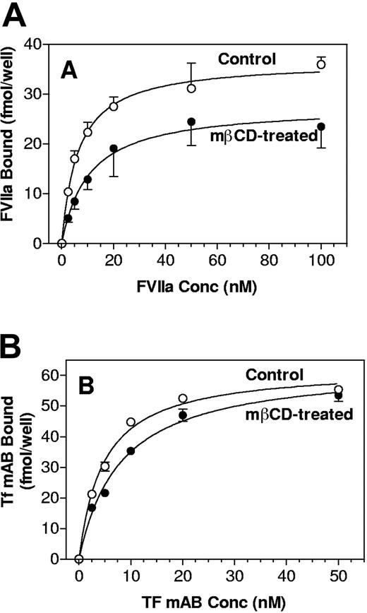
To determine whether the reduced VIIa binding to TF in cholesterol-depleted cells represents the loss of TF receptors on the cell surface, we determined whether cholesterol depletion reduces the total number of TF receptors available on the cell surface. Monolayers of fibroblasts were treated with a control vehicle or 10 mM mβCD for 30 minutes at 37°C and then incubated with varying concentrations of 125I-labeled TF mAB (TF9-10H10) for 2 hours at 4°C (Figure 4B). Analysis of TF mAB binding curves revealed that cholesterol depletion had no significant effect on the total number of TF mAB molecules associated with cells and their affinity to TF (Bmax [binding maximum]: control, 71 ± 1.5 fmole/well; mβCD-treated 65 ± 1.0 fmole/well; Kd [kinetically determined dissociation constant]: control, 5.1 ± 0.4 nM; mβCD-treated, 7.9 ± 0.4 nM, n = 3). Thus, it is unlikely that cholesterol depletion affects the total number of TF receptors, per se, on the cell surface.
VIIa and TF mAB binding to cholesterol-depleted cells. Control and cholesterol-depleted cells were incubated with varying concentration of 125I-VIIa in the presence and absence of anti-TF IgG (A) or 125I-TF mAB (B) for 2 hours at 4°C. At the end of the 2-h incubation, the unbound ligands were removed, cells were washed, and the cell-associated radioactivity was counted. Specific VIIa binding, shown in panel A, was determined by subtracting the nonspecific binding (VIIa binding to cells in the presence of anti-TF IgG) from the total binding (VIIa binding in the absence of anti-TF IgG) (n = 4 to 6, mean ± SE).
VIIa and TF mAB binding to cholesterol-depleted cells. Control and cholesterol-depleted cells were incubated with varying concentration of 125I-VIIa in the presence and absence of anti-TF IgG (A) or 125I-TF mAB (B) for 2 hours at 4°C. At the end of the 2-h incubation, the unbound ligands were removed, cells were washed, and the cell-associated radioactivity was counted. Specific VIIa binding, shown in panel A, was determined by subtracting the nonspecific binding (VIIa binding to cells in the presence of anti-TF IgG) from the total binding (VIIa binding in the absence of anti-TF IgG) (n = 4 to 6, mean ± SE).
If the depletion of cholesterol has no effect on the total number of antibody-reactive TF sites on fibroblast cell membranes, but decreases VIIa binding and thus reduces functional activity, then at least 2 possibilities exist: cholesterol depletion either reduces the number of TF receptors that could support VIIa binding or alters the receptor from high- to low-affinity binding sites for VIIa without changing the number of binding sites. We examined these possibilities by performing dose-dependent VIIa binding (TF-specific) studies with control and cholesterol-depleted cells (Figure 4A) to determine Kd and Bmax for VIIa. Analysis of VIIa binding curves with curve-fitting program (Prism; GraphPad, San Diego, CA) revealed that cholesterol depletion reduced the TF affinity to VIIa (Kd: control, 6.0 ± 0.4 nM; mβCD-treated, 13.2 ± 3.8 nM; n = 6; P = .04). The total number of factor VIIa associated with TF in cholesterol-depleted cells is slightly lower than that was observed in control cells, but the difference was not statistically significant (Bmax: control, 35.8 ± 2.2 fmole/well; mβCD-treated, 30.0 ± 4.6 fmole/well; P = .24). A change in the Kd without a change in the number of binding sites (Bmax) suggests that cholesterol affects the affinity state of TF for VIIa. Although the change in Kd documented here is small and this change alone may not fully explain the 50% reduction in TF-VIIa activity in cholesterol-depleted cells, it does account for at least a 30% reduction in TF-VIIa activity.
Disruption of caveolae is not responsible for impairment of TF activity in cholesterol-depleted cells
As discussed (Figure 1F), cholesterol depletion disrupts caveolar structure. To determine whether the loss of caveolae or the cholesterol depletion per se is responsible for reduced TF functional activity, we treated fibroblasts with filipin, which does not remove cholesterol from the membrane but forms filipin-cholesterol complexes in the membrane and thereby alters the physical distribution of the cholesterol and disrupts caveolae.48 Ultrastructural analysis of control and filipin-treated cells by electron microscopy showed, as expected, filipin treatment reduced the number of caveolae on fibroblasts by about 60% (Figure 5). Quantitative analysis of 19 to 26 sections showed the following: number of caveolae/section for control, 15.1 ± 5.9, and for filipin-treated cells, 6.0 ± 2.9. Immunogold analysis of TF antigen showed no significant differences in the number of gold particles associated with cells in control and filipin-treated cells (gold-particles/section: control, 12.7 ± 1.1; filipin-treated, 11.0 ± 0.97).
Next, we investigated the effect of filipin treatment on VIIa binding to cell surface TF and TF-VIIa activity. As shown in Figure 5C, filipin treatment slightly enhanced VIIa binding to fibroblasts but increased TF-VIIa activation of factor X markedly. These data serve as indirect evidence that the disruption of caveolae in cholesterol-depleted cells is not the cause for impaired TF functional expression observed in these cells. The increased TF-VIIa functional activity observed in filipin-treated cells could have been the result of increased concentration of cholesterol in membranes patches since filipin treatment is shown to result in cholesterol aggregation in the membrane48 or movement of TF from inactive glycosphingolipid-rich microdomains to active anionic phospholipid region of the membrane.
Tissue factor is localized in Brij 58 detergent-resistant membrane domains (DRMs)
To investigate whether TF is localized in cholesterol-sphingolipid rafts, fibroblasts were lysed in Triton X-100 and fractionated on a 5% to 30% sucrose gradient by ultracentrifugation. Fractions were subjected to SDS-PAGE and Western blot analysis using anti-human TF IgG and anti-caveolin IgG. The data revealed that less than 5% of TF was fractionated into low-density Triton X-100–insoluble complexes (as indicated by the presence of caveolin in these fractions). Solubility of a protein in Triton X-100 and/or inability to float after detergent extraction does not exclude a possibility that the protein is actually associated with cholesterol-sphingolipid rafts. Weak interaction of a protein with rafts may lead to its solubilization by the detergent. Further, cell type, detergent type, detergent/lipid ratio, and potential adhesion to the cytoskeleton may influence the raft protein association with DRMs and its migration to low density during sucrose gradient centrifugation.49,50 For example, T-cell antigen receptor51 and epidermal growth factor (EGF) receptor34 were shown to be associated with lipid rafts by fluorescence microscopy, but this interaction is not preserved during Triton X-100 extraction. Studies indicate other nonionic detergents, such as Brij 58 and Lubrol WX, are more suitable in preserving the interaction of receptors with cholesterol-sphingolipid rafts.34,52
Therefore, we next investigated the solubility of TF in the nonionic detergents Brij 56 and Brij 58. (Brij 58 has a higher hydrophilic-lipophilic balance than Triton X-100, whereas Brij 56 is similar to Triton X-100.52 ) Extraction of fibroblasts with Brij 58 resulted in a substantial amount of TF in the pellet, whereas minimal or no TF was found in the pellet when fibroblasts were extracted with Brij 56 or Triton X-100 (Figure 6A, top). Caveolin-1 was found exclusively in the pellet after lysis with both Brij 58 and Brij 56. Insolubility of TF in Brij 58 indicates that TF is localized in lipid rafts; however, the interaction between TF with lipid rafts may be weak.
Tissue factor is associated with Brij 58 detergent-resistant, cholesterol-based membrane domains. (A). WI-38 cells were lysed in 0.5% of the indicated detergent for 30 minutes at 4°C, and the postnuclear supernatants were fractionated into pellets (P) and supernatants (S) by centrifugation at 16 000g for 30 minutes and analyzed by immunoblotting. (B). WI-38 cells were cholesterol-depleted with mβCD (10 mM for 30 minutes) at 37°C and subsequently lysed in Brij 58. The samples were analyzed for TF by Western blotting, and the signals were quantitated by densitometry (n = 5, mean ± SE).
Tissue factor is associated with Brij 58 detergent-resistant, cholesterol-based membrane domains. (A). WI-38 cells were lysed in 0.5% of the indicated detergent for 30 minutes at 4°C, and the postnuclear supernatants were fractionated into pellets (P) and supernatants (S) by centrifugation at 16 000g for 30 minutes and analyzed by immunoblotting. (B). WI-38 cells were cholesterol-depleted with mβCD (10 mM for 30 minutes) at 37°C and subsequently lysed in Brij 58. The samples were analyzed for TF by Western blotting, and the signals were quantitated by densitometry (n = 5, mean ± SE).
Next, to investigate whether the association of TF with Brij 58-DRMs is cholesterol dependent, fibroblasts were cholesterol-depleted with mβCD (10 mM) for 45 minutes at 37°C, lysed with cold Brij 58, fractionated, and subjected to SDS-PAGE followed by immunoblotting for TF. As shown in Figure 6B, cholesterol depletion shifted TF presence into the supernatant. Although the shift is modest, it is reproducible and statistically highly significant (P < .0001). Further, such moderate change in TF distribution is expected since only a fraction of TF is associated with DRMs. Thus, these data provide evidence that the presence of TF in Brij 58-insoluble membrane domains is cholesterol dependent. In filipin-treated cells, the shift in TF distribution is subtle (TF is distributed equally between the pellet and the supernatant).
Discussion
In the present study, we show that the cholesterol content in the plasma membrane regulates TF functional expression by regulating TF interaction with ligand VIIa without altering TF levels at the cell surface. Data presented herein also show that in fibroblasts only a minor fraction of TF receptors is localized in caveolae, whereas a substantial portion of TF is localized in noncaveolar lipid rafts (DRMs) that are sensitive to extraction with Triton X-100 but not to extraction with Brij 58. The association of TF with these DRMs appears to be cholesterol dependent. Overall these data suggest that membrane cholesterol positively regulates TF coagulant function at the cell surface, probably by maintaining TF in a high-affinity state for VIIa binding.
Cholesterol, which plays an important role in the structure of biologic membranes, is known to modulate the activity of various membrane-embedded receptor proteins, including the transferrin receptor,53 the nicotinic acetylcholine receptor,54 insulin receptor,31 EGF receptor,34 and several G-coupled protein receptors24 (reviewed in Burger et al2 ). There are at least 2 defined mechanisms by which cholesterol is shown to modulate receptor function: (1) changes in membrane fluidity or (2) specific interaction between cholesterol and the receptor. Since cholesterol is essential in maintaining the rigidity of cell membranes, removal of cholesterol from the plasma membrane by mβCD treatment increases the membrane fluidity.24 Changes in membrane fluidity associated with cholesterol depletion was shown to be responsible for modulating cholecystokinin binding to cholecystokinin receptors in isolated plasma membranes and in intact cells.24 Altering membrane fluidity by other approaches was also shown earlier to influence ligand-binding, as shown in the case of β-androgenic receptor55,56 and serotonin receptor.57 However, for many other receptors, direct molecular interaction between cholesterol and the receptor but not changes in membrane fluidity is thought to play a role in cholesterol modulation of receptors function.24,26,27 These data presented herein do not permit drawing a firm conclusion on whether change in the membrane fluidity or the loss of structure-specific interaction of cholesterol with TF is responsible for reduced TF activity in cholesterol-depleted cells.
Since changes in lipids regulate membrane fluidity, fluidization of membrane by cholesterol depletion may alter phospholipid distribution of the cell membrane. Earlier studies from others58,59 and us45,60 showed that the increased exposure of phosphatidylserine (PS) at the outer cell membrane enhances TF functional expression. If mβCD treatment results in reduced PS at the outer plasma membrane, then it could reduce TF functional expression. However, this possibility seems unlikely since we found no differences in annexin V, a highly selective PS binding protein, binding to control cells and cholesterol-depleted cells. Further, PS was shown not to affect VIIa binding to TF at steady-state levels,45 whereas cholesterol depletion reduced VIIa binding to TF under similar steady-state conditions.
Comparison of VIIa binding in control and mβCD-treated cells suggests that the cholesterol depletion results in a 2- to 3-fold reduction in VIIa binding affinity to its receptor TF at the cell surface. Similar changes in affinities were observed for galanin receptor27 (3-fold increase in Kd value) or serotonin transporter26 (2-fold increase in Kd value) after cholesterol depletion. The observation that the reduction in membrane cholesterol only affects the affinity of VIIa for TF and not the number of TF molecules on the cell surface suggests that the site of cholesterol action on TF is at the plasma membrane. These data also suggest that cholesterol modulation does not affect synthesis or transport of TF to the plasma membrane, or its internalization. Consistent with this hypothesis, the ratio of internalized and surface-bound VIIa remained similar before or after mβCD treatment (internalized/surface at 30 minutes: control, 0.33; mβCD treated, 0.42; an average of 2 experiments). At present, it is unclear how cholesterol affects VIIa-TF interactions at the cell surface. A number of studies suggest that TF may exist at the cell surface as dimers.61-63 Replacement of the transmembrane region of TF with an unrelated hydrophobic transmembrane segment was found to disrupt self-association of TF.63 One can speculate that cholesterol, which is highly hydrophobic and resides within the membrane bilayers, probably interacts with specific amino acid residues in the transmembrane region of TF, allowing dimerization of TF. It had been suggested that the association of VIIa to the first site would enhance the binding of the second ligand to the receptor.61 Depletion of membrane cholesterol may disrupt the dimeric structure of TF, and this could decrease VIIa affinity for the receptor. In contrast to the well-established dogma that the dimerization of a receptor enhances its function, Bach and Moldow62 suggested that TF dimers were inactive, whereas monomeric TF was procoagulant. If so, dimerization of TF would reduce its functional activity. It is unlikely that the above-stated mechanisms are responsible for the impairment of VIIa interaction with TF on the depletion of membrane cholesterol since the analyses of our binding data showed that the binding isotherms in both control and mβCD-treated cells were similar (ie, hyperbolic and not sigmoidal). Hill plots of the binding data revealed no significant difference in slopes, which is less than 1. Further, we found no evidence for the existence of significant amounts of TF dimers in fibroblasts in our chemical cross-linking studies (L.V.M.R., unpublished data, April 1999). Alternatively, direct interaction of cholesterol with a specific polypeptide region of TF may be essential in maintaining TF in a VIIa binding conformational state. Further studies are needed to address this possibility.
While the present manuscript was being prepared, a manuscript describing data that contrast the present data has been published online.64 These data show that treatment of HEK293 cells and dermal fibroblasts with mβCD increased the TF procoagulant activity by 2- to 3-fold. It is unclear why mβCD treatment elicited the opposing effect in these cells. It is possible that different cell lines may respond differently to mβCD treatment. In the present study, mβCD treatment did not alter PS exposure on the outer plasma membrane, whereas mβCD treatment increased the exposure of PS in HEK293 cells.64 It is interesting to note that the TF activity in HEK293 cells was increased in response to 5 and 10 mM mβCD treatment, whereas 1 and 5 mM but not 10 mM mβCD treatment increased the TF activity in dermal fibroblasts. Since there was no information on measurements of cholesterol levels in these cells following mβCD treatment, it was difficult to judge whether increased TF activity resulted from mβCD treatment in these studies correlates to decreased membrane cholesterol levels. In our studies we found 1 mM mβCD treatment barely depletes membrane cholesterol, whereas 10 mM mβCD treatment reduced the cholesterol content by about 60%.
Ultrastructural localization of TF in smooth muscle cells (SMCs) showed that about 20% of TF in these cells was associated with caveolae.37 On the basis of increased TF activity and enlargement of caveolar structures in SMCs following their detachment, Mulder et al37 speculated that caveolae-associated TF might function as a latent pool of procoagulant activity, which can rapidly be activated at sites in which vessel wall integrity is lost.37 In recent years, cholesterol depletion by mβCD treatment is widely used to disrupt caveolae to investigate the role of caveolae in modulating various cellular functions.30,32,65,66 As expected, removal of cholesterol in fibroblasts by mβCD treatment in the present study completely disrupted caveolar structures. However, mβCD treatment did not increase TF activity at the cell surface of fibroblasts. These data suggest that caveolar localization of TF in itself may not act as a regulator of TF activity at the cell surface. However, since mβCD treatment not only disrupts caveolae but also removes cholesterol from the membrane, which is essential for the optimal expression of TF, we cannot completely rule out the role of caveolae in down-regulating TF functional activity. Increased TF activity in cells treated with filipin, which disrupts caveolae without removing cholesterol from the membrane, suggest that caveolae may act as negative regulators provided that cholesterol was not depleted in the process.
Advances suggest that cholesterol exerts many of its actions mainly by maintaining sphingolipid rafts, which function to segregate and concentrate specific membrane proteins.67 Studies showed that raft-associated proteins, based on the raft structures, their interaction with raft lipids, or other proteins within the same raft, might exhibit differential sensitivity to extraction with different detergents.34,51,52 Consistent with this hypothesis, we found that TF in fibroblasts was soluble in Triton X-100 and Brij 56 (a detergent that is similar to Triton X-100) but partly resistant to extraction with Brij 58, a detergent with a higher hydrophilic-lipophilic balance than Triton X-100. In contrast to TF, caveolin-1 is associated completely with insoluble membrane domains on extraction with all 3 detergents. At present, it is not entirely clear whether differential behavior of caveolin-1 and TF during Triton X-100 extraction is caused by their localization on different membrane domains or dissociation of TF from caveolar membrane domains. Since ultrastructural localization of TF clearly indicated that only a minor fraction of TF present at the cell surface is associated with caveolae, it is reasonable to conclude that differential behavior of caveolin-1 and TF in Triton X-100 reflects TF association with noncaveolar cholesterol-rich membrane domains. The observation that depletion of cholesterol increased the solubility of TF in Brij 58 supports the notion that cholesterol is an integral part of these membrane domains.
In conclusion, the data presented in the manuscript demonstrate for the first time that membrane cholesterol modulates interaction of TF receptor with VIIa and subsequently TF-VIIa activation of factor X. These data may provide an additional explanation on how therapeutic intervention to lower cholesterol reduces the incidence of acute coronary events associated with atherosclerosis. Since studies show that TF-VIIa, in addition to triggering blood coagulation, plays a role in many pathophysiological processes, it is interesting to examine how cholesterol modulates other functions of TF-VIIa. These and similar studies in the future may provide clues in understanding the unexplained benefits of cholesterol-lowering drugs and may stimulate new studies in evaluating potential benefits, in addition to reducing atherosclerosis, associated with therapeutic intervention of lowering cholesterol.
Prepublished online as Blood First Edition Paper, August 24, 2004; DOI 10.1182/blood-2004-03-0990.
Supported by grants from National Institute of Health (HL58869) and American Heart Association, Texas Affiliate (0355096Y).
The publication costs of this article were defrayed in part by page charge payment. Therefore, and solely to indicate this fact, this article is hereby marked “advertisement” in accordance with 18 U.S.C. section 1734.
We acknowledge the excellent technical assistance provided by Mylinh Ngyuen. We are thankful for Dr Ronald Dodson's laboratory at the Health Center for helping in electron microscopy.


This feature is available to Subscribers Only
Sign In or Create an Account Close Modal