Abstract
Cross-linking FcϵRI on mast cells by immunoglobulin E (IgE) and antigen (Ag) initiates cascades leading to antiparasitic or allergic responses. It was recently reported that IgE without antigen, IgE(-Ag), actively promotes mast cell survival. Although we have demonstrated that the immunoreceptor tyrosine-based activation motif within FcRγ is essential for IgE(-Ag)–induced mast cell survival, the underlying mechanism remains still unclear. Here, we investigated the mechanism of IgE(-Ag)–induced survival using mast cells lacking several downstream molecules. Lyn and Syk were essential, whereas Fyn, Gab2, and the phosphoinositide 3-kinase–Akt pathway were not critical for survival. Failure of survival in FcRγ-/- bone marrow mast cells (BMMCs) was rescued by coculture with IgE-treated wild-type BMMCs, suggesting that survival is induced not directly through FcϵRI signals. We found that the survival is predominantly mediated by high production of interleukin 3 (IL-3), evidenced by severe impairment of survival by anti–IL-3 and in IL-3-/- BMMCs. The up-regulation of Bcl-xL/Bcl-2 by IgE was abrogated in IL-3-/- BMMCs, whereas the expression of histidine decarboxylase was normally induced. These results indicate that IL-3 plays a crucial role for IgE(-Ag)–induced mast cell survival, functioning in an autocrine manner by inducing the Bcl-xL/Bcl-2 via signal transducer and activator of transduction 5. We further suggest that IgE(-Ag)–mediated gene expression in mast cells is regulated at least 2 mechanisms: autocrine IL-3 dependent and independent.
Introduction
The high-affinity IgE receptor, FcϵRI, exists as a tetramer composed of α and β monomers and a γ homodimer in rodent cells.1 The β and γ chains contain an immunoreceptor tyrosine-based activation motif (ITAM) within their cytoplasmic domains. FcϵRI can bind to IgE in the absence of antigen (Ag). This step has been viewed as a passive “sensitization” prior to subsequent Ag cross-linking. However, several works revealed that IgE alone can actively promote up-regulation of cell surface FcϵRI2-4 and mast cell survival.5,6 FcϵRI up-regulation by IgE is caused by stabilization of FcϵRI on the plasma membrane,7,8 which requires the FcϵRIα chain9,10 but not FcϵRIγ-ITAM.11
On the other hand, we have recently shown that IgE-induced mast cell survival is mediated through ITAM within the FcRγ chain, similar to degranulation by cross-linking by Ag plus IgE, IgE(+Ag).11 Furthermore, we have also shown that a weak but sustained FcRγ signal is sufficient for mast cell survival by varying the strength of stimulation via a CD8/FcRγ chimera.12 However, the precise signaling pathway elicited by IgE(-Ag) toward survival remains unclear, compared with well-characterized IgE(+Ag)–induced signaling. Cross-linking of FcϵRI by IgE(+Ag) initiates an activation signaling cascade via tyrosine phosphorylation of ITAMs by Src family protein tyrosine kinase, Lyn. Syk is then recruited to phospho-ITAMs within FcRγ, where it is activated to phosphorylate several key substrates such as linker for activation of T cells (LAT) (protein) and SLP-76 (SH2 domain containing leukocyte protein of 76 kDa).13 But the involvement of these signaling molecules in IgE(-Ag)–mediated survival has yet to be clarified.
IgE may induce survival signals directly through FcϵRI. In addition to the classical pathway of FcϵRI-mediated signaling described, Fyn-mediated signaling is also reported to be involved in FcϵRI signal transduction,14 through positively regulating the phosphorylation and function of Gab2. The absence of Gab2 severely impairs mast cell responses to IgE(+Ag).15 Gab2 also induces recruitment and activation of phosphoinositide 3-kinase (PI3K).16 In most cells, PI3K has a role in cell survival via activation of PI3K-dependent kinase 1 (PDK1) and Akt,17 but the contribution of these pathways to IgE-induced survival remains undefined.
As for which effector molecule induces survival, Bcl-xL may play a role because IgE alone prevents down-regulation of Bcl-xL by interleukin 3 (IL-3) withdrawal.6 Whether signaling through FcϵRI directly regulates Bcl-xL, however, is still unclear.
Alternatively, IgE-induced survival was recently reported to be mediated by soluble factors, but it is still controversial.18 It is possible that cytokines could contribute to this event, either individually or synergistically, in an autocrine manner.6 Although IL-3 and IL-4 are potent growth and survival factors for mast cells,18 whether these cytokines are produced on IgE(-Ag) at a level that is for supporting mast cell survival is unknown.
Here, we investigated the signaling pathway that mediates IgE-induced survival using bone marrow mast cells (BMMCs) or fetal liver-derived mast cells (FLMCs) lacking several downstream molecules. Lyn and Syk were essential, whereas the Fyn, Gab2, PI3K p85α, and Akt pathway were dispensable. We further demonstrated that high level of autocrine production of IL-3 on IgE(-Ag) stimulation and subsequent IL-3 signals are critical for the induction of Bcl-xL/Bcl-2 and survival in mast cells.
Materials and methods
Mice
C57BL/6 mice were purchased from SLC (Hamamatsu, Japan). FcRγ-/- C57BL/6 mice were established as we have previously described.19 Gab2-/- mice were provided by Dr T. Hirano (Osaka University, Osaka, Japan and RIKEN, Yokohama, Japan).20 Fyn-/- mice were provided by Dr T. Yagi (Osaka University) through the courtesy of Dr S. Yuasa (Chiba University, Chiba, Japan).21 Lyn-/- mice were provided by Dr T. Yamamoto (University of Tokyo, Tokyo, Japan).22 These 3 mice were C57BL/6 × 129 mixed background. PI3K p85α-/- C57BL/6 mice were provided by Dr S. Koyasu (Keio University, Tokyo, Japan).23 Syk-/- BALB/c mice24 were obtained through the courtesy of Dr H. Yamamura (Kobe University, Kobe, Japan). IL-3–deficient mice were generated by gene targeting in embryonic stem cells, which introduced a frameshift mutation into exon 1 of the IL3 gene and replaced part of exon 3 with a PGK-neo cassette (V. Tybulewicz and P. Mee, unpublished data). Mice bearing the disrupted IL3 gene were backcrossed to BALB/c for 10 generations and intercrossed to generate IL-3-/- BALB/c mice.
Mast cell preparation
Bone marrow (BM) cells or fetal liver (FL) cells of each mouse were cultured in the presence of 30 ng/mL IL-3.12 Four weeks after induction, more than 95% of cells were c-kit+ and FcϵRI+ except for cells from FcRγ-/- mice.
Construct
cDNA for wild-type (WT) and dominant–negative forms of Akt (WT, T308/473A and K179D) were kindly provided by Dr W. Ogawa (Kobe University). WT, constitutively active (CA; H299R, S711F)25 signal transducer and activator of transcription 5a (STAT5a) cDNA was provided by Dr H. Wakao (RIKEN). Bcl-xL cDNA was provided by Dr K. Eshima (Kitasato University, Sagamihara, Japan). Each fragment was subcloned into pMX-puro or pMX-internal ribosome entry site-green fluorescent protein (IRES-GFP; Dr T. Kitamura, University of Tokyo). Retroviral-mediated gene transfer to BM cells and mature BMMCs was performed as previously described.11,12
Antibodies
Antimouse IL-2 receptor (IL-2R; PC61) and antimouse IL-4 (11B11) antibodies (Abs) were purchased from BD Bioscience PharMingen (San Diego, CA). Antimouse IL-3 polyclonal Ab was obtained from R&D Systems (Minneapolis, MN). Anti-Akt polyclonal Ab and anti-Erk Ab were from Cell Signaling Technology (Beverly, MA) and Promega (Madison, WI), respectively. Anti-dinitrophenyl (DNP) mouse IgE mAb (DNP H1-ϵ-26)26 provided by Dr F.-T. Liu (University of California, Davis, CA) was used in all assays unless otherwise described. Anti-trinitrophenyl (TNP) mouse IgE mAbs, C38-2 and C48-2, were purchased from BD Bioscience (San Jose, CA). Each IgE was ultracentrifuged at 100 000g for 10 minutes to exclude aggregates just before use for assays. The IL-3 enzyme-linked immunosorbent assay (ELISA) kit was from BD Bioscience PharMingen.
Survival assays
Survival assays of BMMCs were performed as described previously.12 Briefly, 4 × 10⁵ mast cells were cultured in 400 μL IL-3–free medium in the presence or absence of 1 μg/mL IgE (DNP H1-ϵ-26) on flat-bottom 48-well plates (Falcon, Lincoln Park, NJ). At indicated days, cells were stained with propidium iodide (PI) and live cells (PI- cells) counted by FACScalibur (BD Bioscience).
RT-PCR
Total RNA was isolated from BMMCs and reverse transcribed using the SuperScript first-strand synthesis system (Invitrogen, Carlsbad, CA). Real-time reverse transcription-polymerase chain reaction (RT-PCR) was performed as previously described.11 Primers were as follows; Bcl-xL, (+)5′-AAT GAA CTC TTT CGG GAT GG-3′; (-)5′-CAG CCG CCG TTC TCC TGG AT-3′; Bcl-2, (+)5′-CCG GGA GAA CAG GGT ATG AT-3′; (-)5′-GCA CAG CGG GCA TTG GGT TG-3′; IL-3, (+)5′-ATA GGG AAG CTC CCA GAA CCT GAA CTC-3′; (-)5′-AGA CCC CTG GCA GCG CAG AGT CAT TC-3′; HDC, (+)5′-AGT CTG GCG AGA AGG GAA GG-3′; (-)5′-TCT GGG CAC TCA TAG GCA CA-3′; GAPDH, (+)5′-GCC GGT GCT GAG TAT GTC GT-3′; (-)5′-AGA TGA TGA CCC GTT TGG CT-3′.
Results
FcϵRI signaling pathway required for the IgE(-Ag)–induced survival
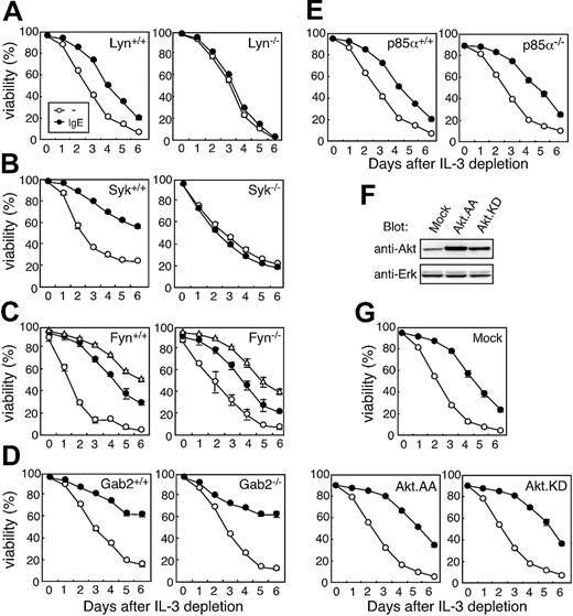
To examine if FcϵRI signaling pathways differ between IgE(+Ag) and IgE(-Ag) stimulation, we analyzed BMMCs lacking a series of signaling molecules that are known to be involved in IgE(+Ag)–induced degranulation. We have recently reported that the ITAM within FcRγ is essential for IgE(-Ag)–induced survival as well as IgE(+Ag)–dependent responses.11 ITAMs within the β and γ chains of the FcϵRI receptor complex are phosphorylated by Lyn on IgE(+Ag) stimulation and provide binding sites for Syk.27 Lyn and Syk were recently shown as essential kinases for IgE (DNP-H1-ϵ-26)–induced mast cell survival in the absence of Ag.28 We also confirmed that IgE did not induce survival in mast cells lacking Lyn and Syk throughout 6 days after IL-3 depletion (Figure 1A-B).
IgE-induced survival in BMMCs or FLMCs lacking Lyn, Syk, Fyn, Gab2, and PI3K p85α. Mast cell survival assay. Mast cells were prepared from bone marrow cells of mice deficient in (A) Lyn, (C) Fyn, (D) Gab2, and (E) PI3K p85α and fetal liver cells from a (B) Syk-/- embryo (E13.5). Mast cell survival was compared with that of cells from littermate WT control mice that were prepared and cultured simultaneously as described in “Materials and methods.” (F-G) Effect of dominant–negative Akt on IgE-induced BMMC survival. BMMCs retrovirally expressing dominant–negative Akt (Akt.AA or KD) were blotted with anti-Akt and anti-Erk Abs as a control (F) and tested for survival (G). Relative intensity of AA and KD were 6.4- and 3.9-fold higher than that of pMX-puro vector (Mock), respectively. Data were means ± SD for triplicate assays. Similar results were obtained from at least 2 independent mice pairs. IgE concentration was 0 μg/mL (○), 1 μg/mL (⬡), and 10 μg/mL (▵).
IgE-induced survival in BMMCs or FLMCs lacking Lyn, Syk, Fyn, Gab2, and PI3K p85α. Mast cell survival assay. Mast cells were prepared from bone marrow cells of mice deficient in (A) Lyn, (C) Fyn, (D) Gab2, and (E) PI3K p85α and fetal liver cells from a (B) Syk-/- embryo (E13.5). Mast cell survival was compared with that of cells from littermate WT control mice that were prepared and cultured simultaneously as described in “Materials and methods.” (F-G) Effect of dominant–negative Akt on IgE-induced BMMC survival. BMMCs retrovirally expressing dominant–negative Akt (Akt.AA or KD) were blotted with anti-Akt and anti-Erk Abs as a control (F) and tested for survival (G). Relative intensity of AA and KD were 6.4- and 3.9-fold higher than that of pMX-puro vector (Mock), respectively. Data were means ± SD for triplicate assays. Similar results were obtained from at least 2 independent mice pairs. IgE concentration was 0 μg/mL (○), 1 μg/mL (⬡), and 10 μg/mL (▵).
Recently, Parravicini et al showed that Fyn positively controls the phosphorylation and function of Gab2,14 which is required for FcϵRI signaling by IgE(+Ag).15 Because Gab2 is associated with PI3K on phosphorylation,16 it is possible that the Fyn-Gab2 pathway may contribute to the IgE(-Ag)–induced survival through PI3K-Akt activation. However, as shown in Figure 1C-D, mast cell survival was not impaired by the lack of Fyn or Gab2.
Next, we investigated the involvement of the PI3K-Akt pathway because this is involved in cell survival in various systems. p85α is a major PI3K expressed in mast cells, and class IA PI3K activity is almost abrogated in p85α-/- mast cells.29 Although it is known that stem cell factor (SCF)–induced survival is impaired in p85α-/- BMMCs,29,30 IgE-mediated mast cell survival was not altered (Figure 1E). Because nonclass IA PI3Ks might be involved in this process, the contribution of Akt as their downstream molecule was further investigated by introducing dominant–negative forms of Akt (AA and KD)31 into differentiated BMMCs by retroviral infection. Although mutant Akts were expressed at a several-fold excess compared to endogenous protein (Figure 1F), all these transfected cells showed similar levels of survival in response to IgE (Figure 1G). It should be noted both dominant–negative forms of Akt suppressed IL-2 production induced by IgE(+Ag) (data not shown), consistent with previous report.32 These results indicate that Fyn, Gab2, PI3K, and Akt are not essential for IgE(-Ag)–induced mast cell survival.
IgE(-Ag)–mediated survival is mediated by soluble factors
Next, we examined whether IgE(-Ag)–induced survival operates through a direct intracellular signal through FcϵRI or is mediated by secreted soluble factors. Figure 2A shows that FcRγ-/- BMMCs did not survive on stimulation with IgE(-Ag) as we have previously described.11 However, when we added supernatant from IgE-treated FcRγ+/+ BMMCs to FcRγ-/- BMMCs, significant survival was observed compared to cells given nontreated supernatant (Figure 2B).
IgE-induced mast cell survival is mediated by soluble factors. (A-B) Survival of FcRγ-/- BMMCs supplemented with supernatant of IgE(-Ag)–treated FcRγ+/+ BMMCs. BMMCs from FcRγ+/+ and FcRγ-/- mice were cultured in the absence (A) and presence (B) of 100% supernatant from IgE-treated (1 μg/mL, Sup(+)[▴]) or untreated (Sup(-)[⬡]; ○ indicates no supernatant) WT BMMCs for 12 hours, and then tested in the survival assay. (C) IgE-induced survival of FcϵRI- BMMCs when cocultured with FcϵRI+ BMMCs. BMMCs were infected with pMX-FcRγ-IRES-GFP and the viability of the bulk population was analyzed for both FcϵRI+ (GFP+) and FcϵRI- (GFP-) populations. IgE concentration was 0 μg/mL (○), 1 μg/mL (⬡), and 10 μg/mL (▴). (D) Coculture of FcRγ-/- and FcRγ+/+ BMMCs in transwell chambers. Either FcRγ-/- or WT (FcRγ+/+) BMMCs were cultured in upper and lower chambers with indicated combinations in the presence (▪) or absence (□) of 1 μg/mL IgE. The viability of cells in the upper chamber was analyzed 4 days after IL-3 depletion. Data are expressed as the mean ± SD for triplicate assays. Similar results were obtained from 3 independent experiments.
IgE-induced mast cell survival is mediated by soluble factors. (A-B) Survival of FcRγ-/- BMMCs supplemented with supernatant of IgE(-Ag)–treated FcRγ+/+ BMMCs. BMMCs from FcRγ+/+ and FcRγ-/- mice were cultured in the absence (A) and presence (B) of 100% supernatant from IgE-treated (1 μg/mL, Sup(+)[▴]) or untreated (Sup(-)[⬡]; ○ indicates no supernatant) WT BMMCs for 12 hours, and then tested in the survival assay. (C) IgE-induced survival of FcϵRI- BMMCs when cocultured with FcϵRI+ BMMCs. BMMCs were infected with pMX-FcRγ-IRES-GFP and the viability of the bulk population was analyzed for both FcϵRI+ (GFP+) and FcϵRI- (GFP-) populations. IgE concentration was 0 μg/mL (○), 1 μg/mL (⬡), and 10 μg/mL (▴). (D) Coculture of FcRγ-/- and FcRγ+/+ BMMCs in transwell chambers. Either FcRγ-/- or WT (FcRγ+/+) BMMCs were cultured in upper and lower chambers with indicated combinations in the presence (▪) or absence (□) of 1 μg/mL IgE. The viability of cells in the upper chamber was analyzed 4 days after IL-3 depletion. Data are expressed as the mean ± SD for triplicate assays. Similar results were obtained from 3 independent experiments.
We then reconstituted FcRγ-/- BMMCs with a retrovirus vector encoding FcRγ-IRES-GFP. After infection, we stimulated the mixed-bulk population cells with IgE in the absence of IL-3 and traced the viability of GFP- (FcRγ-/-) and GFP+ (FcRγ+) cells. If IgE transmits the survival signal directly into FcϵRI-bearing cells, GFP- cells should not survive. However, both GFP- and GFP+ cells exhibited comparable survival (Figure 2C), indicating that survival was mediated indirectly by a factor derived from IgE-bound cells, as recently suggested.6,28 To test whether these factors require cell–cell contact to induce survival, a transwell assay was performed. BMMCs were cultured in a transwell plate divided into upper and lower chambers by a permeable membrane. IgE was added in the lower chambers and the survival of cells in upper chambers was analyzed as indicated in Figure 2D. IgE-treated FcRγ+/+ BMMCs clearly supported survival of FcRγ-/- BMMCs through the permeable membrane (Figure 2D left panel), indicating that some kinds of soluble factors cause BMMC survival in the absence of cell–cell contact.
Anti–IL-3 blocked IgE(-Ag)–induced mast cell survival
To investigate the soluble factors responsible for mast cell survival induced by IgE, blocking Abs against various cytokines were added during the survival assay. Although the addition of anti–IL-2Rα and anti–IL-4 showed no inhibition, anti–IL-3 exhibited significant inhibition of IgE-induced survival in a dose-dependent manner (Figure 3A). This inhibition was not due to any direct cytotoxic effect because anti–IL-3 did not affect IL-4–induced survival (data not shown). The inhibitory effect of anti–IL-3 was observed throughout the culture periods (Figure 3B).
IL-3 is crucial for IgE-induced mast cell survival. (A) Inhibition of IgE(-Ag)–induced survival by anti–IL-3. The survival assay was performed with 1 μg/mL IgE using WT BMMCs at day 3 of culture in the presence of graded amounts of anti–IL-3, anti–IL-2Rα, or anti–IL-4. (B) Inhibition of IgE(-Ag)–induced survival by anti–IL-3 throughout the culture periods. Survival assays were performed with 1 μg/mL IgE for 4 days in the presence or absence of 1 μg/mL anti–IL-3 (▴) and 10 μg/mL anti–IL-4 Abs (▵). ○ indicates absence of IgE and blocking Abs; ⬡, presence of IgE alone. (C) The levels of IL-3 on stimulation by IgE(-Ag) and IgE(+Ag). WT BMMCs were cultured with 1 μg/mL IgE (IgE; □) or 10 ng/mL DNP-HSA after sensitization with 10 μg/mL IgE at 4°C for 1 hour (IgE + Ag; ▪). At the times indicated, the concentration of IL-3 in the culture supernatants was determined by high-sensitivity ELISA. (D) BMMC survival by exogenous IL-3. WT BMMCs were cultured in the presence of graded amount of recombinant IL-3 for 3 days. (E) Induction of IL-3 mRNA on IgE(-Ag) stimulation. mRNA levels of IL-3 were determined by real-time RT-PCR and expressed as fold-induction over the value of IgE(+Ag) at 1 hour after normalization with the values for glyceraldehyde-3-phosphate dehydrogenase (GAPDH). Data are expressed as means ± SD for triplicate assays. Similar results were obtained from at least 3 independent experiments.
IL-3 is crucial for IgE-induced mast cell survival. (A) Inhibition of IgE(-Ag)–induced survival by anti–IL-3. The survival assay was performed with 1 μg/mL IgE using WT BMMCs at day 3 of culture in the presence of graded amounts of anti–IL-3, anti–IL-2Rα, or anti–IL-4. (B) Inhibition of IgE(-Ag)–induced survival by anti–IL-3 throughout the culture periods. Survival assays were performed with 1 μg/mL IgE for 4 days in the presence or absence of 1 μg/mL anti–IL-3 (▴) and 10 μg/mL anti–IL-4 Abs (▵). ○ indicates absence of IgE and blocking Abs; ⬡, presence of IgE alone. (C) The levels of IL-3 on stimulation by IgE(-Ag) and IgE(+Ag). WT BMMCs were cultured with 1 μg/mL IgE (IgE; □) or 10 ng/mL DNP-HSA after sensitization with 10 μg/mL IgE at 4°C for 1 hour (IgE + Ag; ▪). At the times indicated, the concentration of IL-3 in the culture supernatants was determined by high-sensitivity ELISA. (D) BMMC survival by exogenous IL-3. WT BMMCs were cultured in the presence of graded amount of recombinant IL-3 for 3 days. (E) Induction of IL-3 mRNA on IgE(-Ag) stimulation. mRNA levels of IL-3 were determined by real-time RT-PCR and expressed as fold-induction over the value of IgE(+Ag) at 1 hour after normalization with the values for glyceraldehyde-3-phosphate dehydrogenase (GAPDH). Data are expressed as means ± SD for triplicate assays. Similar results were obtained from at least 3 independent experiments.
IL-3 is preferentially produced on IgE(-Ag) stimulation
Although there is a report that a low level of IL-3 mRNA is induced after IgE(+Ag) stimulation,33 whether IgE(-Ag) also induces substantial levels of IL-3 has not been known. In our assay, IgE(+Ag) induced limited amounts of IL-3, whereas IgE(-Ag) induced substantial amounts of IL-3 (Figure 3C) sufficient to support BMMC survival alone (Figure 3D). Although large amounts of IL-6 and tumor necrosis factor (TNF) were also induced by IgE, as previously reported,6 neither of these were sufficient to support mast cell survival (data not shown). The IL-3 production seemed mainly to be regulated at the transcriptional level because IL-3 mRNA was also rapidly induced and reached a peak level as early as 1 hour after IgE(-Ag) stimulation (Figure 3E). These kinetics are consistent with the previous report showing that a 4-hour exposure to IgE is sufficient for enhancing survival.6
IgE-induced survival is impaired in the absence of IL-3
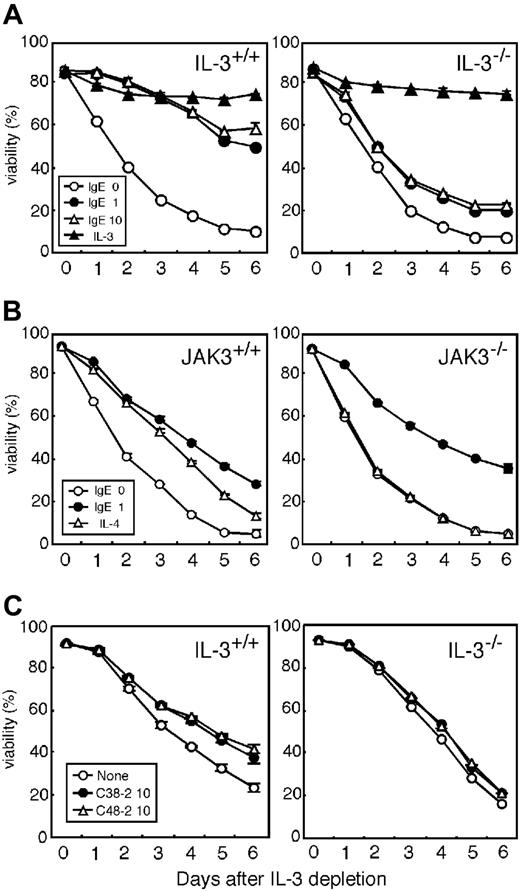
To directly assess the role of endogenous IL-3 in mast cell survival, IL-3-/- BMMCs were examined. IgE-induced survival was severely impaired in IL-3-/- BMMCs, although exogenous IL-3 supported survival (Figure 4A). The expression levels of surface FcϵRI and secretion of β-hexosaminidase by IgE(+Ag) was not reduced in IL-3-/- BMMCs (data not shown), as previously reported.34 We next examined the roles of the IL-4 and IL-9, which can act as potent mast cell growth factors in vitro35,36 and in vivo.37,38 Because the receptors for these cytokines use a common γ chain (γc), we analyzed BMMCs lacking Jak3, an essential protein tyrosine kinase (PTK) associated with the γc. We have previously established Jak3-/- mice and reported the failure of cytokines that use the γc, including IL-4 and IL-9, to function in these mice.19,36 As shown in Figure 4B, although exogenous IL-4 failed to induce mast cell survival in Jak3-/- BMMCs, IgE induced normal survival comparable to littermate control. These results indicated that IL-3 but not “γc cytokines” such as IL-2, IL-4, IL-7, IL-9, IL-15, and IL-21 is responsible for the IgE-induced survival. We also examined another IgE clones, C38-2 and C48-2, which are reported as poor cytokine inducers.28 Survival was significantly induced, albeit weak, by these IgE clones and was impaired in IL-3-/- BMMCs (Figure 4C).
IL-3-/- BMMCs show impaired survival on IgE stimulation. (A) IgE-induced survival of IL-3-/- BMMCs. Survival of BMMCs from WT (IL-3+/+) and IL-3-/- mice was assayed in the presence of 10 ng/mL IL-3 (▴) or IgE (DNP-H1-ϵ-26). IgE concentrations of 0 μg/mL (○), 1 μg/mL (⬡), and 10 mg/mL (▵) are also included. (B) IgE(-Ag)–induced survival in Jak3-/- BMMCs. WT (Jak3+/+) and Jak3-/- BMMCs were tested for survival in the presence of IgE or 10 ng/mL IL-4 (▵). (C) Survival of IL-3-/- BMMCs induced by other IgE clones. Survival induced by 10 μg/mL anti–TNP IgE (C38-2; ⬡) and anti–TNP IgE (C48-2 ▵) was assayed as described in panel A. Data are expressed as means ± SD for triplicate assays. Similar results were obtained from 3 independent pairs.
IL-3-/- BMMCs show impaired survival on IgE stimulation. (A) IgE-induced survival of IL-3-/- BMMCs. Survival of BMMCs from WT (IL-3+/+) and IL-3-/- mice was assayed in the presence of 10 ng/mL IL-3 (▴) or IgE (DNP-H1-ϵ-26). IgE concentrations of 0 μg/mL (○), 1 μg/mL (⬡), and 10 mg/mL (▵) are also included. (B) IgE(-Ag)–induced survival in Jak3-/- BMMCs. WT (Jak3+/+) and Jak3-/- BMMCs were tested for survival in the presence of IgE or 10 ng/mL IL-4 (▵). (C) Survival of IL-3-/- BMMCs induced by other IgE clones. Survival induced by 10 μg/mL anti–TNP IgE (C38-2; ⬡) and anti–TNP IgE (C48-2 ▵) was assayed as described in panel A. Data are expressed as means ± SD for triplicate assays. Similar results were obtained from 3 independent pairs.
IgE-induced up-regulation of Bcl-xL and Bcl-2 was abrogated in IL-3-/- BMMCs
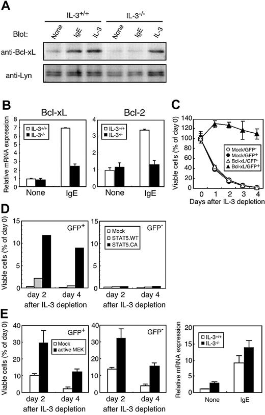
IgE stimulation alone maintains the level of antiapoptotic protein Bcl-xL after IL-3 withdrawal6 and we also confirmed this in WT BMMCs (Figure 5A lanes 1-3). However, IgE did not induce Bcl-xL expression in IL-3-/- BMMCs (Figure 5A lane 5), although exogenous IL-3 did this normally (Figure 5A lane 6). We observed that IgE also up-regulated mRNA for Bcl family including Bcl-xL and Bcl-2 in WT BMMCs but not in IL-3-/- BMMCs (Figure 5B). These results indicated that the up-regulation of Bcl family by IgE is mediated through IL-3.
IgE(-Ag)–induced expression of Bcl family is impaired in IL-3-/- BMMCs. (A) Expression of Bcl-xL protein on IgE(-Ag) stimulation in IL-3+/+ or IL-3-/- BMMCs. BMMCs were cultured with IL-3–depleted medium alone (None), 1 μg/mL IgE (IgE), or 10 ng/mL IL-3 (IL-3) for 24 hours. Cell lysates were analyzed by Western blotting with anti–Bcl-xL (upper panel) and anti-Lyn (lower panel) Abs as a control. (B) Expression of Bcl-xL and Bcl-2 on IgE(-Ag) stimulation in IL-3+/+ or IL-3-/- BMMCs. WT BMMCs (□) and IL-3-/- BMMCs (▪) were cultured in the absence (none) or presence of 1 μg/mL IgE (IgE) for 2 hours. Relative mRNA levels of Bcl-xL and Bcl-2 were determined by real-time RT-PCR and expressed as fold induction over untreated cells after normalization with the values for GAPDH. (C) Bcl-xL expression is sufficient for inducing survival in IL-3-/- BMMCs. Mature IL-3-/- BMMCs were infected using pMX-IRES-GFP retroviral vector either alone (Mock) or together with Bcl-xL (Bcl-xL) as described in “Materials and methods.” ○ indicates mock/GFP-; ⬡, mock/GFP+; ▵, Bcl-xL/GFP-; and ▴, Bcl-xL/GFP+. One day after infection, cells were cultured in the absence of IL-3 and cell viability analyzed for GFP+ or GFP- negative population at the indicated periods. (D) STAT5 activation is sufficient for inducing mast cell survival. Mature WT BMMCs were infected with vector alone (Mock, □), wild-type STAT5a (STAT5a.WT, ▦), or constitutively active STAT5a (STAT5a.CA, ▪). Cell viability was analyzed for GFP+ (left panel) or GFP- negative (right panel) population at indicated periods. (E) Indirect effect of active MEK on BMMC survival. WT BMMCs were infected with vector alone (Mock, □) or MEK.dSESE (active MEK, ▪). Cell viability was analyzed as described in panel D. (F) Expression of HDC mRNA on IgE(-Ag) stimulation in IL-3+/+ (□) or IL-3-/- (▪) BMMCs. The BMMCs were cultured as described in panel B. Relative mRNA expression was determined as indicated in panel B.
IgE(-Ag)–induced expression of Bcl family is impaired in IL-3-/- BMMCs. (A) Expression of Bcl-xL protein on IgE(-Ag) stimulation in IL-3+/+ or IL-3-/- BMMCs. BMMCs were cultured with IL-3–depleted medium alone (None), 1 μg/mL IgE (IgE), or 10 ng/mL IL-3 (IL-3) for 24 hours. Cell lysates were analyzed by Western blotting with anti–Bcl-xL (upper panel) and anti-Lyn (lower panel) Abs as a control. (B) Expression of Bcl-xL and Bcl-2 on IgE(-Ag) stimulation in IL-3+/+ or IL-3-/- BMMCs. WT BMMCs (□) and IL-3-/- BMMCs (▪) were cultured in the absence (none) or presence of 1 μg/mL IgE (IgE) for 2 hours. Relative mRNA levels of Bcl-xL and Bcl-2 were determined by real-time RT-PCR and expressed as fold induction over untreated cells after normalization with the values for GAPDH. (C) Bcl-xL expression is sufficient for inducing survival in IL-3-/- BMMCs. Mature IL-3-/- BMMCs were infected using pMX-IRES-GFP retroviral vector either alone (Mock) or together with Bcl-xL (Bcl-xL) as described in “Materials and methods.” ○ indicates mock/GFP-; ⬡, mock/GFP+; ▵, Bcl-xL/GFP-; and ▴, Bcl-xL/GFP+. One day after infection, cells were cultured in the absence of IL-3 and cell viability analyzed for GFP+ or GFP- negative population at the indicated periods. (D) STAT5 activation is sufficient for inducing mast cell survival. Mature WT BMMCs were infected with vector alone (Mock, □), wild-type STAT5a (STAT5a.WT, ▦), or constitutively active STAT5a (STAT5a.CA, ▪). Cell viability was analyzed for GFP+ (left panel) or GFP- negative (right panel) population at indicated periods. (E) Indirect effect of active MEK on BMMC survival. WT BMMCs were infected with vector alone (Mock, □) or MEK.dSESE (active MEK, ▪). Cell viability was analyzed as described in panel D. (F) Expression of HDC mRNA on IgE(-Ag) stimulation in IL-3+/+ (□) or IL-3-/- (▪) BMMCs. The BMMCs were cultured as described in panel B. Relative mRNA expression was determined as indicated in panel B.
Introduction of Bcl-xL into IL-3-/- BMMCs in a form of IRES-GFP expression vector conferred survival only in a bicistronic GFP+ cells, suggesting that Bcl-xL has the potential to induce mast cell survival (Figure 5C). Consistent with this, STAT5, a known downstream component of the IL-3R signaling pathway, can activate Bcl-xL transcription.39-41 We found that the expression of CA but not WT STAT5a induced mast cell survival only in GFP+ cells (Figure 5D). We have previously shown that introduction of sustained Erk activation by active MAPK/ERK kinase (MEK) is sufficient for the survival.12 However, survival induction by active MEK was observed not only in bicistronic GFP+ (active MEK+) cells but also in GFP- (active MEK-) cells (Figure 5E), indicating that Erk activation is required for the secretion of IL-3 but not for the signaling downstream of IL-3R. These results are highly suggestive of an up-regulation of the Bcl-xL/Bcl-2 by IgE, mediated through autocrine IL-3 and IL-3R signaling rather than through a direct intracellular signal from FcϵRI.
IgE(-Ag) also induces IL-3–independent pathway
Histidine decarboxylase (HDC), an enzyme for histamine synthesis, is also reported to be induced by IgE alone.42 We asked whether the induction of HDC is also mediated through autocrine IL-3 similarly to Bcl-xL/Bcl-2 and survival. We found that the HDC mRNA was induced normally in IL-3-/- BMMCs (Figure 5F), indicating that HDC induction does not require IL-3, in clear contrast to Bcl-xL/Bcl-2 induction and survival.
Discussion
In the present study, the molecular mechanism of IgE-induced mast cell survival was investigated using gene-targeted mice.
By analogy with the response to IgE(+Ag),43 IgE(-Ag)–induced survival also requires FcRγ-ITAM11 and Syk.28 Lyn is also essential for the IgE-induced survival despite the observation that Lyn-/- BMMCs show normal or increased responses to IgE(+Ag).31,44,45 This has been interpreted to indicate that other Src family PTKs such as Fyn, can compensate Lyn function by phosphorylating ITAM on IgE(+Ag) stimulation.44 The reason for the differential requirement of Lyn in response to IgE(-Ag) and IgE(+Ag) is unclear. Recent reports suggested that IgE may cause cross-linking of FcϵRI even in the absence of Ag.12,28 One possible explanation is that “weak” cross-linking of FcϵRI by IgE may be insufficient to enable Fyn to phosphorylate FcRγ-ITAM instead of Lyn. This is consistent with the recent report that SPE-7, an IgE mAb that induces strong aggregation of FcϵRI, could induce partial survival even in the absence of Lyn.28
Fyn is reported to phosphorylate Gab2,14 which is essential for the FcϵRI signaling by IgE(+Ag) through the recruitment of PI3K.15 Activation of PI3K is known to be involved in cell survival via recruitment and activation of Akt in various cells.17 However, our data demonstrated that Fyn, Gab2, PI3K p85α, and Akt were dispensable for IgE-induced survival.
We confirmed that survival is mediated mainly by a secreted soluble factor rather than direct intracellular signaling from FcϵRI, as recently suggested.6,28 Kalesnikoff et al reported that a mixture of possible cytokines including IL-2, IL-3, IL-4, IL-6, IL-13, and TNF-α could partially induce mast cell survival.6 However, among candidate factors, γc cytokines, including IL-4 or IL-9, were dispensable because Jak3-/- BMMCs exhibited normal survival in response to IgE (Figure 4B). We found IL-3 to be secreted immediately after IgE treatment and that it supported survival of BMMCs.
Because the effect of IgE(-Ag) has recently been reported to be largely dependent on the individual clone of IgE, in addition to 3 IgE clones described here, we also examined the survival effect of SPE-7, which is reported to induce strong FcϵRI aggregation and mast cell activation.28 Survival promotion in the presence of SPE-7 was also impaired in IL-3-/- mice (M.K. et al, unpublished observation, 2003), suggesting that the role of IL-3 in IgE(-Ag)–induced survival is not limited to particular IgE mAbs.
IgE(-Ag) is reported to induce sustained Erk activation.6 We have recently reported, using a CD8/FcRγ chimera, that weak but sustained Erk activation favors survival.12 Subsequently, we have found that a large amount of IL-3 is secreted under the condition inducing survival in this system (S.Y. et al, unpublished observation, 2003). In T cells, prolonged Erk activation is suggested to be critical for IL-2 production through c-Rel activation.46 Similarly, sustained Erk signaling may be transmitted to intensive IL-3 transcription in IgE(-Ag)–mediated survival of mast cells. Indeed, sustained Erk activation by active MEK induced enhancement of BMMC survival in a soluble factor-dependent manner. In addition, MEK inhibitor, PD098059, suppressed IgE-induced transcription of IL-3 and Bcl-xL but not IL-3–mediated Bcl-xL transcription (M.K. et al, unpublished observation, 2004).
IgE(-Ag) stimulation maintains Bcl-xL protein expression.6 We found that IgE(-Ag) induces Bcl-xL expression through an indirect autocrine IL-3 pathway rather than through direct intracellular downstream signaling of FcϵRI.
In the downstream signaling pathway of the IL-3R, inactivation of Bad and transcription of Bcl-xL are regulated by Akt and STAT5, respectively.39,47,48 We found Akt activity is not required for IgE(-Ag)–induced survival. In addition, IL-3–induced mast cell survival as well as Bcl-xL/Bcl-2 expression was severely impaired in STAT5a/b-deficient mice.49 Consistent with this, Bcl-xL or constitutively active STAT5a is sufficient for the induction of mast cell survival. Thus, it is highly conceivable that IgE(-Ag) induces IL-3 production, which leads to up-regulation of Bcl-xL through STAT5 activation.
Recently, it has been shown that IgE also triggers HDC induction, which is required for subsequent histamine release on Ag cross-linking. In contrast to survival, we found that HDC up-regulation does not require IL-3, although precise signaling pathway remains to be clarified. Taken from these results together with our previous report,11 IgE-induced mast cell responses appear to involve more than one signaling pathway. Up-regulation of FcϵRI expression following IgE stimulation is independent of FcRγ-ITAM, whereas other responses such as survival and cytokine production are totally dependent on the ITAM.11 FcRγ-ITAM–dependent responses could be further dissected into IL-3–mediated responses such as survival and an IL-3–independent one including HDC induction. Therefore, we suggest that IgE-mediated responses are controlled by multiple mechanisms. Although the present data demonstrate the importance of IL-3 in IgE-induced survival, the contribution of other cytokines cannot be excluded because slight enhancement of survival was still observed in IL-3-/- BMMCs (Figure 4A).
IgE-induced mast cell survival has not been clearly verified in vivo.18 The normal number of mucosal mast cells found in FcϵRIα-/- mice indicates that this mechanism is not essential for normal mast cell homeostasis.50 However, Kitaura et al recently reported that implantation of IgE-producing hybridoma cells in the mouse peritoneal cavity showed slightly increased survival of mucosal mast cells.28 Furthermore, recent works revealed that IgE(-Ag) plays a significant role in immune responses in vivo, possibly through chemokine/cytokine production from mast cells.51 In addition, because infection of helminths or nematodes is also known to induce mastocytosis,52 the response to parasite infection may reflect a more physiologic function of IgE-mediated mast cell survival. On the other hand, parasite infection often promotes a rapid increase of nonspecific IgE in addition to parasite-specific IgE.53 It is possible, therefore, that such IgE may induce mast cell survival in the absence of Ag in specific tissues. Although the role of IgE in parasite-induced mastocytosis is controversial,54-56 splenic mastocytosis induced by primary infection of Trichinella spiralis is greatly impaired in IgE-/- mice.57 Furthermore, Lantz et al reported that mucosal mastocytosis is impaired in IL-3-/- mice during Strongyloides venezuelensis infection.34 Together, these observations imply that IgE-induced autocrine production of IL-3 from mast cells may support mastocytosis by parasite infection, although its importance may differ depending on the type of parasite.56
In humans, the presence of large amounts of total IgE contributes to the exacerbation of atopic diseases,58 possibly by promoting mast cell survival even in the absence of allergen. The in vivo significance of IgE(-Ag)–induced mast cell survival and the contribution of autocrine growth factors to this situation in humans is now under investigation.
Present analyses suggest that IL-3 may become a possible target for the development of new treatments for allergic diseases, provided that further evidence for its significance in these diseases is clarified.
Prepublished online as Blood First Edition Paper, November 12, 2004; DOI 10.1182/blood-2004-07-2639.
M.K. and S.Y. contributed equally to this work.
The publication costs of this article were defrayed in part by page charge payment. Therefore, and solely to indicate this fact, this article is hereby marked “advertisement” in accordance with 18 U.S.C. section 1734.
We would like to thank Dr F-T. Liu for providing DNP H1-ϵ-26. We would like to thank Drs T. Hirano, S. Horikawa, S. Koyasu, K. Nishida, K. Sada, E. Schweighoffer, T. Yamamoto, H. Yamamura, K. Yanagi, and S. Yuasa for providing mice. We thank Dr E. Schweighoffer for excellent technical help. We also thank Drs K. Eshima, D. Sakurai, and K. Funayama for discussion; Ms E. Ishikawa and M. Sakuma for technical help; and Ms H. Yamaguchi for secretarial help.


![Figure 2. IgE-induced mast cell survival is mediated by soluble factors. (A-B) Survival of FcRγ-/- BMMCs supplemented with supernatant of IgE(-Ag)–treated FcRγ+/+ BMMCs. BMMCs from FcRγ+/+ and FcRγ-/- mice were cultured in the absence (A) and presence (B) of 100% supernatant from IgE-treated (1 μg/mL, Sup(+)[▴]) or untreated (Sup(-)[⬡]; ○ indicates no supernatant) WT BMMCs for 12 hours, and then tested in the survival assay. (C) IgE-induced survival of FcϵRI- BMMCs when cocultured with FcϵRI+ BMMCs. BMMCs were infected with pMX-FcRγ-IRES-GFP and the viability of the bulk population was analyzed for both FcϵRI+ (GFP+) and FcϵRI- (GFP-) populations. IgE concentration was 0 μg/mL (○), 1 μg/mL (⬡), and 10 μg/mL (▴). (D) Coculture of FcRγ-/- and FcRγ+/+ BMMCs in transwell chambers. Either FcRγ-/- or WT (FcRγ+/+) BMMCs were cultured in upper and lower chambers with indicated combinations in the presence (▪) or absence (□) of 1 μg/mL IgE. The viability of cells in the upper chamber was analyzed 4 days after IL-3 depletion. Data are expressed as the mean ± SD for triplicate assays. Similar results were obtained from 3 independent experiments.](https://ash.silverchair-cdn.com/ash/content_public/journal/blood/105/5/10.1182_blood-2004-07-2639/6/m_zh80050574870002.jpeg?Expires=1769132472&Signature=wYGE2~AZWkxgNh-C4KMW6RCdgfNu5nPTPyUajiGOgLdjJUmIBE2au4SLV8Hbs6uclbenlgvxT5cczwU7g8Kpvh3QUex7ef374HObbLUAZMphw-WA0YH6Vh1dUU86cbW6~iYQmnHHFApEY-6fUcIk1AFQnUq3aZONsy5kvwqIm3~CmMw89Q1lkt~43g9sF8Hcn45SuTKddoUvrhxdUxu0mkxy1K6BWQZKkOFPmC9tEEDpUDFI8jEnVwYMtZ3dxbbmcf2Q9KrdjhJatlRnCkXeYi7QcjlP5NkgOZ-Whba1o5aCI-nMr3JSntWn-7vVgCxYVwh9NXaByd9~as5NQkdGpA__&Key-Pair-Id=APKAIE5G5CRDK6RD3PGA)


This feature is available to Subscribers Only
Sign In or Create an Account Close Modal