Abstract
18F-labeled Fluorodeoxyglucose Positron Emission Tomography - computed tomography (PET-CT) imaging results from newly diagnosed patients with multiple myeloma (MM) were correlated with serum free light chain (FLC) results from Total Therapy 3 (TT3). Patients had baseline PET-CT scans and serum kappa and lamda FLC analysis at diagnosis and at 2, 14 and 30 days post first cycle of VDTPACE and at pre-transplant 1 of two planned autologous stem cell transplants (TX1), correlating PET-CT response, clonal and nonclonal FCL response, and likelihood of subsequent complete clinical response (CR). Patients were selected with serum clonal FLC levels above the normal range that had been enrolled for sufficient time for a complete response (CR) to be recorded. Correlations between the serum clonal and nonclonal FLC levels and PET-CT response defined by both reduction in number of PET-defined focal lesions (PET-FL) (5 mm or larger areas of circumscribed uptake visually definable above background activity) and reduction in the maximum standardized uptake value normalized to the patients’ lean body mass (max SUV) were performed.
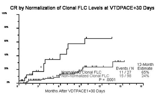
Results: Reduction in FLC and PET-FL and max SUV are correlated, as was normalization of clonal FLC levels with likelihood of subsequent CR (Fig 1). The Spearman’s correlation coefficient between percent reduction in clonal FLC and percent reduction in max SUV was 0.04 (n=49, p=0.79) at 2 days post-VDTPACE, 0.32 (n=48, p=0.028) at 30 days post-VDTPACE, and 0.52 (n=33, p=0.002) prior to TX1. The greatest reduction in max SUV occurred by 2 days post-VDTPACE while the maximum reduction in clonal FLC occurred at 30 days post-VDTPACE, indicating that the PET response lead and predicted the FLC response in near “real time.” While normalization of the clonal FLC levels at 30 days was strongly predictive of subsequent CR (p<0.001, n=125), normalization of the non-clonal FLC levels was not (p=0.41, n=124).
Conclusion: We conclude that normalization of serum clonal FLC correlates both with improvement in FDG PET-CT scanning in both max SUV and number of PET-defined focal lesions and with likelihood of subsequent CR, though it “lags” behind FDG PET response by nearly a month, with FDG PET following the clinical course in near real time.
Kaplan-Meier Analysis Correlating Normalization of Clonal FLC Levels by 30 Days Post Initiation of Treatment with Likelihood of Subsequent Clinical Remission (p=0.0001, n=125).
Kaplan-Meier Analysis Correlating Normalization of Clonal FLC Levels by 30 Days Post Initiation of Treatment with Likelihood of Subsequent Clinical Remission (p=0.0001, n=125).
Author notes
Corresponding author


This feature is available to Subscribers Only
Sign In or Create an Account Close Modal