The functions of the αC domains of fibrinogen in clotting and fibrinolysis, which have long been enigmatic, were determined using recombinant fibrinogen truncated at Aα chain residue 251. Scanning electron microscopy and confocal microscopy revealed that the fibers of α251 clots were thinner and denser, with more branch points than fibers of control clots. Consistent with these results, the permeability of α251 clots was nearly half that of control clots. Together, these results suggest that in normal clot formation, the αC domains enhance lateral aggregation to produce thicker fibers. The viscoelastic properties of α251 fibrin clots differed markedly from control clots; α251 clots were much less stiff and showed more plastic deformation, indicating that interactions between the αC domains in normal clots play a major role in determining the clot's mechanical properties. Comparing factor XIIIa cross-linked α251 and control clots showed that γ chain cross-linking had a significant effect on clot stiffness. Plasmin-catalyzed lysis of α251 clots, monitored with both macroscopic and microscopic methods, was faster than lysis of control clots. In conclusion, these studies provide the first definitive evidence that the αC domains play an important role in determining the structure and biophysical properties of clots and their susceptibility to fibrinolysis.
Introduction
Fibrinogen, which is essential for both platelet aggregation and the fibrin clot formation, is made up of 3 pairs of polypeptide chains, (AαBβγ)2. It is a fibrous protein 45 nm in length with globular regions at each end and in the middle. Each fibrinogen molecule contains 2 αC domains, which are the carboxyl terminal two-thirds of the Aα chains. The αC domains, each made up of a globular and an extended portion, are juxtaposed to one another near the central globular region of fibrinogen where they interact intramolecularly.1-4 During the conversion of fibrinogen to fibrin, there is a large-scale conformational change such that the αC domains dissociate from the central region, allowing them to interact intermolecularly, adding protofibrils to a fiber.2,4,5 Indeed, isolated αC fragments, which were removed proteolytically from fibrinogen, can bind to normal fibrin, competing with the normal αC domain interactions and thereby impairing polymerization.5 Thus, the αC domain interactions appear critical for the enhancement of lateral aggregation during fibrin polymerization.4,6,7
Several dysfibrinogenemias with amino acid substitutions in or truncations of the αC domains showed striking effects on polymerization, clot structure and fibrinolysis.8-10 While the results from these and other dysfibrinogenemias are intriguing and give us many clues to the function of the αC domains, interpretation of the results is still somewhat confused by other accompanying changes to the molecules (such as disruption of disulfides or attachment of albumin) and the fact that most of these cases are heterozygous.11-16 Studies using proteolytic fragments of fibrinogen, which lack the αC domains, are perhaps more straightforward in interpretation, but any preparation of proteolytically modified fibrinogen will have some degree of heterogeneity, again making the results more difficult to interpret.
In the experiments reported here, we examined the structural, physical and biochemical properties of clots made from Aα251 recombinant fibrinogen.17 This recombinant fibrinogen contains Aα chains truncated at residue 251 but otherwise is identical to normal recombinant fibrinogen. Using this recombinant fibrinogen, we were able to circumvent the shortcomings associated with studies of dysfibrinogens and proteolytic fragments.
Materials and methods
Fibrinogen synthesis and purification
Recombinant control fibrinogen has the normal Aα, Bβ, and γ chains of human fibrinogen, whereas recombinant Aα251 fibrinogen contains Aα chains truncated at the residue 251 but is otherwise identical to normal fibrinogen. Normal and Aα251 fibrinogens were synthesized and purified as previously described.17 The fibrinogen concentrations were determined by absorbance at 280 nm, using the extinction coefficient of ϵ= 1.506 for a 1-mg/mL solution of normal fibrinogen and ϵ= 1.610 for Aα251 fibrinogen, as previously described.17 Purity of the proteins was analyzed by sodium dodecyl sulfate–polyacrylamide gel electrophoresis (SDS-PAGE) gel, by the method of Laemmli.18
Preparation of fibrin clots
To a volume of 0.10 mL of recombinant fibrinogen (4.4 μM final concentration in 20 mM HEPES [4-(2-hydroxyethyl)-1-piperazineethanesulfonic acid], pH 7.4, 0.15 M NaCl) was added 10 μL of CaCl2 to a 5-mM final concentration. After one minute of incubation, 10 μL human α-thrombin (Enzyme Research Labs, South Bend, IN) was added to obtain a final concentration of 0.9 IU/mL. Mixing and incubation were conducted in polypropylene tubes at room temperature. This solution was either soaked up into a glass microchamber designed for flow measurements or dropped off between the coverslips of a torsion pendulum. Clotting was carried out for at least 40 minutes in a moist atmosphere at room temperature. In order to obtain cross-linked fibrin clots for flow and torsion pendulum experiments, factor XIII (1000 IU/mL) was activated with 1 IU/mL thrombin for 10 minutes and was added prior to clotting to reach a final concentration of 1 IU/mL factor XIIIa.
Laser scanning confocal microscopy
Fibrin clots in the microchambers were permeated with 20 mM Tris-HCl, pH 7.4, 140 mM NaCl buffer and 5 nm colloidal gold particles (British Biocell International, Cardiff, United Kingdom).19 Labeled specimens were scanned with a Zeiss 510 laser scanning confocal microscope (Carl Zeiss, Thornwood, NY) set up in reflection mode.19 Twenty optical sections were collected at intervals of 1.0 μm in the Z-axis. These sections were then projected at 6 different angles 10 degrees apart and combined into one image, generating 6 different 3-dimensional reconstructed images of the fibrin network. Images were obtained with a Zeiss 63 ×/1.2 water-immersion objective lens, and Zeiss software was used for image acquisition and reconstructions.
Average fibrin fiber diameters (n = 150) were measured from high-magnification reconstructed confocal images using the image analysis software package that came with the microscope work station. Fiber and branching point densities were determined from confocal pictures taken at low magnification.19-21 Branching points were very carefully distinguished from crossing fibers by using the series of reconstructed images at different angles and by the analysis of each scan.
Scanning electron microscopy
Clots were prepared by fixation, dehydration, critical point drying, and sputter coating with gold-palladium as described previously.22 Specimens were observed and photographed digitally using a Philips XL20 scanning electron microscope (Philips Electron Optics, Eindhoven, The Netherlands). Average fibrin fiber diameters (n = 150) were measured from high-magnification images of the clots. Adobe Photoshop software (Adobe, San Jose, CA) was used to adjust brightness and contrast.
Permeation experiments
The permeability index or Darcy constant (Ks) of fibrin clots was measured using the permeation technique. Briefly, clots were formed in thin glass microchambers (250 μm) and were permeated with buffer at different gradients of pressure. The calculated Ks index (in cm2) provides information on the fibrin network architecture (shape and size of the pores) and represents the surface of the gel allowing flow.23 The clot permeability index was calculated as follows: Ks = QLη /AΔPt, where Q is the volume of liquid (in mL) having the viscosity η (10–2 poise), flowing through the fibrin gel with length L (2.2 cm) and cross section A (0.03 cm2) in a given time t (in seconds) under a differential pressure ΔP (ranging from 4000 to 10 000 dyne/cm2).
Viscoelastic experiments
Clots prepared in the same way as those used for permeation experiments were formed between 2 12-mm diameter glass coverslips 1 mm apart in a torsion pendulum device, as previously described.10,24 At 60 minutes after the initiation of clotting, a momentary impulse was carefully applied to the torsion pendulum arm by air pressure, causing free oscillations of this arm with strains less than 3%. The frequency of these free oscillations and the rate at which they are damped are functions of the elastic and viscous properties of the clots and are independent of the amplitude of the initial displacement of the arm. The storage modulus, G′ in dyne/cm2, which reflects the clot's stiffness, and the loss modulus, G″, which reflects the inelastic component, were calculated from recordings of these oscillations on a chart recorder.
Fibrinolysis experiments: microscopic lysis velocity by laser scanning confocal microscopy
A quantity of 10 μL of a solution containing recombinant tissue plasminogen activator (rtPA) (6 nM; Boehringer Ingelheim, Ingelheim, Germany) and Glu-plasminogen (2.5 μg/mL, purified from human plasma; American Diagnostica, Stamford, CT) was loaded at the edge of a fibrin clot formed in a glass microchamber and labeled with colloidal gold. After 15 minutes of incubation in a moist atmosphere during which the rtPA and Gluplasminogen diffused within the fibrin network, the edge of the clot was visualized with the confocal microscope set up in the reflection mode. Scanning was performed at low magnification every 2 minutes. The lysis front velocity and the rate of fiber digestion were determined from reconstructed confocal microscope images. Modifications of the shape of individual fibers while being digested were studied in both types of fibrin.
Fibrinolysis experiments: macroscopic lysis velocity by turbidity
Polymerization of 90 μL normal recombinant and Aα251 fibrinogen (4.4 μM) was initiated by addition of 10 μL thrombin (0.9 IU/mL final concentration) and was monitored at 350 nm for 1 hour in a SpectraMax-340PC 96-well microtiter plate reader at ambient temperatures (Molecular Devices, Sunnyvale, CA). After polymerization was complete, 100 μLofa mixture of Glu-plasminogen and rtPA (see “Fibrinolysis experiments: microscopic lysis velocity by laser scanning confocal microscopy”) was overlaid on each clot and the turbidity was monitored at 350 nm every 7 seconds for 3.5 hours. Two separate experiments were performed in quadruplicate, as previously described.25 The data were normalized to a 1-cm path length by the PathCheck sensor within the instrument. The fibrinolysis curves were normalized with respect to the initial turbidity and the rate of lysis was determined from the slope of the latter part of the fibrinolysis curve, where the slope becomes constant.
Statistical analysis
Statistical analyses were performed with StatView software (version 5.0; Abacus Concepts, Berkeley, CA). Continuous variables were expressed as the mean plus or minus the standard error of the mean (SEM), and differences were determined by analysis of variance (ANOVA). P below .05 was accepted as significant.
Results
Analysis of purified proteins and factor XIIIa–induced cross-linking of fibrin
Sodium dodecyl sulfate–polyacrylamide gel electrophoresis (SDS-PAGE) run under nonreducing conditions showed that normal, recombinant fibrinogen contained the expected bands representing high-molecular-weight and low-molecular-weight fibrinogen and that Aα251 fibrinogen contained a single band at 260 000 (data not shown). SDS-PAGE run under reducing conditions showed that normal, recombinant fibrinogen contained the expected bands representing the Aα, Bβ, and γ chains and that the Aα chain of Aα251 fibrinogen migrated below the band for the γ chains, as expected (Figure 1). The protein preparations were devoid of any detectable contaminants.
The process of cross-linking of fibrin by factor XIIIa was followed by SDS-PAGE as a function of time, as described in the Figure 1 legend. On adding thrombin and factor XIII to control recombinant fibrinogen, the typical pattern was the rapid disappearance of the γ band, concomitant with the appearance of a γ dimer band, followed by a slower disappearance of the α chain accompanied by the appearance of higher-molecular-mass bands near the top of the gel (Figure 1A). In contrast, with addition of thrombin and factor XIII to Aα251 fibrinogen, there was a much slower disappearance of the γ band, together with the appearance of a γ dimer band (Figure 1B). To ensure that the slower and incomplete γ chain cross-linking was not due to slower activation of factor XIII, experiments were also carried out with preactivated factor XIIIa, which showed a similar time course of γ chain cross-linking (data not shown). In all experiments with Aα251 fibrinogen, there was no decrease in the density of the α chain band and no high-molecular-weight band, indicating no α chain cross-linking. Densitometry of the gels confirmed the decrease in γ chain and increase in γ dimer and no change in the density of the α band (data not shown).
Factor XIIIa–induced cross-linking of Aα251 and control fibrinogen. Factor XIII and thrombin were mixed, left on ice for less than 10 minutes, and added to fibrinogen at ambient temperature. The samples with final concentrations of 1 IU/mL factor XIII, 1 IU/mL thrombin, and 1.2 μM fibrinogen were incubated for the indicated times (minutes). The reactions were stopped by addition of SDS (1% final concentration) and incubation in boiling water for 5 minutes; samples were analyzed on 12% SDS-PAGE run under reducing conditions. The 0 time point samples contained only fibrinogen. Positions of molecular weight markers are indicated on the right; positions of bands corresponding to individual fibrin chains are indicated on the left. (A) Control fibrin. (B) α251 fibrin.
Factor XIIIa–induced cross-linking of Aα251 and control fibrinogen. Factor XIII and thrombin were mixed, left on ice for less than 10 minutes, and added to fibrinogen at ambient temperature. The samples with final concentrations of 1 IU/mL factor XIII, 1 IU/mL thrombin, and 1.2 μM fibrinogen were incubated for the indicated times (minutes). The reactions were stopped by addition of SDS (1% final concentration) and incubation in boiling water for 5 minutes; samples were analyzed on 12% SDS-PAGE run under reducing conditions. The 0 time point samples contained only fibrinogen. Positions of molecular weight markers are indicated on the right; positions of bands corresponding to individual fibrin chains are indicated on the left. (A) Control fibrin. (B) α251 fibrin.
Morphologic properties of α251 fibrin
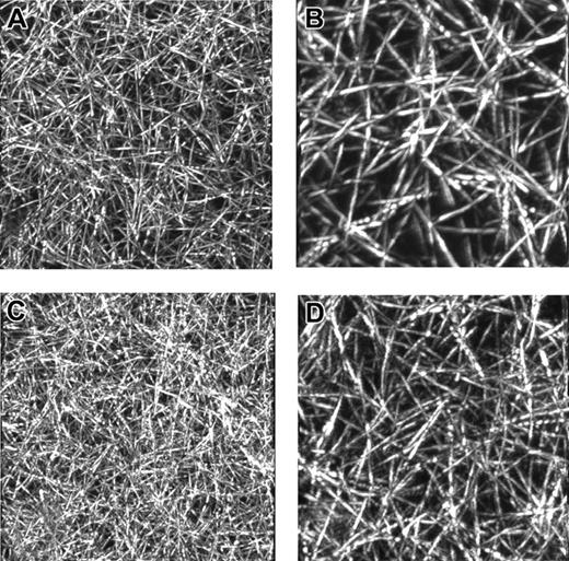
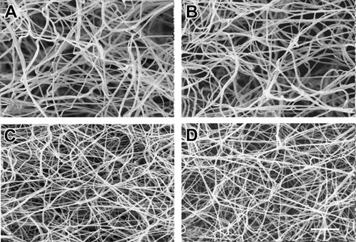
Examination of fibrin clots by both electron and light microscopy showed obvious differences between the morphologic properties of α251 and control clots. Consistent results were obtained with both laser scanning confocal microscopy (Figure 2) and scanning electron microscopy (Figure 3). Quantitative assessments of the clot networks in these images are presented in Table 1. As shown in confocal images, both control and α251 fibrin networks consisted of straight rodlike elements organized in a 3-dimensional network (Figure 2). Although the networks were similar in appearance, α251 fibrin displayed a much tighter 3-dimensional network than control fibrin, with a significantly higher density of both fibrin fibers and branch points as compared with control fibrin (Table 1). Furthermore, individual fibers looked very similar, though the quantitative data show that α251 fibers are significantly thinner as compared with control fibrin, such that the average diameter of α251 fibrin was 40% and 33% of the control fibrin when using electron and confocal microscopy, respectively. The difference in the average fibrin fiber diameter between electron and confocal microscopy is likely due to differences in the hydration of fibrin and to the limited resolution of confocal microscopy, which may overestimate the fibrin diameter. Visual analysis of multiple micrographs showed that the overall spatial organization of the fibrin networks differed, with α251 fibrin having a heterogeneous architecture with compact areas of fibers alternating with looser areas, giving a more disorganized appearance. No differences were observed in the appearance of either α251 or control clots whether or not they were cross-linked with factor XIIIa (Figure 3).
Morphologic properties of control and α251 recombinant fibrin
. | Scanning electron microscopy . | Confocal microscopy . | . | . | ||
|---|---|---|---|---|---|---|
. | Fiber diameter, nm . | Fiber diameter, nm . | Fiber density, 10-3 μm3 . | Branch point density, 10-3 μm3 . | ||
| Control | 120 ± 30 | 323 ± 61 | 7.6 ± 0.2 | 2.2 ± 0.4 | ||
| α251 | 88 ± 21* | 251 ± 42* | 12.1 ± 0.6* | 3.9 ± 0.3* | ||
Data are expressed as mean ± SEM.
Indicates significant differences (P < .05) between control and α251 recombinant fibrin.
Images of α251 and control fibrin clots from laser scanning confocal microscopy. Thrombin at a final concentration of 0.9 IU/mL was added to recombinant control or Aα251 fibrinogen at a 4.4-μM final concentration in 20 mM HEPES, pH 7.4, 0.15 M NaCl, 5 mM CaCl2. Images of 3-dimensional reconstructions of optical sections of control (A, B) and α251 (C, D) fibrin networks obtained by laser scanning confocal microscopy. Original magnifications are as follows: panels A and C: 48 × 48 × 48 μm3; panels B and D: 4 × 24 × 24 μm3.
Images of α251 and control fibrin clots from laser scanning confocal microscopy. Thrombin at a final concentration of 0.9 IU/mL was added to recombinant control or Aα251 fibrinogen at a 4.4-μM final concentration in 20 mM HEPES, pH 7.4, 0.15 M NaCl, 5 mM CaCl2. Images of 3-dimensional reconstructions of optical sections of control (A, B) and α251 (C, D) fibrin networks obtained by laser scanning confocal microscopy. Original magnifications are as follows: panels A and C: 48 × 48 × 48 μm3; panels B and D: 4 × 24 × 24 μm3.
Permeation experiments
Consistent with the morphologic studies, the permeability of cross-linked α251 fibrin was significantly decreased to approximately half that of control fibrin (Table 2). This indicates an increased resistance to pressure-driven permeation related to a higher density of the fibrin fibers and smaller pores. Permeation experiments were not done with α251 fibrin in the absence of factor XIIIa because these clots were too weak for pressure-driven permeation. Nevertheless, we expect that the results with such clots would not have been different, since no significant differences were observed between the morphologic properties of the respective fibrin networks formed without or with factor XIIIa.
Permeation and mechanical properties of α251 and control recombinant fibrin formed with and without factor XIIIa
. | Q/t, mL/h . | Ks, cm2 109 . | G′, dyne/cm2 . | G″, dyne/cm2 . | G″/G′ . |
|---|---|---|---|---|---|
| Control | ND | ND | 164 ± 48 | 10.7 ± 1.2 | 0.067 ± 0.01 |
| α251 | ND | ND | 66 ± 24* | 6.1 ± 1.6* | 0.094 ± 0.01* |
| Control + XIIIa | 4.3 ± 2.2 | 7.9 ± 3.3 | 369 ± 63 | 14.1 ± 2.4 | 0.037 ± 0.01 |
| α251 + XIIIa | 1.9 ± 0.9† | 4.3 ± 2.2† | 106 ± 43† | 7.2 ± 3.9† | 0.063 ± 0.011† |
For each polymerization condition, 2 separate experiments were performed in triplicate. Data are expressed as mean ± SEM.
ND indicates not determined; Q/t, mL/h indicates flow rate.
Indicates significant differences (P < .05) between control and α251 recombinant fibrin that are not cross-linked.
Indicates significant differences (P < .05) between control and α251 recombinant fibrin that are cross-linked.
Scanning electron microscopy of α251 and control fibrin. Thrombin at a final concentration of 0.9 IU/mL was added to recombinant control or Aα251 fibrinogen at a 4.4-μM final concentration in 20 mM HEPES, pH 7.4, 0.15 M NaCl, 5 mM CaCl2. (A) Fibrin made from control fibrinogen. (B) Fibrin made from control fibrinogen in the presence of 1 IU/mL factor XIIIa. (C) Fibrin clot made from Aα251 fibrinogen. (D) Fibrin clot made from Aα251 fibrinogen in the presence of 1 IU/mL factor XIIIa. Magnification bar equals 1000 nm.
Scanning electron microscopy of α251 and control fibrin. Thrombin at a final concentration of 0.9 IU/mL was added to recombinant control or Aα251 fibrinogen at a 4.4-μM final concentration in 20 mM HEPES, pH 7.4, 0.15 M NaCl, 5 mM CaCl2. (A) Fibrin made from control fibrinogen. (B) Fibrin made from control fibrinogen in the presence of 1 IU/mL factor XIIIa. (C) Fibrin clot made from Aα251 fibrinogen. (D) Fibrin clot made from Aα251 fibrinogen in the presence of 1 IU/mL factor XIIIa. Magnification bar equals 1000 nm.
Viscoelastic experiments with and without cross-linking
The viscoelastic properties of α251 fibrin were markedly different from those of control fibrin. Control fibrin was 3 times stiffer than α251 fibrin as shown by the difference in the storage moduli, G′= 66 dyne/cm2 and 164 dyne/cm2 for α251 and control fibrin, respectively (Table 2). The loss modulus G″, which represents the energy dissipated by inelastic viscous processes, was 1.8-fold higher in control fibrin as compared with α251 fibrin. As a consequence, the loss tangent (tan δ= G″/G′), which is a measure of the energy dissipated by viscous processes relative to the energy stored by elastic processes, was 29% lower in control fibrin as compared with α251 fibrin. In summary, the α251 fibrin clots are less stiff with a larger inelastic component than control clots.
As expected, cross-linking with factor XIIIa had a great impact on the overall viscoelastic properties of the normal fibrin network with the storage modulus increased by 2.3-fold. Interestingly, the storage modulus of α251 fibrin increased by only 1.6-fold after cross-linking, as G′ increased from 66 dyne/cm2 to 106 dyne/cm2. The loss moduli increased by 31% in control, from G″= 11 dyne/cm2 to 14 dyne/cm2, versus only 18% in α251 fibrin, from G″= 6.1 dyne/cm2 to 7.2 dyne/cm2. As a consequence, the loss tangent decreased with a similar magnitude in both types of fibrin network, 55% versus 67% for control and α251 fibrin, respectively. In summary, the increase of stiffness of α251 fibrin clots with cross-linking is less than that of the control clots, but both have a similar decrease in the inelastic component.
Analysis of microscopic fibrinolysis by laser scanning confocal microscopy
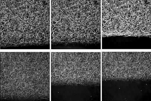
As shown by scanning confocal microscopy (Figure 4), microscopic lysis of fibrin clots progressed as a straight and sharp front moving across the entire area of scanning for both control and α251 clots; these data are similar to those previously reported for clots made from fibrinogen isolated from plasma.19 Because α251 fibrin displayed a tighter fibrin conformation with thinner and more numerous fibers as compared with control fibrin (Figure 2), we expected the α251 clots would lyse more slowly. We found, however, that the rate of fiber lysis in α251 fibrin was 2.2-fold faster than that of control fibrin (Table 3). Cross-linking reduced the rate of fiber lysis by 63% in both types of fibrin (P < .01). As a consequence, cross-linked α251 fibrin displayed a 2.1-fold faster fiber lysis as compared with cross-linked control fibrin (Table 3). In these images we saw the same changes in fiber shape in both types of fibrin. Fibers were progressively disaggregated by transverse cutting with a transient increase in their diameter, as previously described.19 The structural differences between α251 and control fibrin were found with and without factor XIII cross-linking.
Lysis-front velocity and fiber lysis rate in control and α251 fibrin formed with and without factor XIIIa
. | Lysis front velocity, μm/min . | Rate of fiber lysis, fiber/min . |
|---|---|---|
| Control | 4.8 ± 3.3 | 108 ± 125 |
| α251 | 6.4 ± 2.4 | 234 ± 156* |
| Control + XIIIa | 1.9 ± 0.8† | 41 ± 32† |
| α251 + XIIIa | 2.6 ± 0.8† | 86 ± 56*† |
. | Lysis front velocity, μm/min . | Rate of fiber lysis, fiber/min . |
|---|---|---|
| Control | 4.8 ± 3.3 | 108 ± 125 |
| α251 | 6.4 ± 2.4 | 234 ± 156* |
| Control + XIIIa | 1.9 ± 0.8† | 41 ± 32† |
| α251 + XIIIa | 2.6 ± 0.8† | 86 ± 56*† |
Data are expressed as mean ± SEM.
Indicates significant differences (P < .05) between control fibrin and α251 fibrin clots.
Indicates significant differences (P < .05) between fibrin clots that are cross-linked and those that are not cross-linked.
Analysis of macroscopic fibrinolysis by turbidity measurements
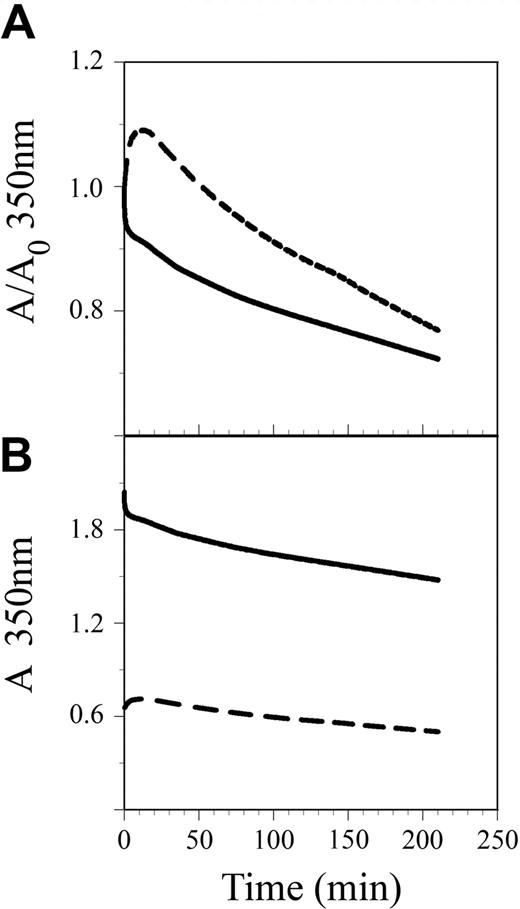
As reported previously,17 α251 fibrin clots reached a lower final turbidity than control fibrinogen, indicating a clot with thinner fibers. Lysis was examined under the same conditions as those for confocal microscopy, so lysis occurred at a very slow rate, and the clots were not fully lysed over the time course monitored (Figure 5). Quantitation of the slope of the fibrinolysis curves (Figure 5A) suggested α251 clots lysed at a slower rate than control fibrinogen (3.5 × 10⁻⁵ ± 0.9 × 10⁻⁵ minute–1 for α251 versus 5.2 × 10⁻⁵ ± 0.7 × 10⁻⁵ minute–1 for control; P = .007, n = 8), in contrast to the confocal microscopy data. When normalized with respect to the initial turbidity (Figure 5B) however, the α251 clots lysed more rapidly than control clots, 4.12 × 10⁻⁵ minute–1 for α251 versus 3.35 × 10⁻⁵ minute–1 for control. We conclude that lysis of α251 fibrin is faster than lysis of control fibrin, whether measured by microscopic or macroscopic methods.
Discussion
The recombinant Aα251 fibrinogen provides a unique opportunity to dissect the functional impact of the αC domains in both clot formation and dissolution, because the Aα251 molecules are homogeneous. Our investigation clearly showed that the αC domains of normal fibrinogen enhance lateral aggregation of protofibrils to produce clots made up of thicker fibers with larger pores. The thinner fibers and the greater density of both fibers and branch points of α251 fibrin clots in comparison to control fibrin clots were associated with the loss of only the αC domains. As expected, these morphologic changes resulted in lower permeation rates for the α251 clots. Taken together, our morphologic and permeation experiments suggest that the αC domains of normal fibrinogen molecules interact with each other to control lateral aggregation and fiber thickness. These findings are consistent with previous studies using either abnormal plasma fibrinogens or the proteolytic fragment that lacks αC, fragment X4 .
Fibrinolysis of α251 and control fibrin clots as visualized by laser scanning confocal microscopy. Series of micrographs showing the dynamic lysis of control (top panels) and α251 (bottom panels) fibrin clots by rtPA (6 nM) and Glu-plasminogen (2.5 μg/mL); clots were prepared as described in Figure 2. Lysis-front motion was visualized by scanning this region of the clot every 2 minutes. The lysis front progressed as a straight and sharp line in both clots with a higher velocity in α251 fibrin as compared with control fibrin. Each micrograph is a reconstruction from optical sections representing a volume of 146 × 146 × 20 μm3. A,D: 2 minutes; B,E: 4 minutes; C,F: 6 minutes.
Fibrinolysis of α251 and control fibrin clots as visualized by laser scanning confocal microscopy. Series of micrographs showing the dynamic lysis of control (top panels) and α251 (bottom panels) fibrin clots by rtPA (6 nM) and Glu-plasminogen (2.5 μg/mL); clots were prepared as described in Figure 2. Lysis-front motion was visualized by scanning this region of the clot every 2 minutes. The lysis front progressed as a straight and sharp line in both clots with a higher velocity in α251 fibrin as compared with control fibrin. Each micrograph is a reconstruction from optical sections representing a volume of 146 × 146 × 20 μm3. A,D: 2 minutes; B,E: 4 minutes; C,F: 6 minutes.
Our studies also showed that the αC domains promote clot stabilization, leading to a stiffer fibrin network. Direct comparison of normal recombinant fibrin with α251 fibrin demonstrated that the αC domains have a greater impact on the mechanical behavior of the fibrin network than on fibrin's morphologic properties. Indeed, α251 fibrin was dramatically lower in stiffness than control fibrin, although only slight differences were found in their 3-dimensional architecture. Of special note, α251 fibrin was lysed more rapidly than normal recombinant fibrin, highlighting the conclusion that the αC domains have a critical role in the clot's stability and its susceptibility to fibrinolysis.
When comparing our results to those obtained from analyses of dysfibrinogenemias with amino acid substitutions or truncations of the αC domains, we find both similarities and discrepancies. In all cases, the natural substitutions have revealed that changes in αC domains impair polymerization. Fibrinogen Caracas II is an abnormal fibrinogen involving an αC domain mutation, Aα 434 serine to N-glycosylated asparagine.11 In this case the αC domains do not interact with each other or with the central domain.12 Clots from this dysfibringenemia have large pores bounded by local fiber networks made up of thin fibers, with increased permeability and normal stiffness. These findings are consistent with the asymptomatic phenotype of individuals with this dysfibrinogenemia. On the other hand, fibrinogen Dusart with the substitution of Aα 554 arginine to reactive cysteine, which becomes disulfide linked to albumin,9 differs from Aα251 fibrinogen. Clots formed from fibrinogen Dusart are extremely stiff and made up of thin fibers with many branch points and very small pore sizes.10 Moreover, these clots have an increased resistance to fibrinolysis, a characteristic that likely is responsible for the dramatic venous thromboembolic complications found in several patients with this mutation.8,26,27 These findings are intriguing but should be interpreted with caution given the accompanying changes to the molecules and the fact that these dysfibrinogenemias are heterozygous.
Fibrinolysis of α251 and control fibrin clots from decrease of turbidity. Clots were prepared and lysis was carried out as described in “Materials and methods.” Representative curves are shown. Solid line, control fibrin; broken line, α251 fibrin. (A) Change of turbidity as measured. (B) Change of turbidity normalized with respect to the initial turbidity of the clot.
Fibrinolysis of α251 and control fibrin clots from decrease of turbidity. Clots were prepared and lysis was carried out as described in “Materials and methods.” Representative curves are shown. Solid line, control fibrin; broken line, α251 fibrin. (A) Change of turbidity as measured. (B) Change of turbidity normalized with respect to the initial turbidity of the clot.
Given the higher density of both fibrin fibers and branching points, one would have expected α251 fibrin to be at least as stiff as control fibrin. The unanticipated results of the viscoelastic experiments highlight the critical role of the αC domains in the mechanical properties of the overall fibrin network as well as in individual fibers. The most likely explanation for the dramatic decrease in stiffness and the increase in plastic deformation of α251 fibrin is that the αC domains in normal fibrin interact with each other to increase rigidity. The loss of the αC domains in α251 fibrin clots would result in the loss of these interactions and thereby decrease stiffness. As expected, after cross-linking with factor XIIIa, the rigidity of the control clots increased and ratio of the energy dissipated in viscous processes to energy stored (G″/G′), most likely related to the slippage of protofibrils, decreased. Factor XIIIa treatment of α251 fibrin clots yielded no cross-linking of α chains, because the αC domains were missing, and less γ chain cross-linking, for unknown reasons. Furthermore, the significant differences between the impact of cross-linking on G′ of control and α251 fibrin are consistent with previous data showing that G′ scales directly with the density of cross-links,28,29 which is dramatically reduced in α251 fibrin.
The results of the fibrinolysis experiments at first appear to be the opposite of what has been previously reported with cross-linked plasma fibrin. Although at the fiber level thinner fibers are lysed faster than thicker fibers, clots with a tight fibrin conformation made of numerous thin fibers have been shown by similar microscopic methods to have a significantly slower lysis rate than clots with a loose fibrin conformation made of thicker fibers with a lower fibrin fiber density.19 The finding that α251 clots are lysed faster than control clots demonstrates that the network conformation and the fiber size are not the only parameters that influence the lysis speed. In particular, the chemistry is different here, since the αC domains, normally important for the initiation of clot lysis, contain tPA and plasminogen binding sites,30 and these domains are substrates for plasmin. We conclude that the αC domains have a critical impact on the process of fibrinolysis at the level of individual fibers, because the lysis front moves more rapidly for the α251 clot. Considering the results of viscoelastic experiments, this conclusion is further supported by previous reports that stiffer clots are digested more slowly than less stiff clots, in general.31,32 An extreme example is fibrinogen Dusart, which produces very stiff clots made of thin fibers with very slow lysis,9,10 partly because of the presence of albumin, which is resistant to digestion.33,34 Mechanistically, lysis might be seen as a series of protease-induced collapse events in which gel stiffness impedes collapse.
Fibrinolysis was assessed at both the macroscopic level by changes in turbidity and the microscopic level by the lysis front velocity and fiber lysis rate. Initial analysis showed contradictory results with these 2 assays, as the turbidity slopes indicated control clots lysed more quickly. When the turbidity data were normalized to the initial absorbance at 350 nm, the lysis rates were inverted, such that the α251 clot lysed more quickly. As the normalized rates measure the percent of clot lysis with time, these values indicate that the lysis of α251 clots would be complete prior to lysis of control clots. This conclusion is consistent with the microscopic lysis data, showing that propagation of the lysis front is faster for the less stiff α251 clots.
It is well known that factor XIIIa cross-linking stabilizes fibrin, making clots stiffer, less plastic, and more resistant to lysis.28,29,31,32 Nevertheless, little is known regarding the relative roles of the α and γ chain cross-linking. Although the γ chains are cross-linked more rapidly, α chain cross-linking starts immediately, so their relative effects cannot be sorted out by examining the time course of the effects. Studies with a specific factor XIII antibody demonstrated that α chain cross-links are more important in establishing clot rigidity than γ chain cross-links.35 In α251 fibrin, only γ chain cross-links are possible, because the normal glutamine acceptors of the α chain are lost. Clots of cross-linked α251 fibrin were stiffer and less plastic than α251 fibrin clots that were not cross-linked, so we conclude that γ chain cross-linking has a significant effect on the mechanical properties of the clot. These results are especially striking since the cross-linking of γ chains is incomplete in the α251 fibrin clots. Consistent with the results of other studies,35 an enhanced effect was evident with cross-linked α chains, in that the increased stiffness and decreased plastic deformation were more pronounced for control fibrin clots than for α251 clots. This conclusion is tempered by our finding that the γ chain cross-linking of α251 fibrin was reduced as compared with control fibrin, a difference that may also affect the other mechanical properties. The mechanism of the reduced γ chain cross-linking of α251 fibrin as compared with control fibrin clots remains to be investigated.
The present investigation provides new insights into the functions of the αC domains of the fibrinogen molecules. We showed that the αC domains have a greater impact on the mechanical properties of the fibrin network than on the morphology of the clot. Our data suggest that these mechanical properties influence clot dissolution, an important physiologic function. In addition, we found that γ chain cross-links alone significantly increase clot stiffness.
Prepublished online as Blood First Edition Paper, August 9, 2005; DOI 10.1182/blood-2005-05-2150.
Supported by the National Institutes of Health grants HL30954 (J.W.W.) and HL 31048 (S.T.L.).
An Inside Blood analysis of this article appears in the front of this issue.
The publication costs of this article were defrayed in part by page charge payment. Therefore, and solely to indicate this fact, this article is hereby marked “advertisement” in accordance with 18 U.S.C. section 1734.
We thank Li Fang Ping and Chandrasekaran Nagaswami for their excellent technical assistance.


This feature is available to Subscribers Only
Sign In or Create an Account Close Modal