Mast cells are the major effector-cell type for immediate hypersensitivity and other forms of allergic reactions. Expression of 4-1BB, a member of the tumor necrosis factor receptor superfamily, is induced at mRNA and protein levels on stimulation through the high-affinity receptor for immunoglobulin E (IgE; FcϵRI). In this study, we present evidence that agonistic anti-4-1BB antibodies can enhance FcϵRI-induced cytokine production and secretion. Consistent with this, 4-1BB-deficient mast cells exhibit reduced degranulation and cytokine production on FcϵRI stimulation. Analysis of 4-1BB ligand (4-1BBL)-deficient cells supported this notion. As a potential mechanism for these defects, we identified a defect in Ca2+ flux induced by FcϵRI stimulation. The defective Ca2+ flux could be accounted for by the reduced activity of Lyn/Btk/phospholipase C-γ2 pathway and constitutive interactions between 4-1BB and Lyn. Therefore, FcϵRI-inducible 4-1BB plays a costimulatory function together with FcϵRI stimulation.
Introduction
Mast cells are the major effector cells for acute and chronic allergic reactions and host defense against certain parasites and bacteria.1 Activated mast cells release preformed proinflammatory mediators (such as histamine, proteases, proteoglycans, and nucleotides) and release or secrete de novo synthesized lipids (such as leukotrienes and prostaglandins) and polypeptides (such as cytokines and chemokines). These substances contribute to the development of allergy and other forms of inflammation.
The high-affinity receptor for immunoglobulin E (IgE; FcϵRI), together with antigen receptors such as T-cell receptor (TCR), belongs to the multichain immune recognition receptor superfamily.2 FcϵRI expressed on murine mast cells consists of 4 subunits (αβγ2): an IgE-binding α subunit, a signal-amplifying, receptor-stabilizing β subunit, and 2 disulfide-bonded γ subunits that are the main signal transducer.3 The aggregation of FcϵRI, induced by stimulation of IgE-sensitized mast cells with multivalent antigen or anti-IgE antibody, leads to the activation of this receptor system: β subunit-associated Lyn, a Src family protein tyrosine kinase (PTK), becomes activated, and phosphorylates tyrosine residues in the immunoreceptor tyrosine-based activation motifs (ITAMs) in the cytoplasmic regions of β and γ subunits. Phosphorylated β and γ ITAMs recruit Lyn and Syk (another PTK with 2 tandem Src homology 2 domains N-terminal to the catalytic domain), respectively. Another Src family PTK, Fyn, was also shown to associate with FcϵRI and to play a complementary role, particularly by activating phosphatidylinositol 3-kinase.4 These PTKs phosphorylate numerous targets and activate several signaling pathways, including the phosphatidylinositol 3-kinase, phospholipase C (PLC)/Ca2+, and several mitogen-activated protein kinase pathways.5,6 These signaling events lead to degranulation and cytokine production.
Activation of T cells requires 2 signals: signal 1 from the TCR by the recognition of antigen presented in the context of major histocompatibility complex molecules by antigen-presenting cells (APCs) and signal 2 from CD28 or other “costimulatory” receptors. Without costimulation, T cells become unresponsive or anergic.7 By contrast, it is not clear whether costimulation is important for FcϵRI-induced activation. CD28 is expressed at low levels in mouse mast cells, and concurrent stimulation of CD28 and FcϵRI can weakly enhance the production of tumor necrosis factor (TNF)-α over that induced by FcϵRI crosslinking alone.8 Despite these in vitro studies, the in vivo significance of the weak costimulatory activity of CD28 is not known.
In addition to B7-CD28 and related ligand-receptor interactions, the TNF family-TNF receptor (TNFR) family interactions are a rich source of costimulation for T-cell activation.9 4-1BB (CD137) is a type I membrane protein of the TNFR family and functions as a costimulatory molecule in T cells (reviewed in Kwon et al10 ). 4-1BB is expressed primarily on activated CD4+ and CD8+ T cells, activated natural killer cells, and activated natural killer T cells. Its expression is inducible in T cells by various mitogens and antigens.11 In contrast, its ligand, 4-1BBL, is a type II membrane protein of the TNF family and expressed primarily on APCs such as mature dendritic cells, activated B cells, and activated macrophages,12,13 although it is also expressed in activated T cells and NK cells.14,15 4-1BB is able to stimulate both T-cell proliferation and production of interleukin 2 (IL-2) when TCR signals are provided simultaneously with 4-1BB stimuli.13,16-19 4-1BB-mediated signaling plays a critical role in preventing activation-induced cell death, promoting the rejection of cardiac allografts and skin transplants, increasing the T-cell cytolytic potential,20 and eradicating established tumors.21
In this study, we provide evidence showing that 4-1BB plays a costimulatory function in FcϵRI-stimulated mast cells. Mast cells can be generated from bone marrow cells of 4-1BB-deficient mice, but the mutant mast cells exhibit reductions in Ca2+ flux, degranulation, and cytokine production in response to FcϵRI crosslinking. The mutant mast cells show an impairment in the pathway composed of Lyn, Btk, and PLC-γ2 that leads to Ca2+ flux. 4-1BB physically interacts with Lyn. Together with the highly FcϵRI-inducible nature of 4-1BB expression, these results strongly suggest an important role of 4-1BB in costimulation of mast cells. Consistent with the role of 4-1BB/4-1BBL interactions in 4-1BB signaling, 4-1BBL-deficient mast cells also showed defects in FcϵRI-induced Ca2+ flux, degranulation, and cytokine production.
Materials and methods
Antibodies and other reagents
IgE antibodies used were described previously.22 Agonistic rat anti-4-1BB monoclonal antibody (mAb) clone 3H3 was described.20 Sources of other antibodies are as follows: agonistic rat anti-4-1BB mAb clone 158321 and isotype control rat IgG2A from R&D systems (Minneapolis, MN); phycoerythrin (PE)-conjugated anti-4-1BB (17B5), its isotype control PE-conjugated Syrian hamster IgG, biotinylated anti-4-1BBL (TKS-1), and its isotype control biotinylated rat IgG2A from BioLegend (San Diego, CA); rabbit anti-mouse 4-1BB from Alexis Biochemicals(San Diego, CA); goat polyclonal anti-4-1BB (T-13), anti-Lyn, anti-Fyn, anti-Btk, and PLC-γ2 from Santa Cruz Biotechnology (Santa Cruz, CA); anti-FcγRII/RIII 2.4G2 mAb, fluorescein isothiocyanate (FITC)-conjugated anti-mouse IgE, and PE-conjugated anti-mouse c-Kit from BD Biosciences Pharmingen (San Diego, CA); FITC-conjugated anti-FCERIα from eBioscience (San Diego, CA); phospho-Src family (Tyr416), phospho-Src (Tyr527), phospho-Btk (Tyr223), phospho-linker of activated T cells (LAT; Tyr191), and phospho-PLC-g2 (Tyr1217) from Cell Signaling Biotechnology (Beverly, MA); phosphotyrosine mAb 4G10 and anti-LAT from Upstate Cell Signaling Solutions (Charlottesville, VA). Mouse recombinant IL-3 and stem-cell factor (SCF) were kindly provided by Kirin Brewery (Tokyo, Japan).
Cells and stimulation
Bone marrow cells from wild-type (129/SvJ and C57BL/6) and mutant mice were cultured in IL-3-containing medium for 4 to 6 weeks to generate mast cells (BMMCs), as described previously.23 The following mutant mice were used: 4-1BB-/- 24 and 4-1BBL-/- 25 mice were backcrossed to C57BL/6 mice at least 7 and 10 generations, respectively. For regular IgE + antigen (Ag) stimulation, BMMCs were sensitized by overnight incubation with 0.5 μg/mL H1-ϵ-dinitrophenyl (DNP)-206 (abbreviated as 206) IgE. BMMCs washed twice with buffer were stimulated with the indicated concentrations of antigen, dinitrophenyl conjugates of human serum albumin (DNP23-HSA), for the indicated periods. In some experiments, IgE-sensitized BMMCs were stimulated with 10 ng/mL DNP23-HSA for 5 hours to induce 4-1BB expression. Then the cells were washed and restimulated with antigen in the presence or absence of anti-4-1BB mAbs. Animal studies were approved by the institutional review board of La Jolla Institute for Allergy and Immunology.
Reverse transcription-polymerase chain reaction (RT-PCR)
Total RNA was extracted from cell suspensions using the TRIzol reagent (Invitrogen Life Technologies, Carlsbad, CA). Total RNAs (2 μg) were used as template for the synthesis of cDNAs using Superscript II reverse transcriptase and oligo (dT)12-18 primers (Invitrogen). PCR primer pairs used are as follows: mouse 4-1BB (5′-AGTGTCCTGTGCATGTGA-3′ and 5′-AGTTATCACAGGAGTTCTGC-3′), mouse 4-1BBL (5′-CGCTTTGGTTTTGCTGCTTCTG-3′ and 5′-CATCTACCTGAGGCTTTGCTTGC-3′), mouse glyceraldehyde-3-phosphate dehydrogenase (GAPDH; 5′-ACCACAGTCCATGCCATCAC-3′ and 5′-TCCACCACCCTGTTGCTGTA-3′), mouse IL-6 (5′-ATGAAGTTCCTCTCTGCAAG-3′ and 5′-GTACTCCAGGTAGCTATGGT-3′), and mouse IL-13 (5′-ATGGCGCTCTGGGTGACTGCAGTCC-3′ and 5′-GAAGGGGCCGTGGCGAAACAGTTGC-3′). PCR was performed under the following conditions: predenaturation at 94°C for 5 minutes, 35 cycles of DNA amplification (4-1BB: 60 seconds at 94°C, 60 seconds at 58°C, 120 seconds at 72°C; 4-1BBL and GAPDH: 60 seconds at 94°C, 60 seconds at 59°C, 60 seconds at 72°C), and final elongation at 72°C for 7 minutes. Reaction products were analyzed by electrophoresis on 1.5% agarose gels.
Flow cytometry
To quantify live cells, BMMCs were incubated with 1 μg/mL FITC-labeled annexin V (BD Biosciences Clontech, Palo Alto, CA) and 2.5 μg/mL propidium iodide (BD Biosciences Clontech) at room temperature for 20 minutes in the dark. Flow cytometric measurement of the stained cells was performed with FACSCalibur (BD Biosciences, San Jose, CA) equipped with a CellQuest software (BD Biosciences), and obtained data were analyzed using a FlowJo version 6.1.1 software (Tree Star, San Carlos, CA). For measurement of cell-surface expression of FcϵRI and c-Kit, BMMCs were incubated first with 10 μg/mL 2.4G2 mAb at 4°C for 10 minutes, then with 5 μg/mL 206 IgE for 50 minutes, and finally with FITC-conjugated anti-mouse IgE (or FITC-conjugated anti-mouse FcϵRIα) and PE-conjugated anti-mouse c-Kit for another 30 minutes before flow cytometric analysis. To detect cell-surface 4-1BB, PE-conjugated anti-mouse 4-1BB mAb clone 17B5 was used. PE-conjugated Syrian hamster IgG isotype served as a control. For 4-1BBL detection, biotinylated anti-mouse 4-1BBL (clone TKS-1) and biotinylated rat IgG2A isotype control (anti-KLH) were used. R-phycoerythrin-conjugated streptavidin (Molecular Probes, Eugene, OR) was used as a secondary reagent.
Measurements of histamine and cytokines
Amounts of histamine in BMMCs or in culture supernatants from BMMCs that had been stimulated through the FcϵRI were measured, as described.26 Supernatants of FcϵRI-stimulated BMMCs were measured by enzyme-linked immunosorbent assay (ELISA) for IL-6, TNF-α (BD Pharmingen) and IL-13 (R&D Systems).
Ca2+ measurement
Anti-DNP IgE-sensitized BMMCs were resuspended in Hanks balanced salt solution (HBSS) containing 1% fetal calf serum (FCS), 1 mM CaCl2, 1 mM MgCl2. The cells were incubated with 30 μM Indo 1-am (Calbiochem, San Diego, CA) at 37°C for 25 minutes. Then the cells were washed twice and resuspended at 1 × 10⁶ cells/mL in HBSS containing 1% FCS, 1 mM CaCl2, 1 mM MgCl2. Continuous monitoring of the fluorescence ratio (525:405 nm) was performed using flow cytometer BD-LSR (BD Biosciences). Baseline fluorescence ratios were collected for 100 seconds before different concentrations of DNP23-HSA were added. Ionomycin was added after 300 seconds of observation of calcium mobilization by IgE + Ag stimulation. Fluorescence-activated cell sorting (FACS) profiles were analyzed using FlowJo (Tree Star).
Immunoblotting and immunoprecipitation
Mast cells were lysed in 1% Nonidet P-40, [Octylphenoxy]polyethoxyethanol [NP-40]-containing lysis buffer (20 mM Tris-HCl [pH 8.0], 0.15 M NaCl, 1 mM EDTA [ethylenediaminetetraacetic acid], 1 mM sodium orthovanadate, 1 mM phenylmethylsulfonyl fluoride, 10 μg/mL aprotinin, 10 μg/mL leupeptin, 25 μM P-nitrophenyl p′-guanidinobenzoate, 1 μM pepstatin, and 0.1% sodium azide). Cell lysates were either directly analyzed by sodium dodecyl sulfate-polyacrylamide gel electrophoresis (SDS-PAGE) followed by electroblotting onto polyvinylidene difluoride membranes (Millipore, Bedford, MA) or first immunoprecipitated with specific antibody and the immune complexes analyzed by SDS-PAGE and blotting. The membranes were incubated with a primary antibody. Proteins reactive with primary antibody were visualized with an horseradish peroxidase-conjugated secondary antibody and enhanced chemiluminescence reagents (PerkinElmer Life Sciences, Boston, MA).
In vitro kinase assays
Immune complexes precipitated with anti-Lyn or anti-Fyn were incubated with buffer containing γ-32[P]ATP (adenosine triphosphate) in the presence or absence, respectively, of acid-denatured enolase (Sigma, St Louis, MO). Reaction products were analyzed by SDS-PAGE followed by electroblotting. Radioactive phosphorylated bands were detected by autoradiography.
Results
Expression of 4-1BB mRNA and protein is induced on FcϵRI stimulation
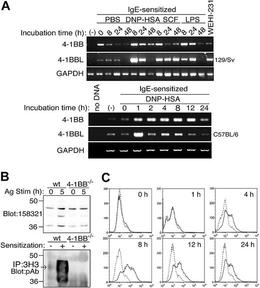
Given the costimulatory role of 4-1BB in T cells,10 we began to study the expression and function of this receptor in mouse mast cells. First, we examined its mRNA expression in resting or FcϵRI-stimulated mast cells. BMMCs derived from 129/SvJ mice were left unstimulated or incubated overnight with anti-DNP IgE 206. The IgE-sensitized cells were washed and incubated with phosphate-buffered saline (PBS), 10 ng/mL DNP23-HSA (the stimulation mode termed IgE + Ag), 100 ng/mL SCF, or 1 μg/mL lipopolysaccharide (LPS) for various periods. Semiquantitative RT-PCR analysis showed that 4-1BB mRNA is not detected in naive mast cells but induced dramatically after overnight sensitization with IgE (Figure 1A). Although its expression steadily decreased over the incubation period without additional stimulation, antigen stimulation induced a further increase in 4-1BB mRNA by 8 hours, and its levels decreased over the 48-hour incubation period. Similar fast induction and subsequent gradual decrease of 4-1BB mRNA expression was observed when 129/SvJ BMMCs were stimulated with 100 ng/mL SCF or 1 μg/mL LPS. Using semiquantitative RT-PCR as well as real-time PCR analyses with BMMCs from C57BL/6 mice, we observed 4-1BB mRNA induction on IgE + Ag stimulation and it peaked around 8 to 12 hours of stimulation (Figure 1A; data not shown).
We next examined the expression of 4-1BB protein in BMMCs. Immunoblotting of mast-cell lysates (100 μg) with anti-4-1BB mAb, 1583321, failed to demonstrate any definitive band specific to 4-1BB in IgE-sensitized C57BL/6 (wt) BMMCs (Figure 1B, top, lane 1). However, the same analysis showed the 40-kDa band of 4-1BB in wt, but not 4-1BB-/-, cells that had been stimulated with IgE + Ag for 5 hours (Figure 1B, top, lanes 2-4). Using 100 times more cell lysates (10 mg) for detection by immunoblotting of immune complexes precipitated with another anti-4-1BB mAb 3H3 with a polyclonal anti-4-1BB antibody (Alexis Biochemicals), we could show low but significant levels of 4-1BB protein in naive cells and much more robust expression in IgE-sensitized BMMCs (Figure 1B, bottom, lanes 1-2). The polyclonal antibody recognized a broad (37-45 kDa) band of 4-1BB with the strongest signal around 40 kDa. Flow cytometric analysis also demonstrated a dramatic induction of 4-1BB expression on the cell surface of IgE + Ag-stimulated wt, but not 4-1BB-/-, cells (Figure 1C; data not shown). Although the timing of peak surface expression of 4-1BB was similar to the expression of 4-1BB mRNA, it is noteworthy that surface expression was undetectably low in resting mast cells by flow cytometry. This low level may reflect low sensitivity of flow cytometry compared with immunoblotting. Alternatively, 4-1BB might be expressed only intracellularly, but not on the surface, in IgE-sensitized cells. Importantly, IL-3 treatment of starved mast cells induced strong surface expression on wt, but not 4-1BB-/-, mast cells (Figure S1, available at the Blood website; see the Supplemental Figures link at the top of the online article). Given the weekly refeeding of the cells with fresh IL-3-containing medium, it was interpreted that 4-1BB is expressed on the cell surface at least intermittently throughout bone marrow cell cultures that leads to the generation of mast cells. The above results confirmed those of Nakajima et al27 that ranked 4-1BB mRNA among the top 10 mRNAs whose expression is most highly induced by IgE + Ag.
Inducible expression of 4-1BB mRNA and protein in mouse mast cells. (A) BMMCs derived from 129/SvJ and C57BL/6 mice were left unstimulated (-) or incubated overnight with anti-DNP IgE 206. The IgE-sensitized cells were washed and incubated with PBS, 10 ng/mL DNP23-HSA, 100 ng/mL SCF, or 1 μg/mL LPS for the indicated periods. RNAs were isolated from these cells and subjected to RT-PCR for the expression of 4-1BB, 4-1BBL, and GAPDH mRNAs. (B) 4-1BB expression was directly analyzed by immunoblotting of lysates of wild-type (wt) and 4-1B-/- BMMCs with anti-4-1BB mAb clone 158321 (top). Alternatively, immune complexes precipitated with anti-4-1BB mAb clone 3H3 were analyzed by immunoblotting with anti-4-1BB polyclonal antibody (bottom). The broad (37-45 kDa) band of 4-1BB with the strongest signal around 40 kDa in both unsensitized and IgE-sensitized wt, but not 4-1B-/-, cells is indicated by an arrow. (C) C57BL/6 BMMCs were sensitized with IgE and stimulated with antigen for the indicated periods as described in panel A. Flow cytometric analysis of surface expression of 4-1BB was performed. Continuous lines represent 4-1BB expression and broken lines isotype control. Results shown in panels A and B are representative of 3 independent experiments and those in panel C are representative of 5 similar experiments.
Inducible expression of 4-1BB mRNA and protein in mouse mast cells. (A) BMMCs derived from 129/SvJ and C57BL/6 mice were left unstimulated (-) or incubated overnight with anti-DNP IgE 206. The IgE-sensitized cells were washed and incubated with PBS, 10 ng/mL DNP23-HSA, 100 ng/mL SCF, or 1 μg/mL LPS for the indicated periods. RNAs were isolated from these cells and subjected to RT-PCR for the expression of 4-1BB, 4-1BBL, and GAPDH mRNAs. (B) 4-1BB expression was directly analyzed by immunoblotting of lysates of wild-type (wt) and 4-1B-/- BMMCs with anti-4-1BB mAb clone 158321 (top). Alternatively, immune complexes precipitated with anti-4-1BB mAb clone 3H3 were analyzed by immunoblotting with anti-4-1BB polyclonal antibody (bottom). The broad (37-45 kDa) band of 4-1BB with the strongest signal around 40 kDa in both unsensitized and IgE-sensitized wt, but not 4-1B-/-, cells is indicated by an arrow. (C) C57BL/6 BMMCs were sensitized with IgE and stimulated with antigen for the indicated periods as described in panel A. Flow cytometric analysis of surface expression of 4-1BB was performed. Continuous lines represent 4-1BB expression and broken lines isotype control. Results shown in panels A and B are representative of 3 independent experiments and those in panel C are representative of 5 similar experiments.
Agonistic anti-4-1BB mAbs augment IgE + Ag-induced production and secretion of cytokines
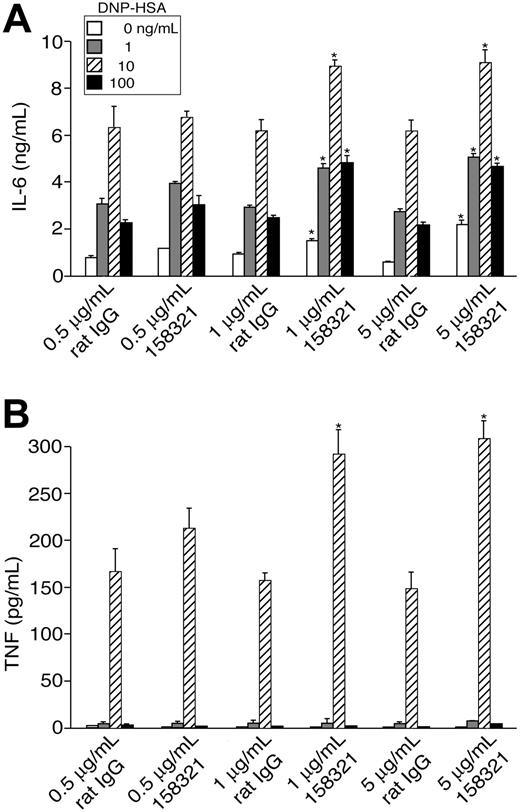
We next tested whether stimulation of 4-1BB affects the effector function of mast cells. We first tested whether agonistic 4-1BB mAbs can induce mast-cell activation. Incubation with agonistic 4-1BB mAbs in the presence or absence of secondary anti-rat IgG antibody induced no significant histamine release or secretion of IL-6, IL-13, or TNF (data not shown). Furthermore, when IgE-sensitized C57BL/6 BMMCs were stimulated with antigen together with an agonistic 4-1BB mAb (with or without secondary anti-rat IgG antibody), no significant effects of the 4-1BB mAbs were observed on the production and secretion of IL-6, IL-13, or TNF measured after 8 hours (data not shown). This may be reasonable, as surface expression of 4-1BB is very low at a resting stage and induced later. Given the up-regulation of 4-1BB surface expression after IgE + Ag stimulation, we stimulated IgE-sensitized C57BL/6 BMMCs with antigen for 6 hours, washed, and restimulated them with different concentrations of antigen in the presence or absence of 0.5, 1, or 5 μg/mL agonistic 4-1BB mAbs. Restimulation with antigen in the absence of 4-1BB mAbs (without secondary antibody) induced the production and secretion of IL-6, IL-13, and TNF in a dose-dependent manner (Figure 2; data not shown). The presence of 1 or 5 μg/mL agonistic 4-1BB mAb significantly enhanced cytokine production/secretion. Two different 4-1BB mAbs, clones 158321 (Figure 2) and 3H3 (data not shown), exhibited similar efficiency at enhancing IgE + Ag-induced cytokine production/secretion. It is noteworthy that TNF production induced by antigen stimulation in the cells that have been prestimulated by IgE + Ag exhibits a very narrow sensitivity to antigen (DNP23-HSA) levels, unlike that induced by antigen stimulation in regular IgE-sensitized mast cells (eg, Figure 4C). The specificity of the 4-1BB mAbs effects was confirmed by the lack of the effects in 4-1BB-/- mast cells (data not shown). These results suggested that 4-1BB plays a costimulatory function in FcϵRI-stimulated mast cells similar to TCR-stimulated T cells.
Agonistic anti-4-1BB antibodies enhance IgE + Ag-induced cytokine production and secretion. IgE-sensitized BMMCs were first stimulated with antigen for 6 hours, washed, and restimulated for 12 hours with different concentrations of antigen in the presence or absence of 0.5, 1, or 5 μg/mL agonistic 4-1BB mAb (clone 158321) or control rat IgG. IL-6 and TNF secreted into culture media were quantified by ELISA. *P < .05 (versus control antibody, Student t test). Results (mean ± SD) shown are representative of 3 similar experiments.
Agonistic anti-4-1BB antibodies enhance IgE + Ag-induced cytokine production and secretion. IgE-sensitized BMMCs were first stimulated with antigen for 6 hours, washed, and restimulated for 12 hours with different concentrations of antigen in the presence or absence of 0.5, 1, or 5 μg/mL agonistic 4-1BB mAb (clone 158321) or control rat IgG. IL-6 and TNF secreted into culture media were quantified by ELISA. *P < .05 (versus control antibody, Student t test). Results (mean ± SD) shown are representative of 3 similar experiments.
We next tested whether agonistic 4-1BB mAbs affect mast-cell proliferation. Although both IL-3 and SCF could induce surface expression of 4-1BB in wt, but not 4-1BB-/-, mast cells (Figure S1), agonistic 4-1BB mAbs failed to affect mast-cell proliferation induced by IL-3 or SCF (data not shown). Incubation with agonistic 4-1BB mAbs alone also did not influence mast-cell proliferation (data not shown).
4-1BB-deficient mast cells exhibit lower growth responses to IL-3 and SCF
In light of the costimulatory potential of 4-1BB in mast-cell activation, we studied roles of 4-1BB in the development and function of mast cells using 4-1BB-/- mice.24 When bone marrow cells from the mutant mice were cultured in IL-3-containing medium for 4 to 6 weeks, immature mast cells were generated with purity as high as that with wt mice: greater than 95% resulting live cells were c-Kit+ and FcϵRI+ in flow cytometric analysis (Figure 3A). Toluidine blue staining of cytospin preparations revealed no discernible differences between wt and 4-1BB-/- cells (Figure 3B). Cellular contents of histamine, a major constituent of secretory granules, were similar between wt and 4-1BB-/- cells (data not shown). We also did not see differences in these measurements on bone marrow cells that had been cultured with IL-3 and SCF for 4 to 5 weeks (data not shown). Therefore, we concluded that 4-1BB deficiency does not substantially affect in vitro mast-cell differentiation.
However, we noticed that the growth of 4-1BB-/- mast cells is much slower during cultures in IL-3 (Figure 3C). This could be due to either reduced proliferation or enhanced cell death in 4-1BB-/- cells. Consistent with the former possibility, DNA synthesis induced by IL-3 or SCF was reduced in 4-1BB-/- cells compared with wt cells (Figure 3D). However, apoptosis induced by growth factor withdrawal was similar between wt and 4-1BB-/- cells (Figure 3E). Together with our observations that IL-3 and SCF induce 4-1BB expression on BMMC surface (Figure S1), these results demonstrate that 4-1BB is required for maximal proliferation of mast cells induced by IL-3 and SCF.
4-1BB deficiency results in reduced degranulation and cytokine production on FcϵRI stimulation
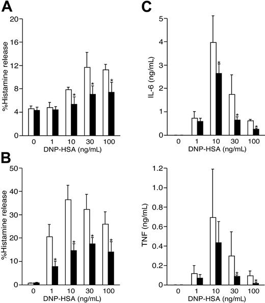
Given the 4-1BB up-regulation after IgE + Ag stimulation, we stimulated IgE-sensitized wt and 4-1BB-/- BMMCs with antigen for 5 hours, washed, and restimulated the cells with different concentrations of antigen. Under these conditions that entailed desensitization of the cells, even wt cells released histamine at low levels: only approximately 12% of the total cellular content of histamine was released at an optimal concentration of 30 ng/mL DNP23-HSA (Figure 4A). By contrast, 4-1BB-/- cells released about 60% of the wt level. Surprisingly, even under nondesensitizing conditions (ie, without prior antigen stimulation), histamine release was 50% to 65% reduced in IgE + Ag-stimulated 4-1BB-/- cells compared with identically stimulated wt cells (Figure 4B).
We also measured cytokines produced and secreted on IgE + Ag stimulation. Under nondesensitizing conditions, amounts of secreted IL-6 and TNF were significantly lower in 4-1BB-/- cells (Figure 4C). IL-13 levels were also lower in 4-1BB-/- cells, but the difference did not reach statistical significance. Semiquantitative RT-PCR analysis demonstrated that reductions of these cytokines in 4-1BB-/- cells were seen at mRNA levels (Figure S2). Therefore, these results demonstrated the ability of 4-1BB to costimulate mast cells with FcϵRI.
Cell-surface phenotypic, morphologic, and growth properties of 4-1BB-/- mast cells. (A) Wt and 4-1BB-/- BMMCs were generated from bone marrow cells in IL-3-containing culture medium. Cohorts of 3 to 5 mice in each group were used to generate BMMCs. Results shown in this figure are representative of at least 3 independent experiments. Surface expression of FcϵRI and c-Kit was analyzed by flow cytometry. (B) Toluidine blue-stained wt and 4-1BB-/- mast cells. Images of the cells at a magnification of × 1000 were acquired with a Nikon Optiphot photomicroscope (Nikon, Tokyo, Japan) with a Plan Apo 100 ×/1.40 NA oil objective, using a DVC-1310 camera (DVC, West Austin, TX) and DVCC-View v2.2 software. (C) Growth curves of bone marrow cells derived from wt (□) and 4-1BB-/- (▪) mice in IL-3-containing medium. Each growth curve was generated from cultures in 6 flasks, each containing bone marrow cells derived from 1 femur (left or right) of 3 wt or 3 mutant mice. Similar results were obtained in 3 independent culture series. *P < .05, **P < .01, ***P < .005 (versus wt control, Student t test). (D) Wt (□) and 4-1BB-/- (▪) mast cells that had been deprived of growth factors for 8 hours were incubated with the indicated concentrations of IL-3 and SCF for 12 hours. DNA synthesis was measured by [3 H]thymidine incorporation during the last 6 hours of culture. *P < .05 (versus wt, Student t test). (E) Wt (□) and 4-1BB-/- (▪) mast cells were incubated without IL-3 or other growth factors for the indicated periods. Live cells were quantified by flow cytometry of annexin V- and propidium iodide-stained cells. Annexin V-negative and propidium iodide-negative cells are plotted as function of incubation time. For panels C-E. Values shown are mean ± SD.
Cell-surface phenotypic, morphologic, and growth properties of 4-1BB-/- mast cells. (A) Wt and 4-1BB-/- BMMCs were generated from bone marrow cells in IL-3-containing culture medium. Cohorts of 3 to 5 mice in each group were used to generate BMMCs. Results shown in this figure are representative of at least 3 independent experiments. Surface expression of FcϵRI and c-Kit was analyzed by flow cytometry. (B) Toluidine blue-stained wt and 4-1BB-/- mast cells. Images of the cells at a magnification of × 1000 were acquired with a Nikon Optiphot photomicroscope (Nikon, Tokyo, Japan) with a Plan Apo 100 ×/1.40 NA oil objective, using a DVC-1310 camera (DVC, West Austin, TX) and DVCC-View v2.2 software. (C) Growth curves of bone marrow cells derived from wt (□) and 4-1BB-/- (▪) mice in IL-3-containing medium. Each growth curve was generated from cultures in 6 flasks, each containing bone marrow cells derived from 1 femur (left or right) of 3 wt or 3 mutant mice. Similar results were obtained in 3 independent culture series. *P < .05, **P < .01, ***P < .005 (versus wt control, Student t test). (D) Wt (□) and 4-1BB-/- (▪) mast cells that had been deprived of growth factors for 8 hours were incubated with the indicated concentrations of IL-3 and SCF for 12 hours. DNA synthesis was measured by [3 H]thymidine incorporation during the last 6 hours of culture. *P < .05 (versus wt, Student t test). (E) Wt (□) and 4-1BB-/- (▪) mast cells were incubated without IL-3 or other growth factors for the indicated periods. Live cells were quantified by flow cytometry of annexin V- and propidium iodide-stained cells. Annexin V-negative and propidium iodide-negative cells are plotted as function of incubation time. For panels C-E. Values shown are mean ± SD.
4-1BB deficiency results in reduced Ca2+ response on FcϵRI stimulation
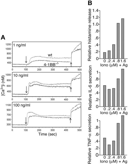
Optimal degranulation and cytokine production require adequate Ca2+ response.28,29 To test whether an impairment in Ca2+ response is responsible for the impaired mast-cell activation, we measured intracellular concentrations of Ca2+, [Ca2+]i, in IgE + Ag-stimulated wt and 4-1BB-/- cells. Consistent with the degranulation and cytokine data, [Ca2+]i was substantially reduced in 4-1BB-/- cells particularly at low concentrations of antigen (Figure 5A). The role of Ca2+ in IgE + Ag-induced degranulation and cytokine production was confirmed by full restoration of histamine release and production of IL-6 and TNF in IgE + Ag-stimulated 4-1BB-/- cells by adding increasing concentrations of ionomycin (Figure 5B). However, it is noteworthy that there is some discordance between Ca2+ levels and histamine release depending on antigen doses. Although Ca2+ flux is required for optimal histamine release, there is not necessarily perfect correlation between Ca2+ levels and histamine release. Thus, this is exemplified by the phenotype of lyn-/- mast cells, whereby Ca2+ flux is dramatically reduced despite robust histamine release.30 We have also experienced a small range of experiment-to-experiment variability in histamine release and Ca2+ concentrations even when we used the same inbred mouse strain.
Defective Lyn/Btk/PLC-γ2 pathway leads to defective Ca2+ response
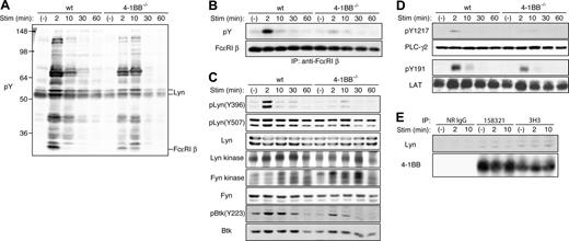
To dissect how 4-1BB deficiency leads to the defective Ca2+ response, we analyzed membrane-proximal signaling events. Immunoblotting with anti-phosphotyrosine mAb revealed some differences between IgE + Ag-stimulated wt and 4-1BB-/- cells (Figure 6A). For example, tyrosine phosphorylation of 56- and 53-kDa bands containing p56lyn and p53lyn was reduced and peaked more slowly in 4-1BB-/- cells; tyrosine phosphorylation of 30-kDa band containing FcϵRI β subunit was reduced in 4-1BB-/- cells. Indeed, probing of cell lysates with phosphospecific antibodies revealed a reduction in phosphorylation of Tyr396 in the activation loop of Lyn, whereas that of Tyr507 in the negative regulatory C-terminal region of Lyn was constitutive and not much affected by 4-1BB deficiency (Figure 6C). Consistent with these results, enzymatic activity of immunoprecipitated Lyn toward an exogenous substrate, enolase, was reduced in 4-1BB-/- cells particularly at early time points. Immunoblotting of immunoprecipitated FcϵRI β subunit confirmed a dramatic reduction in tyrosine phosphorylation of FcϵRI β subunit in 4-1BB-/- cells (Figure 6B), similar to Lyn-deficient mast cells.30 Similar to Lyn-deficient mast cells,31 enzymatic activity of Fyn and tyrosine phosphorylation of Btk were increased and decreased, respectively, in 4-1BB-/- cells (Figure 6C). In agreement with the previous studies showing that phosphorylation and activation of PLC-γ2 are regulated by Btk,32 tyrosine phosphorylation of PLC-γ2 was reduced in 4-1BB-/- cells (Figure 6D). Furthermore, phosphorylation of Tyr191 in LAT, an adaptor molecule involved in the recruitment of PLC-γ, was also reduced in 4-1BB-/- cells (Figure 6D). Therefore, it appears that the established pathway composed of Lyn/Btk/PLC-γ2 is down-regulated, leading to the defective Ca2+ response in 4-1BB-/- cells.
Reduced degranulation and cytokine production/secretion in FcϵRI-stimulated 4-1BB-/- mast cells. (A) Wt (□) and 4-1BB-/- (▪) BMMCs that had been sensitized overnight with 0.5 μg/mL IgE were first stimulated with antigen for 5 hours, washed, and restimulated for 45 minutes with different concentrations of antigen. Histamine released into culture media was measured. Results shown are representative of 2 experiments. (B) Wt and 4-1BB-/- BMMCs that had been sensitized overnight with IgE were stimulated for 45 minutes with different concentrations of antigen. Histamine released into culture media was measured. Results shown are representative of 2 experiments. (C) Wt and 4-1BB-/- BMMCs that had been sensitized overnight with IgE were stimulated for 8 hours with different concentrations of antigen. IL-6 and TNF secreted into culture media was measured. *P < .05 (versus wt control, Student t test). Results (mean ± SD) shown are representative of 5 experiments.
Reduced degranulation and cytokine production/secretion in FcϵRI-stimulated 4-1BB-/- mast cells. (A) Wt (□) and 4-1BB-/- (▪) BMMCs that had been sensitized overnight with 0.5 μg/mL IgE were first stimulated with antigen for 5 hours, washed, and restimulated for 45 minutes with different concentrations of antigen. Histamine released into culture media was measured. Results shown are representative of 2 experiments. (B) Wt and 4-1BB-/- BMMCs that had been sensitized overnight with IgE were stimulated for 45 minutes with different concentrations of antigen. Histamine released into culture media was measured. Results shown are representative of 2 experiments. (C) Wt and 4-1BB-/- BMMCs that had been sensitized overnight with IgE were stimulated for 8 hours with different concentrations of antigen. IL-6 and TNF secreted into culture media was measured. *P < .05 (versus wt control, Student t test). Results (mean ± SD) shown are representative of 5 experiments.
Defective Ca2+ flux in FcϵRI-stimulated 4-1BB-/- mast cells. (A) Wt and 4-1BB-/- BMMCs that had been sensitized overnight with IgE were loaded with Indo-1 am. Continuous monitoring of the fluorescence ratio (525:405 nm) was performed on washed cells using a flow cytometer. Baseline fluorescence ratios were collected for 100 seconds before different concentrations of DNP23-HSA was added (indicated by ↓). Ionomycin was added to all samples after 300 seconds of observation of calcium mobilization by IgE + Ag stimulation (indicated by ↑). Results shown are representative of 3 experiments. (B) Wt and 4-1BB-/- BMMCs that had been sensitized overnight with IgE were stimulated by the indicated concentrations of ionomycin alone or ionomycin and DNP23-HSA for 45 minutes (histamine release) or 8 hours (cytokine production). Ratios of histamine release and cytokine production in 4-1BB-/- versus wt BMMCs are plotted as function of ionomycin concentrations. Results shown are representative of 2 experiments.
Defective Ca2+ flux in FcϵRI-stimulated 4-1BB-/- mast cells. (A) Wt and 4-1BB-/- BMMCs that had been sensitized overnight with IgE were loaded with Indo-1 am. Continuous monitoring of the fluorescence ratio (525:405 nm) was performed on washed cells using a flow cytometer. Baseline fluorescence ratios were collected for 100 seconds before different concentrations of DNP23-HSA was added (indicated by ↓). Ionomycin was added to all samples after 300 seconds of observation of calcium mobilization by IgE + Ag stimulation (indicated by ↑). Results shown are representative of 3 experiments. (B) Wt and 4-1BB-/- BMMCs that had been sensitized overnight with IgE were stimulated by the indicated concentrations of ionomycin alone or ionomycin and DNP23-HSA for 45 minutes (histamine release) or 8 hours (cytokine production). Ratios of histamine release and cytokine production in 4-1BB-/- versus wt BMMCs are plotted as function of ionomycin concentrations. Results shown are representative of 2 experiments.
4-1BB has been observed to interact with Lck, another Src family PTK, in T cells,33 suggesting the possibility that 4-1BB may interact with Lyn. Immunoprecipitation of 4-1BB with anti-4-1BB mAbs, both 158321 and 3H3, coprecipitated Lyn, and this association was irrespective of FcϵRI stimulation (Figure 6E), indicating that 4-1BB and Lyn are constitutively associated. Although both FcϵRI and 4-1BB could coimmunoprecipitate Lyn, FcϵRI could not be coimmunoprecipitated with 4-1BB (data not shown).
Characterization of 4-1BBL expression and function in mast cells
We next investigated whether 4-1BBL is expressed in mast cells. Similar to 4-1BB mRNA, 4-1BBL mRNA was not expressed in naive 129/SvJ BMMCs, but low basal expression was seen in C57BL/6 BMMCs (Figure 1A). 4-1BBL expression was induced by IgE + Ag stimulation in both 129/SvJ and C57BL/6 BMMCs. Interestingly, 4-1BBL mRNA was induced in different kinetics with a faster peak at 1 hour in IgE-Ag-stimulated C57BL/6 mast cells than that of 4-1BB mRNA (Figure 1A). SCF and LPS also induced 4-1BBL mRNA expression. Surface expression of 4-1BBL was also detected in IgE + Ag-stimulated BMMCs by flow cytometry (data not shown).
Unlike 4-1BB-/- bone marrow cells (Figure 3C), growth of bone marrow cells derived from 4-1BBL-/- mice25 was only slightly (with no statistical significance) reduced compared with wt cells (Figure S3). Proliferation induced by IL-3 or SCF was slightly lower in 4-1BB-/- cells, but this reduction did not reach statistical significance. Rates of apoptosis induced by growth factor deprivation were also similar between wt and 4-1BBL-/- BMMCs (Figure S3).
Degranulation was hardly affected, whereas cytokine production/secretion was significantly reduced in IgE + Ag-stimulated 4-1BBL-/- mast cells (Figure 7A-B), like 4-1BB-/- cells. Ca2+ flux was also reduced in IgE + Ag-stimulated 4-1BBL-/- mast cells. However, these reductions were to lower extents compared with those in 4-1BB-/- cells (Figure 7C).
Defective Lyn/Btk/PLC-γ2 pathway in 4-1BB-/- mast cells. (A) Wt and 4-1BB-/- BMMCs that had been sensitized overnight with anti-DNP IgE were stimulated with 100 ng/mL DNP23-HSA for the indicated periods. Cell lysates were directly analyzed by SDS-PAGE followed by immunoblotting with the indicated antibodies (A,C-D) or first immunoprecipitated with specific antibodies and immune complexes analyzed by SDS-PAGE and immunoblotting (B,E). In vitro kinase assays were also performed on immune complexes of Lyn and Fyn (C). (E) IgE-sensitized wt BMMCs were stimulated with 10 ng/mL DNP23-HSA for 6 hours to induce 4-1BB expression, then restimulated with 100 ng/mL DNP23-HSA for the indicated periods. Results shown are representative of at least 2 experiments.
Defective Lyn/Btk/PLC-γ2 pathway in 4-1BB-/- mast cells. (A) Wt and 4-1BB-/- BMMCs that had been sensitized overnight with anti-DNP IgE were stimulated with 100 ng/mL DNP23-HSA for the indicated periods. Cell lysates were directly analyzed by SDS-PAGE followed by immunoblotting with the indicated antibodies (A,C-D) or first immunoprecipitated with specific antibodies and immune complexes analyzed by SDS-PAGE and immunoblotting (B,E). In vitro kinase assays were also performed on immune complexes of Lyn and Fyn (C). (E) IgE-sensitized wt BMMCs were stimulated with 10 ng/mL DNP23-HSA for 6 hours to induce 4-1BB expression, then restimulated with 100 ng/mL DNP23-HSA for the indicated periods. Results shown are representative of at least 2 experiments.
Reduced degranulation and cytokine production/secretion in FcϵRI-stimulated 4-1BBL-/- mast cells. (A) Wt (□) and 4-1BB-/- (▪) BMMCs that had been sensitized overnight with IgE were stimulated for 45 minutes with different concentrations of antigen. Histamine released into culture media was measured. (B) Wt and 4-1BB-/- BMMCs that had been sensitized overnight with IgE were stimulated for 8 hours with different concentrations of antigen. IL-6 and TNF secreted into culture media was measured. *P < .05 (versus wt control, Student t test). (C) Measurement of [Ca2+]i was performed on anti-DNP IgE-sensitized wt and 4-1BBL-/- mast cells, as described in Figure 5. Results (mean ± SD) shown are representative of at least 2 experiments.
Reduced degranulation and cytokine production/secretion in FcϵRI-stimulated 4-1BBL-/- mast cells. (A) Wt (□) and 4-1BB-/- (▪) BMMCs that had been sensitized overnight with IgE were stimulated for 45 minutes with different concentrations of antigen. Histamine released into culture media was measured. (B) Wt and 4-1BB-/- BMMCs that had been sensitized overnight with IgE were stimulated for 8 hours with different concentrations of antigen. IL-6 and TNF secreted into culture media was measured. *P < .05 (versus wt control, Student t test). (C) Measurement of [Ca2+]i was performed on anti-DNP IgE-sensitized wt and 4-1BBL-/- mast cells, as described in Figure 5. Results (mean ± SD) shown are representative of at least 2 experiments.
Discussion
This study demonstrates that the 4-1BB/4-1BBL system acts as a positive signaling pathway for mast cells. 4-1BB expression, which is very low intracellularly or undetectable by flow cytometry in naive mast cells, is induced by FcϵRI (low-level induction by incubation with IgE and high-level induction by IgE + Ag stimulation), IL-3, SCF, and LPS stimulation. Costimulation of 4-1BB with FcϵRI using agonistic mAbs enhances mast-cell-effector functions, particularly production of cytokines. Roles of 4-1BB in FcϵRI-induced mast-cell activation are further demonstrated by the reduced degranulation and the reduced production of multiple cytokines in 4-1BB-/- cells. Very low or undetectable expression of 4-1BB protein on the surface of naive mast cells suggests that these defective phenotypes in 4-1BB-/- cells might reflect a function of intracellular 4-1BB. Mechanistic analysis suggests that the defective Ca2+ signaling pathway composed of Lyn, Btk, and PLC-γ2 is involved in these events. Indeed, degranulation and cytokine production in 4-1BB-/- mast cells were fully restored by supplementing Ca2+ signal with ionomycin. In addition, 4-1BB was required for optimal proliferation of mast cells induced by IL-3 and SCF.
The signaling defects found in FcϵRI-stimulated 4-1BB-/- mast cells can be accounted for largely by the reduced Lyn activity. Importantly, Lyn was shown to be constitutively associated with 4-1BB. This situation is similar to 4-1BB/Lck interactions through the Cys-Arg-Cys-Pro sequence in the cytoplasmic region of 4-1BB.33 This Cys-Xaa-Cys-Pro motif (Xaa indicates any amino acid residue) is present in CD4 and CD8 molecules and used for interactions with Lck. It is likely that Lyn, the most prominent Src family PTK expressed in mast cells, might interact with this motif of 4-1BB in mast cells. Although signaling events triggered by the engagement of 4-1BB as a TNFR member are presumed to involve activation of nuclear factor κB and c-Jun N-terminal kinase 1 and 2, none of these events were substantially impaired in FcϵRI-stimulated 4-1BB-/- mast cells (data not shown).
4-1BBL mRNA expression was also induced by FcϵRI, SCF, and LPS stimulation in mast cells. Similar to 4-1BB-/- cells, 4-1BBL-/- cells exhibited the reduced degranulation and cytokine production as well as the defective Ca2+ responses on FcϵRI stimulation, although these defects were milder in 4-1BBL-/- cells than in 4-1BB-/- cells. These results are consistent with the notion that 4-1BB's role in costimulation with FcϵRI is executed at least in part by 4-1BB/4-1BBL interactions. Unfortunately, our efforts to try to assess the extent of the role of 4-1BB/4-1BBL interactions in FcϵRI-induced degranulation and cytokine production were hampered by the inability of anti-4-1BBL mAb (TKS-1) to inhibit the binding interactions in mast cells; binding of 4-1BB-Fc fusion protein to wt or 4-1BB-/- mast cells was not affected by TKS-1, unlike T cells (data not shown). However, 4-1BBL, unlike 4-1BB, was dispensable for proliferation induced by IL-3 or SCF. These results raise several possibilities, including one that 4-1BB promotes proliferation through a mechanism distinct from 4-1BBL.
Antigen-specific T cells play crucial roles in allergic and autoimmune diseases and in the host defense against various pathogens. Mast cells can occur in close proximity to T cells at sites of allergic and other inflammatory reactions. In vitro studies show that mast cells can directly interact with a TCR to induce antigen-specific clonal expansion of T cells.34,35 Because major histocompatibility complex (MHC) class I molecules are expressed on mouse BMMCs and human mast cells. Mouse BMMCs in vitro can stimulate bacterial antigen-specific T cells by presenting bacterial antigens in the context of MHC class I.36 MHC class II, not expressed on resting mast cells, can be expressed on mast cells that have been stimulated by TNF, IFN-γ, or LPS.35 Granulocyte-macrophage colony-stimulating factor can enhance expression of CD80 and CD86 on mouse BMMCs.37 Human mast cells can express OX40 ligand and 4-1BBL.38,39 Our data, showing that mast cells stimulated with IgE + Ag, SCF, or LPS induce 4-1BBL expression, further support the potential of activated mast cells to induce T-cell activation by direct costimulation via 4-1BB/4-1BBL interactions.
To our knowledge this study is the first to report costimulatory capacity of 4-1BB with FcϵRI-stimulated mast cells. Degranulation and cytokine production are enhanced by the presence of 4-1BB, and the enhanced activities are mediated at least in part by 4-1BB/4-1BBL interactions. 4-1BB signaling appears to depend on its associated Lyn and the regulation of the Lyn/Btk/PLC-γ/Ca2+ pathway. Therefore, our study raises the possibility that 4-1BB and its interactions with its ligands could be a therapeutic target for the treatment of allergic diseases and other IgE-dependent diseases.
Prepublished online as Blood First Edition Paper, August 25, 2005; DOI 10.1182/blood-2005-04-1358.
Supported in part by grants from the National Institutes of Health (AI50209 and AI38348) (T.K.), (AI42944) (M.C.), (R37 AI33068 and CA69381) (C.F.W.) and by the SRC Fund to Immunomodulation Research Center at the University of Ulsan from Korea Science and Engineering Foundation (KOSEF) (B.S.K.).
H.N. performed most of the experiments; S.-W.L. assisted H.N. to design and perform the experiments; H.H. performed the Ca2+ experiments; K.G.P. and T.K. performed some mRNA experiments; M.M.-Y. measured histamine; Y.K. assisted H.N. to design the experiments and analyze the results; R.S.M. and B.S.K. provided key reagents; C.F.W. and M.C. provided reagents and helped to design the experiments and to write the manuscript; T.K. helped H.N. to design the experiments and analyze the results and write the manuscript.
The online version of the article contains a data supplement.
The publication costs of this article were defrayed in part by page charge payment. Therefore, and solely to indicate this fact, this article is hereby marked “advertisement” in accordance with 18 U.S.C. section 1734.
We thank Kirin Brewery Co, Ltd, for their kind donation of recombinant growth factors. This is Publication 697 from the La Jolla Institute for Allergy and Immunology. The authors have no conflicting financial interests.

![Figure 3. Cell-surface phenotypic, morphologic, and growth properties of 4-1BB-/- mast cells. (A) Wt and 4-1BB-/- BMMCs were generated from bone marrow cells in IL-3-containing culture medium. Cohorts of 3 to 5 mice in each group were used to generate BMMCs. Results shown in this figure are representative of at least 3 independent experiments. Surface expression of FcϵRI and c-Kit was analyzed by flow cytometry. (B) Toluidine blue-stained wt and 4-1BB-/- mast cells. Images of the cells at a magnification of × 1000 were acquired with a Nikon Optiphot photomicroscope (Nikon, Tokyo, Japan) with a Plan Apo 100 ×/1.40 NA oil objective, using a DVC-1310 camera (DVC, West Austin, TX) and DVCC-View v2.2 software. (C) Growth curves of bone marrow cells derived from wt (□) and 4-1BB-/- (▪) mice in IL-3-containing medium. Each growth curve was generated from cultures in 6 flasks, each containing bone marrow cells derived from 1 femur (left or right) of 3 wt or 3 mutant mice. Similar results were obtained in 3 independent culture series. *P < .05, **P < .01, ***P < .005 (versus wt control, Student t test). (D) Wt (□) and 4-1BB-/- (▪) mast cells that had been deprived of growth factors for 8 hours were incubated with the indicated concentrations of IL-3 and SCF for 12 hours. DNA synthesis was measured by [3H]thymidine incorporation during the last 6 hours of culture. *P < .05 (versus wt, Student t test). (E) Wt (□) and 4-1BB-/- (▪) mast cells were incubated without IL-3 or other growth factors for the indicated periods. Live cells were quantified by flow cytometry of annexin V- and propidium iodide-stained cells. Annexin V-negative and propidium iodide-negative cells are plotted as function of incubation time. For panels C-E. Values shown are mean ± SD.](https://ash.silverchair-cdn.com/ash/content_public/journal/blood/106/13/10.1182_blood-2005-04-1358/2/m_zh80240588230003.jpeg?Expires=1765925053&Signature=40aaHii28lWYmwb60CbLUXizsL-C-l3O0HHSCVQ6oQkYmmR-PbCCYGcSDfsB-byhhkSiVM1BW0mHHpM~Wu2QmD3jKL7Aps2OJjx5VD-YEZc-HaGzo1lI2Ws1udlv7S0FFqwHsWl3UBrSN7UlRWojzV~ddu8GRVaZKRZCyTBIgztvXI3I6hDc7nSej~cuAkjK-B5jIB5SkRixCUPaupYtcfswVcdwzV8KkpwFBSlJOKlXWgp9L-TKUDB92~V49UT-eVJUanXWvLPuRpF0LvueNeCMPr3kz2v~6qBW6XyiHL4hM2DQmdVtVr5A8SPlAeC15TtVqXaQP0RAD4dwwMhCjg__&Key-Pair-Id=APKAIE5G5CRDK6RD3PGA)
![Figure 7. Reduced degranulation and cytokine production/secretion in FcϵRI-stimulated 4-1BBL-/- mast cells. (A) Wt (□) and 4-1BB-/- (▪) BMMCs that had been sensitized overnight with IgE were stimulated for 45 minutes with different concentrations of antigen. Histamine released into culture media was measured. (B) Wt and 4-1BB-/- BMMCs that had been sensitized overnight with IgE were stimulated for 8 hours with different concentrations of antigen. IL-6 and TNF secreted into culture media was measured. *P < .05 (versus wt control, Student t test). (C) Measurement of [Ca2+]i was performed on anti-DNP IgE-sensitized wt and 4-1BBL-/- mast cells, as described in Figure 5. Results (mean ± SD) shown are representative of at least 2 experiments.](https://ash.silverchair-cdn.com/ash/content_public/journal/blood/106/13/10.1182_blood-2005-04-1358/2/m_zh80240588230007.jpeg?Expires=1765925053&Signature=faWtNFMc1C64DddWAd12N8Kz83wkAIs2OhNBJlBRrcvafjIkrD3S48k3Wzh5maCzcm-6Mpq4VO8XKzqazyrqbzyPRBhRotVpLr5ePQYlnd5GyNJsRPszQQ8aKm07rnheWDbqQD~8SNINWzcxZSsGi5pZgR2jllQoKFittSLMT1XeH55y45bqaPDSKTOPbwIqRm9Db3T0uvdAJTkYHE~k2toQUiOXPCftdJF-YlwGZCNxJ4eXLWI5B9C7LFyqUkAidEJB5Bt71H2wQTMGZgDW2Qu9IfxcrhPyGH4ecHVVr9DBcwpe328zralelgp4YXmnqY2GQVwpXpFHSMQSLsu1dQ__&Key-Pair-Id=APKAIE5G5CRDK6RD3PGA)
This feature is available to Subscribers Only
Sign In or Create an Account Close Modal