Abstract
Mutations resulting in diminished activity of the dimeric enzyme ferrochelatase are a prerequisite for the inherited disorder erythropoietic protoporphyria (EPP). Patients with clinical EPP have only 10% to 30% of normal levels of ferrochelatase activity, and although many patients with EPP have one mutant allele and one “low-expression” normal allele, the possibility remains that, for some, low ferrochelatase activity may result from an EPP mutation that has an impact on both subunits of the wild-type/mutant heterodimer. Here we present data for 12 ferrochelatase wild-type/EPP mutant heterodimers showing that some mutations result in heterodimers with the residual activity anticipated from individual constituents, whereas others result in heterodimers with significantly lower activity than would be predicted. Although the data do not allow an a priori prediction of heterodimeric residual activity based solely on the in vitro activity of EPP homodimers or the position of the mutated residue within ferrochelatase, mutations that affect the dimer interface or [2Fe-2S] cluster have a significantly greater impact on residual activity than would be predicted. These data suggest that some EPP mutations may result in clinically overt EPP in the absence of a low-expression, wild-type allele; this is of potential significance for genetic counseling of patients with EPP.
Introduction
Erythropoietic protoporphyria (EPP) is an inherited disease that may result when there is a defect in one or both alleles of the gene coding for ferrochelatase (protoheme ferrolyase, E.C. 4.99.1.1), the terminal enzyme in the heme synthetic pathway that catalyzes the insertion of ferrous iron into protoporphyrin IX to form protoheme IX (heme).1-4 The most common clinical symptom of the disease is a painful dermatologic photosensitivity that is attributable to the accumulation of protoporphyrin IX in the skin and the subsequent generation of tissue-damaging free radicals on exposure to visible light. Although the disease is generally categorized as a mild photosensitivity, in approximately 5% of patients the excess protoporphyrin drastically affects the solubility of bile, leading to the formation of gallstones and eventually to marked cholestasis, which may result in cirrhosis and liver failure.5,6 Although the gene defect results in lowered ferrochelatase activity in all tissues, it is in the erythrocyte precursors in bone marrow that quantitatively the largest amount of protoporphyrin is produced. Given that the progeny of these cells contain the largest amount of organismal heme and have relatively short life spans, it is apparent that EPP is an erythroid rather than a hepatic porphyria. Ferrochelatase activity in the livers and kidneys of patients with EPP is diminished, but only under special conditions may there be an impact on the metabolic function of the liver.5,6 Indeed, even in patients with EPP in whom liver cirrhosis necessitates transplantation, the damage is caused by biliary occlusion composed of free protoporphyrin crystals that come from circulating erythrocytes and that are not produced by the liver itself. This results in the unfortunate sequelae of potential repeat liver transplantations because the healthy donor liver may be damaged in a fashion similar to the original EPP liver.5,6
Although EPP is primarily an autosomal dominantly inherited disease with only one mutant ferrochelatase allele found in affected patients, a few homozygous patients with recessive disease have been reported.7,8 Interestingly, in vivo residual ferrochelatase activity in patients is only 10% to 30% of normal. Patients with 50% residual activity are usually not symptomatic. During the past 2 decades, several hypotheses have been forwarded to explain this observation, including a 3-allele, autosomal recessive hypothesis.9,10 Data currently available support a model by which clinical EPP and the 10% to 30% residual ferrochelatase activity result when a patient has an EPP mutant allele along with one wild-type allele that is a low-expression allele. Thus, decreased activity results from a diminished synthesis of normal ferrochelatase mRNA along with a normal level of EPP transcript. This low-expression allele is associated with a -251G/IVS1-23T marker.11,12 Although good data support this model for many published clinical EPPs, additional possibilities must be considered for some other EPPs.
One hypothesis raised previously and considered herein is based on the homodimeric structure of human ferrochelatase.13,14 The crystal structure of human ferrochelatase reveals that the enzyme is a homodimeric protein with a dimeric molecular weight of 86 000 and that it contains one [2Fe-2S] cluster per monomeric subunit.13 The nuclear-encoded and cytoplasmically synthesized precursor of the enzyme is translocated, proteolytically processed, and bound to the matrix side of the inner mitochondrial membrane.1,15,16 Assuming an equivalent level of transcription and translation of normal and EPP ferrochelatase alleles, it would be expected that the ratio of homodimeric normal to heterodimeric normal/EPP to homodimeric EPP ferrochelatases would be 1:2:1. Studies on a number of recombinant homodimeric EPP ferrochelatases reveal that enzyme activity for these ferrochelatases vary from 0% to more than 50%.2,17 No data, however, are available on the catalytic efficiency of any normal/EPP heterodimer. If a given EPP mutation has an effect on only the mutated subunit and not on its partner in the heterodimer, the residual cellular activity for any heterozygous person with EPP would be at least 50%. However, if EPP mutations that have an impact on their heterodimeric wild-type subunit exist, then residual cellular ferrochelatase activity may be less than 50% (possibly as low as 25%), which may be low enough to result in clinical EPP in the absence of a low-expression normal allele.
Here we describe a technique for the isolation of EPP/normal heterodimeric ferrochelatase. Twelve previously identified EPP mutants were examined: Q139L,8 Y191H,18,19 C236Y,8 F260L,8 S264L,20 M267I,21 M288K,18 P334L,18 H386P,22 K379N,8 C406S,23 and F417S.24 The data presented demonstrate that some EPP mutations yield heterodimers with unexpectedly low levels of activity. In particular, mutations near the dimer interface or [2Fe-2S] cluster have a more profound effect on residual activity than would be predicted. These data suggest that for a certain class of EPP mutations, an EPP clinical phenotype may be found in the absence of a low-expression normal allele.
Materials and methods
Construction of heterodimeric mutant human ferrochelatase expression vectors
Heterodimeric mutant human ferrochelatase (an enzyme containing one normal and one mutant subunit) was produced using 2 ferrochelatase expression vectors. One vector, pHisTF20E, contains a 6-histidine (His) tag and an ampicillin resistance gene along with a ferrochelatase cDNA containing an EPP mutation under control of the Tac (Roche, Indianapolis, IN) promoter. The construction of this vector has been described previously.14 The second vector was designed to contain the wild-type ferrochelatase cDNA under the control of the TrcHis promoter (Invitrogen, Carlsbad, CA) with a calmodulin-binding protein (CBP) tag and a kanamycin resistance gene. The vector was constructed in multiple steps from plasmid pTrcHisA (Invitrogen) by replacing the ampicillin resistance gene with the kanamycin resistance gene. The His-tag was replaced with a CBP-tag cassette that had been produced by polymerase chain reaction (PCR). This vector was then subjected to restriction digestion to insert the cDNA coding for human ferrochelatase R115L, a mutant that exhibits wild-type kinetics but is more stable than the wild-type enzyme as purified. The resultant plasmid was named pTrc-CBP-kanamycin-human ferrochelatase. For simplicity, the enzyme produced using this plasmid will be referred to as wild-type.
Construction of His-tag mutant human ferrochelatase
To create different His-tag mutant human ferrochelatase (hFc) mutations, the plasmid pHisTF20E containing the R115L mutation was used as the template in site-directed mutagenesis. Site-directed mutagenesis was performed using the QuikChange Site-Directed Mutagenesis kit (Stratagene, La Jolla, CA) with synthetic oligonucleotides containing the desired specific base changes corresponding to the EPP mutations investigated in this study. Mutated plasmids were sequenced by the Molecular Genetics Instrumentation Facility (MGIF) at the University of Georgia for confirmation that the desired mutation was present.
Expression of His/CBP-tag and His-tag wild-type and mutant human ferrochelatases
Expression of recombinant human ferrochelatase was carried out in Escherichia coli JM109 containing the appropriate antibiotic(s), either ampicillin (final concentration, 0.1 mg/mL), kanamycin (final concentration, 0.2 mg/mL), or both. Cultures were started by inoculating one colony containing His-tag, CBP-tag, or both into 100 mL Circlegrow (QBiogene, Carlsbad, CA). Cultures were grown at 30°C in 500-mL Pyrex flasks (Corning, Corning, NY) with shaking at 225 rpm. After 6 hours, the 100-mL culture was transferred into 1 L Circlegrow containing the appropriate antibiotic(s), and cultures were grown at 30°C for 18 to 20 hours in 2800-mL Fernbach flasks with shaking at 225 rpm. Wild-type and mutant human ferrochelatase homodimers were purified as described previously.14
Purification of mutant heterodimeric human ferrochelatase
For the purification of expressed heterodimeric ferrochelatases, all buffers, solutions, and purification steps were conducted at 4°C. Cells were harvested by centrifugation, and the pellet was resuspended in 60 mL solubilization buffer (50 mM Tris-MOPS, pH 8.0, 0.1 M KCl, 1.0% sodium cholate). Cells were sonicated and centrifuged, as was described for the homodimers.14 The supernatant was expected to contain a mixture of dimeric proteins containing His-tag homodimeric ferrochelatase with 2 mutated subunits, CBP-tag homodimeric ferrochelatase with 2 wild-type subunits, and His-tag/CBP-tag heterodimeric ferrochelatase where one subunit (with the CBP-tag) is wild type and the second subunit (with the His-tag) has the EPP mutation. Separation of the heterodimers from the homodimers involved 4 column chromatography steps. The first column was a 1.5-mL bed volume of TALON matrix (Clontech, Palo Alto, CA) equilibrated with 5 mL solubilization buffer. After loading the supernatant, the column was washed with 25 mL solubilization buffer and eluted with an elution buffer (6 mL solubilization buffer containing 300 mM imidazole). The eluted solution from this column contained the His-tag homodimer human ferrochelatase and the His-tag/CBP-tag heterodimer human ferrochelatase. The second column contained 40 mL Sephadex G-25 (Pharmacia, Uppsala, Sweden) equilibrated with the CBP binding buffer (50 mM Tris-HCl, pH 8.0, 150 mM NaCl, 10 mM β-mercaptoethanol, 1.0 mM magnesium acetate, 1.0 mM imidazole, 2 mM CaCl2, 0.1% Tween 20). This column was used to eliminate the imidazole present in the His-tag elution buffer. The eluted protein (mixture of His-tag homodimer and His-tag/CBP-tag heterodimer human ferrochelatases) from the Sephadex G-25 was loaded into a column containing 10 mL calmodulin affinity resin (Stratagene) equilibrated with the CBP binding buffer. The column was washed with 30 mL CBP binding buffer. Heterodimeric human ferrochelatase was eluted with 30 to 40 mL elution buffer (50 mM Tris-HCl, pH 8.0, 10 mM β-mercaptoethanol, 2 mM EGTA [ethyleneglycotetraacetic acid], 1 M NaCl, 0.1% Tween 20) and concentrated by centrifugation at 1600 g for approximately 1 hour in an Ultrafree centrifugal filter device (Millipore, Billerica, MA). Finally, a 10-mL Sephadex G-25 column was used to eliminate the EGTA present in the CBP elution buffer. This column was equilibrated with the ferrochelatase solubilization buffer (50 mM Tris-MOPS, pH 8.0, 0.1 M KCl, 1.0% sodium cholate). The purity of ferrochelatase was assessed by ultraviolet (UV)–visible spectroscopy and sodium dodecyl sulfate–polyacrylamide gel electrophoresis (SDS-PAGE) using 12% Tris-HCl Ready Gel (Bio-Rad, Richmond, CA) following the manufacturer's instructions.
Characterization of homodimeric and heterodimeric human ferrochelatases
Characterization by UV-visible spectroscopy. UV-visible spectroscopy was conducted using a Cary-G1 spectrophotometer. Wild-type and mutant ferrochelatase concentrations were determined using the extinction coefficient 46 900 M-1 · cm-1 at 278 nm or with the bicinchoninic acid (BCA) procedure (Pierce Chemical, Rockford, IL). The presence of the [2Fe-2S] cluster was determined using the characteristic spectra at 330 nm, 460 nm, and 550 nm.25
Western blot. Western blot analysis was performed to confirm the presence of mutant ferrochelatase heterodimers. The purified protein was separated by PAGE on a 12% Tris-HCl gel (Bio-Rad, Hercules, CA), including a prestained molecular weight marker (Kaleidoscope prestained standards; Bio-Rad), and was electrotransferred for 1 hour at 4°C onto a nitrocellulose membrane. The membrane was blocked with 1% bovine serum albumin (BSA) in TBSC buffer (20 mM Tris-HCl, pH 7.5, 150 mM NaCl, 1 mM CaCl2), then divided into 2 pieces, one for His-tag detection and the other for CBP-tag detection.
To detect the CBP tag (the wild-type subunit), the nitrocellulose membrane was probed with 300 ng of bio-CaM (biotinylated calmodulin) in 1 mL TBSC for 1 hour at room temperature. The membrane was washed twice with TBST (TBSC + 0.05% Tween 20) and once with TBSC and was then incubated, with shaking, in the appropriate dilution of Streptavidin–alkaline phosphatase (Stratagene) 1:2000 in TBSC for 1 to 1.5 hours at room temperature. The nitrocellulose membrane was washed again with TBST and TBSC and was immersed in nitroblue tetrazolium and 5-bromo-4-chloro-3-indilyl phosphate (NBT-BCIP; Stratagene) color development solution (0.3 mg/mL NBT and 0.15 mg/mL BCIP). The color development reaction was terminated by immersing the nitrocellulose membrane in the stop solution (20 mM Tris-HCl, pH 2.9, 1 mM CaCl2).
To detect the His-tag (the mutant subunit), the nitrocellulose membrane was first probed with a 1:1000 dilution of the primary antibody (Penta-His antibody mouse immunoglobulin G [IgG]; Qiagen, Valencia, CA) in 1 × TBST (10 mM Tris-HCl, pH 8.0, 150 mM NaCl, 0.5% Tween 20) for 30 minutes. The nitrocellulose membrane was washed 3 times with TBST to eliminate unbound antibody, then transferred to a TBST solution containing the appropriate dilution of the secondary antibody (rabbit anti–IgG alkaline phosphatase conjugate; Pierce) and incubated for 30 minutes. The membrane was again washed 3 times in TBST and transferred to an NBT-BCIP color development solution. Rinsing the membrane in deionized water stopped the color development reaction.
Kinetic analysis of wild-type, homodimeric, and heterodimeric mutant ferrochelatases
Ferrochelatase activity was assayed aerobically at room temperature by continuous spectrophotometric monitoring of the disappearance of the porphyrin substrate by following the decrease in the absorbance of the porphyrin substrate at 496 nm. The extinction coefficient of 7.5 mM-1 · cm-1 was used to determine the amount of heme produced. Freshly prepared 10 to 100 μM ferrous ammonium sulfate and 10 to 100 μM mesoporphyrin IX (Frontier Scientific, Logan, UT) were used as substrates. Mesoporphyrin stock solution was made by mixing 2.5 mg mesoporphyrin IX, 30 μL 2N NH4OH, and 500 μL 10% Triton X-100 in 4.5-mL double-distilled water. Mesoporphyrin quantitation was calculated using the extinction coefficient of 445 mM-1 · cm-1 at 399 nm in 0.1 N HCl. The concentration of ferrous iron in the stock solution was quantitated with the ferrous iron chelator ferrozine using the extinction coefficient (278 mM-1 · cm-1) of the complex at 562 nm. When 10 mL ferrous ammonium sulfate substrate solution was prepared, 10 mg ascorbic acid was added to stabilize the ferrous iron.
One milliliter of ferrochelatase assay mixture contained 100 mM Tris-HCl (pH 8.0), 0.5% Tween 20, 1 mM mesoporphyrin, 1 mM ferrous ammonium sulfate, and 10 mM β-mercaptoethanol (added to provide a reducing environment). Ferrochelatase (1 nmol/mL) was added last to start the reaction. Ferrochelatase activity from purified samples was expressed as nanomoles of heme per nanomoles of ferrochelatase per minute.
Results
Expression and purification of homodimeric and heterodimeric ferrochelatases
Twelve human ferrochelatase EPP missense mutant expression vectors (Q139L, Y191H, C236Y, F260L, S264L, M267I, M288K, P334L, K379N, H386P, C406S, and F417S) were constructed. When expressed as homodimers or heterodimers, 6 mutants had decreased expression levels relative to wild-type human ferrochelatase. For those 6 mutants, 1-L cultures were grown, harvested, and processed to obtain sufficient protein. Heterodimeric ferrochelatases were purified as described in “Materials and methods.” Formation of the dimeric wild-type ferrochelatase (1 His-tagged monomer + 1 CBP-tagged monomer) and the heterodimeric mutant ferrochelatases (1 normal CBP-tagged monomer + 1 mutated His-tagged monomer) was evaluated using Western blot analysis. These analyses demonstrated that the heterodimeric ferrochelatase mutants H386P and M288K could not be isolated as pure proteins, even though Western blot analysis showed that wild-type and mutant proteins were expressed (data not shown). All other mutants examined could be isolated as homodimers and heterodimers.
Complementation of Escherichia coli ΔhemH by F417S, C406S, H386P, M288K, and S264L
The ferrochelatase-deficient E coli strain (ΔhemH) grows poorly in the absence of heme supplementation or complementation by a functional ferrochelatase.26 The level of enzyme activity necessary to complement these mutants is low and at the lower limit of in vitro detection in crude cell extracts. Plasmids carrying ferrochelatase cDNA with the EPP mutations that did not yield either measurable enzymatic activity or isolatable heterodimers were electroporated into ΔhemH cells to determine whether any of these mutant ferrochelatases could restore a normal growth phenotype. Data from this experiment demonstrated that whereas the F417S and C406S mutant ferrochelatases complemented the ferrochelatase-deficient ΔhemH E coli, ferrochelatases with mutations at H386P, M288K, and S264L did not.
Dynamic light scattering
Dynamic light scattering (DLS) was performed on mutants that did not appear to have a cluster. Data for the homodimeric mutant of C406S showed the presence of 2 different molecular sizes (data not shown). The first had a hydrodynamic radius of 4.09 nm, corresponding to a molecular weight of approximately 90 kDa and suggesting that the C406S ferrochelatase is still able to form a dimer in the absence of its [2Fe-2S] clusters. However, the stability of the dimer is questionable. The second molecular size exhibited a hydrodynamic radius of 10.6 nm, corresponding to a higher molecular weight of almost 1000 kDa and suggesting that ferrochelatase is possibly susceptible to aggregation because of this mutation.
Characterization of recombinant human ferrochelatases
To verify that the substitution of one or both His-tags on the wild-type ferrochelatase by a CBP-tag did not have any effect on the characteristics of the wild-type protein, the His/CBP-tagged, His/His-tagged, and CBP/CBP-tagged wild-type ferrochelatases were expressed, purified, and characterized. All 3 forms of recombinant wild-type ferrochelatase (His/His-tag, CBP/CBP-tag, and His/CBP-tag) had the same characteristic spectra (data not shown). Kinetic analysis showed that the ferrochelatases had similar apparent Michaelis constant (Km) for iron and mesoporphyrin substrates (Table 1), though the maximal velocity (Vmax) of the wild-type heterodimer is approximately 25% lower than that of the homodimer wild type. This may be explained by the differences in the purification procedures. Ferrochelatase with both His and CBP tags was purified through a procedure involving 4 different columns and was concentrated for approximately 1 hour before use in kinetic analysis. In contrast, the homodimeric wild type with His-tag on both subunits was purified through a single column and did not have to be concentrated (see “Materials and methods”). The lack of, or diminished, activity in some heterodimers for which the homodimers have significant activity, such as P334L, may reflect protein denaturation occurring during the longer isolation procedure for heterodimers.
Effect of EPP mutations on homodimeric and heterodimeric human ferrochelatases kinetic parameters
. | Homodimers . | . | . | Heterodimers . | . | . | ||||
|---|---|---|---|---|---|---|---|---|---|---|
| Mutant . | KmFe, μM . | KmMeso, μM . | kcat, min-1 . | KmFe, μM . | KmMeso, μM . | kcat, min-1 . | ||||
| WT | 11.9 ± 2.3 | 12.1 ± 2.0 | 3.4 ± 0.2 | 11.4 ± 1.6 | 9.4 ± 1.3 | 2.6 ± 0.1 | ||||
| Q139L | 26.2 ± 4.1 | 26.7 ± 4.3 | 0.6 ± 0.1 | 21.9 ± 3.8 | 9.2 ± 1.4 | 1.6 ± 0.1 | ||||
| Y191H | 25.1 ± 4.7 | 12.9 ± 2.5 | 2.6 ± 0.6 | 38.0 ± 8.7 | 17.2 ± 1.9 | 1.8 ± 0.2 | ||||
| C236Y | 14.1 ± 2.6 | 30.2 ± 6.9 | 0.9 ± 0.1 | 17.9 ± 3.9 | 12.9 ± 1.3 | 0.7 ± 0.1 | ||||
| F260L | 76.3 ± 14.7 | 14.6 ± 2.1 | 1.8 ± 0.1 | 44.1 ± 9.0 | 9.7 ± 1.5 | 0.9 ± 0.1 | ||||
| S264L | ND | ND | 0 | 23.5 ± 4.3 | 13.8 ± 2.8 | 1.7 ± 0.1 | ||||
| M267I | 11.7 ± 1.2 | 12.3 ± 2.0 | 3.2 ± 0.1 | 8.3 ± 1.3 | 6.1 ± 1.1 | 2.5 ± 0.1 | ||||
| P334L | 48.8 ± 14.0 | 18.2 ± 3.3 | 0.6 ± 0.1 | ND | ND | 0 | ||||
| K379N | 110.1 ± 26.1 | 22.4 ± 3.4 | 2.0 ± 0.1 | 23.2 ± 4.3 | 25.3 ± 3.4 | 1.6 ± 0.1 | ||||
Data for each parameter are from 10 assays, and reported values represent the average of 3 replications of each assay.
WT indicates wild type; ND, not done.
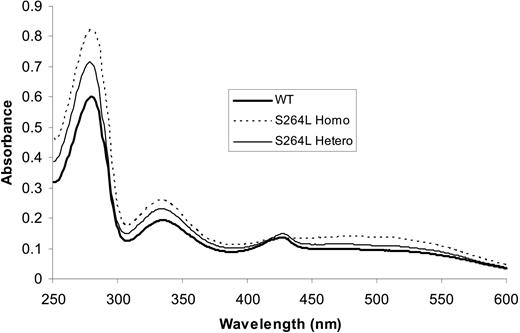
The spectra of recombinant homodimeric and heterodimeric mutant (C236Y, Y191H, K379N, F260L, Q139L, S264L, P334L, and M267I) ferrochelatases were the same as the wild type, showing a feature at approximately 330 nm indicative of a [2Fe-2S] cluster (Figure 1). In contrast, the spectra of the homodimeric ferrochelatases with the mutations F417S, C406S, H386P, or M288K lacked the characteristic spectra of the [2Fe-2S] cluster, indicating that the clusters were not assembled or that they were assembled but were not stable during isolation. Similar results were obtained with the heterodimeric forms of these mutants except for F417S, which looked pale red during purification but which precipitated as eluted from the last column.
Kinetic parameters of the wild-type and mutant ferrochelatases for iron and mesoporphyrin are summarized in Table 1. There was no detectable enzyme activity for the homodimers or heterodimers of M288K, C406S, F417S, or H386P, so they are not included in the table.
Discussion
Ferrochelatase is a membrane-associated homodimeric enzyme that exists at the convergence of the porphyrin synthetic pathway and iron supply.1,14,27 It has recently been demonstrated that the myosin-dependent, transferrin-endosome movement is essential for the efficient use of transferrin-borne iron for heme synthesis in erythroid cells,28 and it has been suggested that iron delivery to ferrochelatase in the mitochondrion results from direct contact with iron-loaded frataxin.29 In addition, it has been proposed, based on kinetic and structural approaches, that ferrochelatase interacts at least transiently with protoporphyrinogen oxidase (PPO), the penultimate enzyme in the pathway.15,30,31 Thus, the spatial orientation of an EPP mutation might have an impact on enzyme catalysis, structural stability, or interactions with frataxin or PPO. The phenotype and clinical presentation of these various classes of mutations could be profoundly different and may explain some of the diverse published observations for EPP patients.2-5
UV-visible spectra for purified wild-type and mutant human ferrochelatases. The key identifies the proteins examined. All other mutant proteins showed similar spectra except for the proteins with the mutations M288K, H386P, C406S, and F417S (see “Results”). WT indicates wild type.
UV-visible spectra for purified wild-type and mutant human ferrochelatases. The key identifies the proteins examined. All other mutant proteins showed similar spectra except for the proteins with the mutations M288K, H386P, C406S, and F417S (see “Results”). WT indicates wild type.
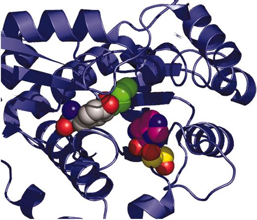
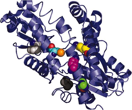
All the EPP mutations examined in the present study have been published previously, and the residual catalytic activities for the recombinant homodimeric EPP ferrochelatases have been reported. However, in no mutation has the heterodimeric normal/EPP enzyme been characterized, and for only a limited number have any kinetic data been presented for homodimeric EPP enzymes. The EPP mutations examined in the present study can be grouped based on their spatial localization in ferrochelatase. One set—Y191H,18,19 S264L,20 and P334L18 —is located within the active site pocket (Figure 2). A second set—M267I,21 M288K,18 H368P,22 C406S,23 and F417S24 —is near the dimer interface, or [2Fe-2S] cluster (Figure 3). A third set—Q139L,8 C236Y,8 F260L,8 and K379N8 —is not clearly associated with any catalytic or intermolecular interaction (Figure 4).
Active site–located EPP mutations. The catalytically essential H263 (which is not an identified EPP mutation) is shown as a CPK space-filling model with carbon atoms colored violet as a landmark for the central portion of the active-site pocket. EPP active-site mutant residues described in the present study are at positions P334 (carbons in green), Y191 (carbons in light gray), and S264 (carbons in yellow). All structure figures were generated with PyMOL.46
Active site–located EPP mutations. The catalytically essential H263 (which is not an identified EPP mutation) is shown as a CPK space-filling model with carbon atoms colored violet as a landmark for the central portion of the active-site pocket. EPP active-site mutant residues described in the present study are at positions P334 (carbons in green), Y191 (carbons in light gray), and S264 (carbons in yellow). All structure figures were generated with PyMOL.46
Positions of mutations near the dimer interface and the [2Fe-2S] cluster. Dimer from one side. Residues at the dimer interface are M267 (carbons in orange) and M288 (carbons in violet). Residues in proximity to the [2Fe-2S] cluster are C406 (carbons in yellow) and F417 (carbons in light gray). In this figure and in Figure 4, the [2Fe-2S] cluster is shown in black. One subunit is rendered in green and the other in slate blue.
Positions of mutations near the dimer interface and the [2Fe-2S] cluster. Dimer from one side. Residues at the dimer interface are M267 (carbons in orange) and M288 (carbons in violet). Residues in proximity to the [2Fe-2S] cluster are C406 (carbons in yellow) and F417 (carbons in light gray). In this figure and in Figure 4, the [2Fe-2S] cluster is shown in black. One subunit is rendered in green and the other in slate blue.
Explanations of the observed effects of mutations can, in some cases, be best understood in the context of residue side chain interactions within the spatial structure of ferrochelatase. In general, it can be stated that mutations other than those at or near the dimer interface (including those associated with the [2Fe-2S] cluster) have effects that do not grossly differ from what might be anticipated from kinetic analysis of the mutant homodimers. For the 3 active-site EPP mutations, some understanding of their detrimental effect can be surmised from what is currently known about human ferrochelatase. For the active-site–located Y191H, the hydroxyl group of Y191 participates in a hydrogen bond with the guanido of R164, a residue previously implicated in iron delivery during catalysis.19 The imidazole of Y191H may still be able to participate in a hydrogen bond with R164, but its physical characteristics will be altered. Y191H exhibits slightly diminished catalytic capabilities, and there is no evidence of any cooperativity between subunits in the heterodimeric enzyme.
The data for S264L homodimers and heterodimers appear consistent for an enzyme homodimer that has 2 distinct and independent active sites. S264 is adjacent to H263, which, it has been suggested, is key in catalysis as the pyrrole proton acceptor from the entering substrate porphyrin.1,19,27 Substitution of the bulkier leucine side chain in S264L may cause sufficient distortion of the backbone structure and move the imidazole of H263 out of position. P334 is highly conserved and is located at the bottom of the active site. For the mutation P334L, it might be anticipated that at least local structures are deranged because of the insertion of the aliphatic side chain and the altered backbone orientation resulting from the change from an imino acid to an amino acid.
One clear conclusion from the data presented here is that mutations affecting the stability of the [2Fe-2S] cluster (C406S and F417S) or on the formation/stability of the dimer (M288K) exhibit these effects in homodimers and heterodimers. The EPP mutation M267I has previously been shown by our group to impart a temperature-sensitive phenotype on ferrochelatase.32 Conditions used in the present study favor this mutant protein's stability. When the M288K EPP mutation was first identified and expressed in vitro, the recombinant homodimeric mutant was reported to have residual activity less than 19% of the wild type.18 The residue M288, which is present in the dimer interface, is highly conserved among all eukaryotic ferrochelatases that are dimeric proteins. In our hands, recombinant M288K human ferrochelatase was inactive when it was assayed in vitro; when it was expressed in vivo, it failed to complement the ferrochelatase-deficient ΔhemH E coli strain. DLS examination of the homodimeric mutant M288K revealed a highly polydisperse size distribution, indicating that ferrochelatase with the mutation M288K was subject to aggregation. A recombinant heterodimeric mutant form of this enzyme could not be isolated using the technique described herein. Because the M288K homodimer could be isolated with the more rapid single metal-affinity column procedure, we believe that the heterodimer may be formed in vivo but is poorly stable.
Neither recombinant homodimeric nor heterodimeric H386P ferrochelatases could be isolated. The human ferrochelatase tertiary structure13 shows that this residue is located in the helix α-15, close to the C-terminal end of the protein. The substitution of H386 by the imino acid proline would be expected to disrupt the helical structure and the stability of the carboxyl terminal extension. Western blot analysis of the crude extract and the eluate from the last purification column showed that the heterodimeric mutant enzyme was not formed, suggesting that this missense mutation affects the ability of the protein to dimerize.
C406S and F417S are mutations spatially close to the [2Fe-2S] cluster. C406S, which was identified by Schneider-Yin et al23 in an EPP patient, was previously shown by Crouse et al33 to lack the [2Fe-2S] cluster and to have no detectable activity as a homodimer. The homodimeric mutant does complement the ferrochelatase-deficient ΔhemH E coli strain, demonstrating that in vivo the protein has minimal activity in the absence of the cluster or that a small amount of the mutant protein might have a cluster. Given that the heterodimer has only one mutated monomer, it might have been anticipated that the protein would have one cluster bound to the normal monomer. However, the heterodimeric mutant preparations lacked the typical reddish color of the [2Fe-2S] cluster during purification, and the purified protein had neither the spectral features of the cluster nor measurable enzyme activity.
The human EPP mutation F417S, initially identified by Brenner et al,24 has been shown to have low enzyme activity as a homodimer33 and to lack the [2Fe-2S] cluster.25 However, it was found to complement the ferrochelatase-deficient ΔhemH E coli strain, indicating that this mutant had sufficient activity to enable cell growth. The heterodimeric mutant F417S was slightly red during purification, indicative of the presence of the [2Fe-2S] cluster and suggesting that the heterodimeric protein might have had one cluster bound to the normal monomer, thus explaining the reddishness. However, the protein precipitated as it eluted from the last purification column, and it was not possible to obtain a spectrum of the purified protein.
Location of other EPP mutations. Single monomeric subunit. The surface presented to the viewer is the backside of the protein that faces toward the mitochondrial matrix and is opposite the active-site pocket face. Residues shown are Q139 (carbons in yellow), K379 (carbons in violet), C236 (carbons in orange), F260 (carbons in light gray), and H386 (carbons in green).
Location of other EPP mutations. Single monomeric subunit. The surface presented to the viewer is the backside of the protein that faces toward the mitochondrial matrix and is opposite the active-site pocket face. Residues shown are Q139 (carbons in yellow), K379 (carbons in violet), C236 (carbons in orange), F260 (carbons in light gray), and H386 (carbons in green).
These data make it clear that not all EPP mutations have similar effects on enzyme structure or activity. Mutations present in the dimer interface or near the [2Fe-2S] cluster have a more profound impact on residual activity in the heterodimeric form of the enzyme. Because the data imply that residual in situ ferrochelatase activity in patients with mutations in these regions may be approximately 25%, it would be valuable to know whether patients with F417S, C406S, M288, or H386P have clinical symptoms in the absence of the low expression allele. Unfortunately, these data are unavailable in the literature. It would also be of interest to determine whether EPP patients with liver damage are represented by mutations in the dimer interface or the cluster region. Such information, along with the recognition that this class of EPP mutations has distinctly profound structural and kinetic implications, may be of value for genetic counseling.
An interesting feature of EPP is the usual lack of anemia and ringed sideroblasts2,3 (erythroblasts containing iron-overloaded mitochondria), as is found with X-linked sideroblastic anemia,34 another disease resulting from a defect in heme biosynthesis. During differentiation of erythrocyte precursors, iron uptake is induced to satisfy the need for chelation into protoporphyrin to form heme, but it is also essential for translational up-regulation of erythroid 5-aminolevulinate synthase (ALAS) through an iron-responsive element.35,36 Interestingly, in some EPP patients, the disease is exacerbated by oral iron therapy.37-39 In EPP, the erythroid program functions normally except that diminished levels of ferrochelatase result in less than stoichiometric conversion of protoporphyrin to heme. Because anemia is generally not a feature of EPP, it is clear that sufficient levels of heme are eventually synthesized.
There are several reports of sideroblastic anemia in patients with clinical EPP, though not all patients had diminished ferrochelatase activity.40-44 In one report of patients with decreased ferrochelatase activity, the nature of the defect was not reported.42 However, it would be expected that for the 2 patients with a reported chromosome 18 deletion, the lowered activity results from the loss of one ferrochelatase allele because the gene is located on 18q21.3. This was shown for one patient with ringed sideroblasts and a chromosome 18 deletion.44 As suggested by these authors, it would be anticipated that the remaining allele is somehow defective, given that clinical EPP is generally observed only when ferrochelatase activity is approximately 25%. On the basis of our own findings above and those reported previously,45 we suggest that for the 3 patients with sideroblastic anemia identified with EPP-like symptoms, including decreased ferrochelatase activity, the specific ferrochelatase mutations may have impact on aspects of the enzyme related to iron acquisition and utilization. Thus, we suggest from the present work that the nature of the EPP mutation in ferrochelatase may have an impact on the clinical presentation of the patient.
Prepublished online as Blood First Edition Paper, April 14, 2005; DOI 10.1182/blood-2004-12-4661.
Supported by grant DK 32303 from the National Institutes of Health (H.A.D.).
The publication costs of this article were defrayed in part by page charge payment. Therefore, and solely to indicate this fact, this article is hereby marked “advertisement” in accordance with 18 U.S.C. section 1734.


![Figure 3. Positions of mutations near the dimer interface and the [2Fe-2S] cluster. Dimer from one side. Residues at the dimer interface are M267 (carbons in orange) and M288 (carbons in violet). Residues in proximity to the [2Fe-2S] cluster are C406 (carbons in yellow) and F417 (carbons in light gray). In this figure and in Figure 4, the [2Fe-2S] cluster is shown in black. One subunit is rendered in green and the other in slate blue.](https://ash.silverchair-cdn.com/ash/content_public/journal/blood/106/3/10.1182_blood-2004-12-4661/6/m_zh80150582160003.jpeg?Expires=1765893232&Signature=rYIpkSt7wW7m8at-bngBrFdN6ADZkw7K~IvT~jOAxO0vkZ8c67KaY3wsWHmzWlt3~QWPcVEgH7wqEJLdn~wtnJLq6tbEIqnC4cfW3mcnU9v8n2h8QPNQyhYEGpWITcjXyfu2iZBiOTAVSmTPKgMg8h-m5~8V-ThMVpbywncs-rRfHEnxgMzFqIGyisvNQI55hwbqVFJSv-L4CcserBDi3EN8RhirQBrFC0pdUN6jgCAwgUwKgp5PEjASDK2xvMK44uRz-rc-6mnGy6tJZfALCYnAcKOK2eY0IGISveJhSn~mrr2Gl-EflNz6O4wkpn06XxH95AcHnWuK-aq~snvprw__&Key-Pair-Id=APKAIE5G5CRDK6RD3PGA)

This feature is available to Subscribers Only
Sign In or Create an Account Close Modal