We identified 243 patients with Philadelphia (Ph) chromosome–positive chronic myeloid leukemia (CML) who had BCR-ABL transcripts monitored by quantitative reverse transcriptase–polymerase chain reaction (RT-PCR) after allogeneic stem cell transplantation for a median of 84.3 months. Individual patients were regarded as having achieved molecular relapse (MR) if the BCR-ABL/ABL ratio exceeded 0.02% on 3 occasions or reached 0.05% on 2 occasions. Patients were allocated to 1 of 4 categories: (1) 36 patients were “persistently negative” or had a single low-level positive result; (2) 51 patients, “fluctuating positive, low level,” had more than 1 positive result but never more than 2 consecutive positive results; (3) 27 patients, “persistently positive, low level,” had persisting low levels of BCR-ABL transcripts but never more than 3 consecutive positive results; and (4) 129 patients relapsed. In 107 of these, relapse was based initially only on molecular criteria; in 72 (67.3%) patients the leukemia progressed to cytogenetic or hematologic relapse either prior to or during treatment with donor lymphocyte infusions. We conclude that the pattern of BCR-ABL transcript levels after allograft is variable; only a minority of patients with fluctuating or persistent low levels of BCR-ABL transcripts satisfied our definitions of MR, whereas the majority of patients who did so were likely to progress further.
Introduction
It is generally accepted that chronic myeloid leukemia (CML) begins when a single hematopoietic stem cell acquires a BCR-ABL fusion gene in association with a Philadelphia (Ph) chromosome, and this leads in due course to inappropriately regulated expansion of the myeloid tissue in the body. At least in some patients there is evidence that the lymphoid lineage is also involved at an early stage. Without effective treatment, the chronic phase progresses inexorably to an advanced phase, which ultimately proves fatal. Allogeneic stem cell transplantation (SCT) was first used to treat patients with CML in chronic phase (CP) in the early 1980s1-4 ; with clinical experience over 25 years, it is clear that some patients have survived for long periods after transplantation without evidence of relapse and may be regarded as “cured,”5 although the mechanism underlying such cures is not well defined. Moreover, occasional patients who were treated by allogeneic stem cell transplantation in chronic phase have relapsed more than 10 years after an otherwise “successful” transplantation,6,7 and data collated by the Center for International Blood and Marrow Transplant Research show that the cumulative incidence of relapse at 15 years for patients in remission at 5 years after transplantations involving a matched sibling donor for CML in chronic phase was 17%.8 Thus the issue whether some or conceivably all patients continue to harbor in their body small quantities of residual leukemia after allogeneic SCT (allo-SCT) remains open.
We started in 1990 to monitor CML patients who received an allogeneic SCT at the Hammersmith Hospital in London by a qualitative reverse transcriptase–polymerase chain reaction (RT-PCR) for BCR-ABL transcripts and we introduced a quantitative RT-PCR (Q-PCR) in 1992.9,10 The use of real-time quantitative PCR (RQ-PCR) was introduced in 2000. The availability now of serial BCR-ABL transcript data on a relatively large number of patients has enabled us to reassess the significance and possible need for early therapeutic intervention for patients who have BCR-ABL transcripts identified in their blood at relatively low levels after transplantation. The data also provide a basis for speculating about the possible significance of the survival of BCR-ABL–positive “stem” cells in some patients after allografting.
Patients, materials, and methods
Patients
We identified a cohort of 243 patients with Ph chromosome–positive CML who had received an allogeneic SCT in chronic or advanced phase between November 1981 and April 2003 and had BCR-ABL transcripts measured on a minimum of 5 occasions starting more than 6 months after transplantation. The study cohort comprised 139 males and 104 females (Table 1). The median interval from diagnosis to transplantation was 1.1 years (range, 0.1-8.2 years). Their median age at time of transplantation was 34.3 years (range, 6.5-59.1 years). Two hundred thirteen patients were in CP at the time of transplantation and 30 patients were in advanced phase (23 in accelerated phase, 1 in blast crisis, 5 in second CP, and 1 in third CP). Approval was obtained from the Hammersmith Hospital Research Ethics Committee for these studies. Informed consent was provided in accordance with the Declaration of Helsinki.
Demographic data
. | Category . | . | . | . | . | ||||
|---|---|---|---|---|---|---|---|---|---|
| Characteristic . | 1, Persistently negative . | 2, Fluctuating positive . | 3, Persistently positive . | 4, Relapse . | Total . | ||||
| No. patients | 36 | 51 | 27 | 129 | 243 | ||||
| Patient age, y, median | 35.9 | 32.7 | 36.9 | 34.3 | 34.3 | ||||
| Patient sex | |||||||||
| Female | 13 | 20 | 11 | 60 | 104 | ||||
| Male | 23 | 31 | 16 | 69 | 139 | ||||
| Duration of disease before SCT, y | |||||||||
| Overall | 1.16 | 0.86 | 0.76 | 1.16 | 1.07 | ||||
| Siblings | 0.83 | 0.60 | 0.67 | 0.73 | 0.72 | ||||
| Alternate | 2.1 | 1.80 | 1.6 | 1.7 | 1.69 | ||||
| Disease status at transplantation | |||||||||
| CP | 33 | 39 | 24 | 117 | 213 | ||||
| Advanced | 3 | 12 | 3 | 12 | 30 | ||||
| Donor | |||||||||
| Identical sibling | 24 | 33 | 20 | 52 | 129 | ||||
| Alternative | 12 | 18 | 7 | 77 | 114 | ||||
| GvHD prophylaxis | |||||||||
| Non-TCD | 24 | 31 | 20 | 43 | 114 | ||||
| TCD | 12 | 20 | 7 | 86 | 125 | ||||
| Follow-up, mo | |||||||||
| Range | 21-271 | 15-230 | 20-212 | 16-257 | 15-271 | ||||
| Median duration | 125 | 86 | 54 | 77 | 84.3 | ||||
| PCR determinations/patient | |||||||||
| Median no. | 9 | 14 | 20 | 25 | 18 | ||||
| Range | 5-20 | 6-39 | 5-43 | 5-35 | 5-55 | ||||
SCT indicates stem cell transplantation; and TCD, T-cell depletion.
Transplantation procedure
All patients received conventional conditioning before transplantation with high-dose cyclophosphamide and total body irradiation4,11,12 ; patients treated with nonmyeloablative conditioning were excluded from this analysis. The stem cell donors were HLA-identical siblings (n = 129), HLA-matched (n = 106) or mismatched (n = 5) unrelated volunteers, or phenotypically HLA-matched family members (n = 3; Table 1). In vivo T-cell depletion with alemtuzumab (CD52 antibody) followed by cyclosporine and methotrexate was used was as graft-versus-host disease (GvHD) prophylaxis in 114 transplantations (106 from alternative donors),12 and cyclosporine and methotrexate alone were used in 114 cases, of which 111 transplantations involved a sibling donor. Ex vivo T-cell depletion was undertaken in 11 procedures (6 sibling donors, 5 alternative donors). Cyclosporine alone as GvHD prophylaxis was used in 4 transplantations involving a sibling. In 93 procedures, both donor and recipient were males; 46 transplantations involved male patients with female donors, 60 transplantations involved female patients with male donors, and 44 procedures involved female patients with female donors. Acute GvHD was classified according to criteria proposed by Glucksberg et al13 ; chronic GvHD when present was categorized as limited or extensive.
Measurement of BCR-ABL transcript numbers
All patients had BCR-ABL transcript numbers measured by Q-PCR on at least 5 occasions during follow-up in the period starting 6 months after transplantation (median number of measurements, 26; range, 5-55). Twenty milliliters of heparinized peripheral blood was collected from each patient and a cell pellet prepared following red-cell lysis. Total RNA was extracted from the guanidine thiocyanate (GTC) cell lysate containing β-mercaptoethanol. Until 1997, the total RNA was extracted using the cesium chloride ultracentrifuge method as previously reported.14 Thereafter, total RNA was isolated by ion-exchange chromatography using a commercially available kit (Qiagen, Crawley, United Kingdom) in accordance with the manufacturer's instructions. The cDNA was synthesized using random hexamers as previously described.9,15
All cDNA samples with BCR-ABL transcripts undetectable by multiplex PCR were subjected to nested primer PCR as previously reported.9 Until 2000, the cDNA samples positive by nested primer PCR were then subjected to competitive PCR for BCR-ABL and ABL quantification as reported previously.9,15 Since 2000, BCR-ABL and ABL transcripts in cDNA were measured by RQ-PCR following multiplex PCR (Taqman; Applied Biosystems, Foster City, CA). This assay detects e13a2 (b2a2) and e14a2 (b3a2) transcripts in a single reaction. The BCR-ABL transcript quantification was performed in triplicate to minimize sampling error. The ABL RQ-PCR was performed in duplicate. A standard curve was generated for each assay using serial dilutions of linearized plasmid containing a BCR-ABL insert. Hence, the same plasmid was used for quantification of BCR-ABL and ABL, which should control for any variation in plasmid quantitation efficiency between BCR-ABL and ABL.
Definitions of molecular relapse
We have previously established criteria to define molecular relapse (MR) in patients who were monitored by Q-PCR after allogeneic SCT. We originally suggested that therapeutic intervention might not be required for patients with transcript levels below 100 μg/RNA,16 which is equivalent to a BCR-ABL/ABL ratio of 0.02%.17 Subsequently we suggested that a patient should be regarded as in molecular relapse if his or her BCR-ABL/ABL ratio remained at or exceeded a level thought to reflect a level approximately 50-fold lower than that seen in early MR18 ; the criteria we adopted were designed to avoid classifying as relapse the occasional patient who had fluctuating low levels of BCR-ABL transcripts. Thus the criteria for MR used in this study were specified in 200119 : a patient satisfied criteria for MR if they had (1) 3 PCR results over a minimum period of 4 weeks with a BCR-ABL/ABL ratio in excess of 0.02%, or (2) 2 results higher than 0.05% over a minimum period of 4 weeks. The finding of a positive PCR within the first 6 months after allogeneic SCT was not regarded as indicating relapse and so such results were discounted. Any patient who did not achieve relapse at the molecular, cytogenetic, or hematologic level was regarded as remaining in molecular remission.
Administration of DLI
Patients who met criteria for relapse were considered for treatment by donor lymphocyte infusions (DLIs) if lymphocytes from the original transplant donor were available and if the patient was not undergoing treatment for extensive GvHD. Lymphocytes were administered as a single bulk dose until 1995 and thereafter on an escalating dose schedule.20,21
Comparison of nested primer PCR and real-time quantitative PCR (RQ-PCR)
We considered it important to compare results obtained with RQ-PCR with those obtained with nested primer PCR. We therefore used nested primer PCR to quantitate BCR-ABL transcripts in cDNA samples determined to have between 0 and 10 copies by real-time PCR and we used Q-PCR to measure transcripts in cDNA samples that were negative by nested PCR. Only those samples with control ABL copies greater than 1 × 10⁴ in the same volume of cDNA used to detect BCR-ABL were included in these comparisons, thus limiting the comparison to specimens with high-fidelity RNA. Samples with less than 1.0 BCR-ABL copies by Q-PCR were reported as negative. In each case, 5 μL cDNA was used.
Statistical methods
The probabilities of relapse and disease progression were calculated using the cumulative incidence procedure. Curves were compared using the log-rank test.
Results
Patient outcome after transplantation
For this patient population, the median follow-up after transplantation was 84.3 months (range, 15-271 months). Two hundred twenty patients were alive at the time of this analysis, while 23 patients have died. The primary cause of death was leukemia relapse or progression in 12 cases, GvHD in 8 cases, infection in 1 case, and secondary malignancies in 2 cases.
Representative patterns of BCR-ABL/ABL transcript values from individual patients from the 3 patient groups who did not satisfy criteria for MR. (A) Persistently negative; (B) fluctuating positive, low level; and (C) persistently positive, low level (but not satisfying criteria for molecular relapse). The upper broken horizontal line shows the 2.0% level (corresponding to cytogenetic relapse). The lower 2 broken horizontal lines show the 0.02% and 0.05% levels, respectively. UPN indicates unique patient number.
Representative patterns of BCR-ABL/ABL transcript values from individual patients from the 3 patient groups who did not satisfy criteria for MR. (A) Persistently negative; (B) fluctuating positive, low level; and (C) persistently positive, low level (but not satisfying criteria for molecular relapse). The upper broken horizontal line shows the 2.0% level (corresponding to cytogenetic relapse). The lower 2 broken horizontal lines show the 0.02% and 0.05% levels, respectively. UPN indicates unique patient number.
Patterns of BCR-ABL transcripts after SCT
The patterns of serial BCR-ABL transcript numbers differed appreciably in the 243 patients included in this study. One hundred fourteen patients remained in molecular remission, of whom some never had transcripts detectable and others on one occasion only. Some patients had transcripts detectable intermittently only at low levels. Some patients had transcripts detected “persistently” at low levels. One hundred twenty-nine patients were classified as having relapsed. We were thus able to classify individual patients into 1 of 4 major categories.
Category 1. Thirty-six patients (14.8%) were classified as “persistently negative” after transplantation; of these, 16 (6.6%) were always negative on the basis of 5 to 19 separate results and 20 (8.2%) had just one single low-level positive result; of these 20, 15 had fewer than 5 transcripts detectable on just 1 occasion. The median follow-up after transplantation for these 36 patients was 123.4 months (range, 21.5-303.3 months; Figure 1A)
Category 2. Fifty-one patients (21.0%) had more than 1 positive result during the period of follow-up but never more than 2 consecutive positive results. These patients were classified as “fluctuating, low level.” The median follow-up after transplantation for these 51 patients was 86.0 months (range, 15.2-245.1 months; Figure 1B).
Category 3. Twenty-seven patients (11.1%) had 3 or more consecutive positive results during the period of follow-up but their transcript numbers never satisfied our criteria for MR. These patients were classified as “persistently positive, low level.” The median follow-up after transplantation was 54.3 months (range, 19.9-211.9 months; Figure 1C).
Category 4. One hundred twenty-nine (53.1%) patients satisfied our criteria for relapse. The diagnosis of relapse was based on molecular criteria alone in 107 patients and on cytogenetic or hematologic criteria in 22 (Table 2). Of these 129 patients, 115 (89.1%) proceeded to receive DLIs. Fourteen patients had not received DLIs at the time of this analysis, of whom 6 maintained stable transcript levels over a median period of 102 months after MR diagnosis, despite having received no specific treatment. The median time from transplantation to relapse was 9.8 months (range, 1.1-93.2 months). The median times to relapse for patients who received a transplant in chronic and advanced phases were 10.6 months (range, 2.3-93.2 months) and 3.7 months (range, 1.1-14.3 months), respectively.
Incidence and severity of acute and chronic graft-versus-host disease in the 137 patients who did not satisfy criteria for molecular relapse
Severity of GvHD . | Persistently negative . | Fluctuating positive . | Persistently positive . |
|---|---|---|---|
| No. patients | 36 | 51 | 27 |
| aGvHD, no. (%) | |||
| Grades 0-1 | 15 (42) | 30 (59) | 19 (70) |
| Grades 2-4 | 21 (58) | 21 (41) | 8 (30) |
| cGvHD, no. (%) | |||
| Nil | 9 (25) | 22 (44) | 8 (30) |
| Limited or extensive | 27 (75) | 28 (56) | 19 (70) |
The relationship between severity of aGvHD and the pattern of molecular positivity is significant (P = .039, chi-square trend test), but there is no significant relationship between severity of cGvHD and pattern of molecular disease (P = .71). Numbers in parentheses are percentages.
Influence of graft-versus-host disease
The incidence of serious acute GvHD (grades 2 to 4) was relatively high (60%) in patients categorized as persistently negative, and relatively low (33%) in those classified as persistently positive (P = .039; Table 2). However there was no discernible relationship between incidence of chronic GvHD and the pattern of transcript numbers after transplantation. There was no apparent relationship between the method of T-cell depletion (in vivo vs ex vivo) and the pattern of residual disease after transplantation.
Pattern of disease prior to diagnosis of MR
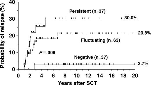
Of the 129 patients who satisfied our criteria for relapse, it was possible to analyze the results of molecular monitoring in the period from 6 months after transplantation until diagnosis of relapse in only 23 cases. The remaining 106 either relapsed early after transplantation or had insufficient serial PCR data to permit classification. Of these 23 patients, 1 patient had been persistently negative, 12 were classifiable as fluctuating positive, and 10 were persistently positive at low level before recognition of relapse. By grouping patients in each of these categories together with patients in the same categories who did not satisfy our criteria for relapse, it was possible to calculate the risk of relapse for patients in each category (Figure 2). It is apparent that the risk of relapse is significantly related to the pattern of BCR-ABL transcripts in the period preceding recognition of relapse (P = .009).
Events after diagnosis of relapse and results of administering DLI
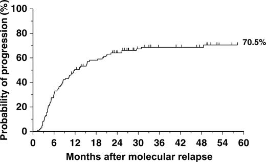
Of the 129 patients who satisfied our criteria for relapse, 117 were in chronic phase and 12 were in advanced phases at time of transplantation. Of the 107 patients who were diagnosed with MR, 72 showed evidence of progression to cytogenetic relapse (or its molecular equivalent) or hematologic relapse either prior to or after starting treatment with DLI. The projected incidence of progression at 5 years from recognition of MR was 70.5% (Figure 3). In 26 of the 72 patients, cytogenetic relapse was not documented by bone marrow analysis and a BCR-ABL ratio of 2% or greater was taken to be equivalent to cytogenetic relapse.17 Forty-five patients had Ph chromosome–positive marrow metaphases identified before initiation of DLI and 28 had hematologic features of CML (Table 3). Progression of the 72 patients into cytogenetic relapse occurred at a median of 5.9 months (range, 1.0-49.2 months). Thirty-five patients did not progress beyond molecular relapse. Of these, 28 received DLIs, with 15 regaining molecular remission (defined as 2 consecutive studies with undetectable BCR-ABL transcripts), 11 continue in molecular relapse without further progression, and in 2 patients insufficient follow-up data are available. Seven patients were not treated in any way for leukemia for a variety of reasons.
Disease progression after diagnosis of relapse
Earliest identified criteria for relapse . | Total . | Did not progress . | Cytogenetic relapse . | Hematologic relapse . |
|---|---|---|---|---|
| Molecular | 107 | 34 | 45 | 28 |
| Cytogenetic | 19 | 5 | NA | 14 |
| Hematologic | 3 | 3 | NA | NA |
| Total | 129 | 42 | 45 | 42 |
Earliest identified criteria for relapse . | Total . | Did not progress . | Cytogenetic relapse . | Hematologic relapse . |
|---|---|---|---|---|
| Molecular | 107 | 34 | 45 | 28 |
| Cytogenetic | 19 | 5 | NA | 14 |
| Hematologic | 3 | 3 | NA | NA |
| Total | 129 | 42 | 45 | 42 |
NA indicates not applicable.
Probability of relapse according to Q-PCR category. Patients in this study who met criteria for MR whose prior pattern of molecular monitoring could be assessed were considered in conjunction with patients in the same molecular remission category who did not relapse. It was possible thereby to derive projected risks of relapse for patients in each category. The respective risks in the 3 categories were significantly different (P = .009).
Probability of relapse according to Q-PCR category. Patients in this study who met criteria for MR whose prior pattern of molecular monitoring could be assessed were considered in conjunction with patients in the same molecular remission category who did not relapse. It was possible thereby to derive projected risks of relapse for patients in each category. The respective risks in the 3 categories were significantly different (P = .009).
Probability of disease progression after diagnosis of molecular relapse (n = 107). Progression was defined by finding evidence of cytogenetic or hematologic relapse in a patient who lacked such evidence when molecular relapse was diagnosed. Vertical lines represent patients in MR who had not progressed.
Probability of disease progression after diagnosis of molecular relapse (n = 107). Progression was defined by finding evidence of cytogenetic or hematologic relapse in a patient who lacked such evidence when molecular relapse was diagnosed. Vertical lines represent patients in MR who had not progressed.
Prior to 1997, DLIs were given in a single “bulk” dose that contained variable numbers of CD3+ T cells; after 1995, an escalating dose regimen was used.21 Because in most cases arrangements had to be made to collect lymphocytes from the original transplant donor, it was not usually possible to administer DLI immediately after the recognition of molecular relapse. In practice, DLIs were initiated at a median of 9.4 months (range, 1-72 months) from relapse.
One hundred fifteen patients received DLIs. Seventy-three patients (63%) went into molecular remission after DLI with 2 consecutive negative PCR results. Twenty-seven patients (23%) are early in follow-up but 10 of them have at least 1 negative PCR result. Twelve patients (10.4%) have had no response, and of these 9 have died whereas 3 are alive and currently receiving alternative treatment. In 3 cases (2.6%) there were not enough data to assess their response.
Comparison of nested primer PCR and RQ-PCR
Our data show that of 216 samples negative by nested PCR, 51.9% were positive by RQ-PCR, whereas only 6.3% of samples negative by RQ-PCR were positive by nested PCR (Table 4). This would suggest that in our laboratory, the RQ-PCR is more sensitive than nested primer PCR, though the clinical significance of these low copy numbers is presently unclear. Furthermore these data suggest that it might be inappropriate to report samples with 1 to 10 BCR-ABL copies by RQ-PCR as negative or undetectable, particularly if the endogenous control gene copy number is within acceptable limits.
Comparison of PCR results from nested and RQ-PCR analyses on the same samples
Nested PCR result . | RQ-PCR negative . | RQ-PCR, 1-10 copy nos. . | Total . |
|---|---|---|---|
| Nested, negative | 104 | 112 | 216 |
| Nested, 1-10 copy nos. | 7 | 27 | 34 |
| Total | 111 | 139 | 250 |
Nested PCR result . | RQ-PCR negative . | RQ-PCR, 1-10 copy nos. . | Total . |
|---|---|---|---|
| Nested, negative | 104 | 112 | 216 |
| Nested, 1-10 copy nos. | 7 | 27 | 34 |
| Total | 111 | 139 | 250 |
RQ-PCR indicates real-time quantitative reverse-transcriptase-polymerase chain reaction.
Discussion
Leukemia-specific transcripts may be detected in the blood and bone marrow of individual CML patients for some months following allogeneic stem cell transplantation9,22,23 and their level may predict the risk of relapse.24,25 Thus we showed previously that patients with no BCR-ABL transcripts detected between 3 and 5 months after transplantation had a probability of relapse at 3 years of 16.7%, whereas patients with low level and relatively high transcript levels had risks of relapse of 42.9% and 86%, respectively.25 When patients were categorized at 5 years after transplantation on the basis of prior transcript pattern, the subsequent risk of relapse was related to the transcript pattern in the interval preceding the 5-year point.19 However, we observed some patients who had transcripts detectable at low level for long periods after SCT without obvious progression. Because we did not know the clinical significance of finding these transcripts at low level, we were reluctant to regard them as having relapsed and thus in need of further therapy. We therefore proposed criteria for MR designed to exclude patients in whom transcripts were present only at low levels, regardless of how long such low levels might persist.19
We show in this paper that patients who fail to satisfy our criteria for MR during prolonged periods of observation can be classified into 3 categories, some persistently negative for BCR-ABL transcripts, some intermittently positive at low level, and some persistently positive at low level. There was a significant relationship between the allocated category of transcripts and incidence of acute GvHD (grades 2 or more). Of the patients who did eventually achieve MR, some could be categorized on the basis of their transcript pattern before relapse; those persistently positive at low level were significantly more likely to relapse than those who were persistently negative, and for those intermittently positive before relapse the risk was intermediate.
The longest periods of observation for 78 patients classified as fluctuating low-level positive or persistently low-level positive (without achieving MR) were 13.7 years and 13.1 years, respectively, from first positive result. Conversely, the 107 patients who did satisfy our criteria for MR were advised to accept treatment with DLI if this were feasible. In practice, the median interval from diagnosis of MR to starting DLI was 9.5 months. During this waiting period, 43 patients (46%) progressed to cytogenetic and/or hematologic relapse and a further 22 patients (24%) progressed after starting DLI. The fact that 72 (67.3%) patients showed evidence of disease progression after diagnosis of MR provides some support for the validity of the criteria for MR that we have adopted.
Donor lymphocyte infusions have proved remarkably effective for treating patients who relapse in CP after allogeneic SCT.20,21,26,27 Of the 115 patients in this series treated by DLI, 73 responded and the response has been maintained hitherto in 67. In 12 patients there was no response, whereas in 30 patients the treatment continues and is thus too early to evaluate. The durability of the response in this and other series provides further support for the generally accepted concept that a graft-versus-leukemia (GvL) effect plays a pivotal role in eradication of residual leukemia after allogeneic SCT.28 It may do this by targeting transcriptionally silent “stem cells” as well as leukemia progenitor cells expressing the Bcr-Abl oncoprotein.
It is possible that the GvL effect is required on a continuing basis for control of CML after allogeneic SCT and thus that the persistence of very low-level disease, recognizable at times by Q-PCR techniques, is of no clinical significance. Conversely, the demonstration that BCR-ABL transcripts can persist in an individual patient at even very low level could reflect the obligatory presence of small numbers of residual leukemia stem cells, some transcriptionally active but others presumably quiescent and perhaps transcriptionally silent. If leukemia cells in either category were susceptible to acquisition of some of the new molecular changes that cause CML to progress, it would mean that the discovery of any BCR-ABL transcripts after transplantation should be a cogent indication for therapeutic intervention. However, the issue whether such intervention should comprise use of an Abl tyrosine kinase inhibitor, DLI, or some combination of the 2 is not yet resolved. It seems clear however from our data that all CML patients need to be monitored indefinitely after transplantation by sensitive molecular techniques, perhaps at 3- or 6-month intervals. Such monitoring should help to define the ultimate clinical significance of BCR-ABL transcripts that remain detectable at low level for long periods of time.
Supported in part by the Leukaemia Research Fund (United Kingdom). J.M.S. is a Fogarty Scholar at the National Heart, Lung, and Blood Institute in Bethesda, MD.
J.K. supervised the day-to-day running of the Minimal Residual Disease laboratory and assembled the molecular data for further analysis; D.O'S. collated clinical data and wrote the original draft of the paper; R.M.S. devised the categorization of the patients and carried out all statistical analyses; E.O. supervised patient care and contributed to writing the manuscript; F.D. coordinated the DLI program and collated DLI data for this paper; D.M. supervised patient care and helped to collate clinical data; S.S. performed many of the molecular studies; J.S.K. performed some of the molecular studies; N.C.P.C. designed the original quantitative PCR and provided laboratory guidance over many years; J.M.G. radically revised the manuscript; and J.F.A. was responsible for coordinating the transplant program and made constructive suggestions for improving the manuscript.
J.K. and D.O'S. contributed equally to this paper.
An Inside Blood analysis of this article appears at the front of this issue.
The publication costs of this article were defrayed in part by page charge payment. Therefore, and solely to indicate this fact, this article is hereby marked “advertisement” in accordance with 18 U.S.C. section 1734.
Prepublished online as Blood First Edition Paper, February 7, 2006; DOI 10.1182/blood-2005-08-3320.
We thank the nursing staff who collaborated in the collection of samples from patients included in this study. We also thank staff in the Minimal Residual Disease laboratory.


This feature is available to Subscribers Only
Sign In or Create an Account Close Modal