Abstract
ICAM-2 has been implicated in leukocyte transmigration in vitro, but there is little in vivo evidence to support this. To address this, neutrophil migration was investigated in ICAM-2–deficient mice (KO) and in wild-type (WT) mice treated with an anti–ICAM-2 blocking monoclonal antibody (mAb) (3C4). In a peritonitis model, IL-1β–induced accumulation of neutrophils was significantly reduced in mice treated with 3C4 (51% inhibition) and in KO mice (41% inhibition). In contrast, TNF-α– or thioglycolate-induced responses were not suppressed in KO mice. Analysis of IL-1β–induced leukocyte responses in cremasteric venules of KO animals by intravital microscopy indicated a defect in transmigration (44% inhibition) but not rolling or adhesion. As found before, TNF-α–induced leukocyte transmigration was unaltered in the KO mice. WT mice treated with the anti–ICAM-2 mAb also exhibited a selective reduction in leukocyte transmigration in response to IL-1β while an anti–ICAM-1 mAb inhibited both leukocyte adhesion and transmigration. Interestingly, mAb 3C4 significantly suppressed IL-1β–induced neutrophil transmigration in PE-CAM-1 KO animals in the peritonitis model but not in the cremaster muscle. The findings provide direct evidence for the involvement of ICAM-2 in neutrophil transmigration in vivo, though this role appears to be stimulus specific. Furthermore, ICAM-2 appears capable of mediating PECAM-1–independent leukocyte transmigration.
Introduction
The emigration of leukocytes from blood vessels into tissues is a crucial process both during immunosurveillance and inflammation. This response is thought to involve sequential steps of leukocyte rolling, activation, firm adhesion and, finally, migration through endothelial cells and the vascular basement membrane. These cellular events all require adhesive interactions between leukocytes and endothelial cells/extracellular matrix proteins with the initial intravascular phases being largely mediated by selectins and integrins. The mechanisms that mediate migration through venular walls are less well understood though there is now good evidence for the involvement of a number of junctional molecules in the process of leukocyte migration through endothelial cells.1-3 In this context, molecules such as PECAM-1 (CD31), members of the JAM family (in particular, JAM-A, -B, and -C), CD99, ESAM, ICAM-1, and ICAM-2 have been implicated.2,3 Although there is much in vivo evidence for the involvement of PECAM-1 in leukocyte transmigration4-7 and increasing in vivo evidence for the involvement of the other listed endothelial cell junctional molecules in leukocyte migration in inflammatory models,8-11 there is to date no direct evidence for the involvement of ICAM-2 in leukocyte transmigration in vivo, a critical issue that is addressed in the present study.
ICAM-2 is a 55-kDa member of the immunoglobulin superfamily of adhesion molecules composed of 2 N-terminal Ig domains with 35% homology to ICAM-1, a transmembrane domain, and a short cytoplasmic tail of 27 amino acids. This molecule is constitutively expressed on endothelial cells, platelets, and most leukocytes. The expression of ICAM-2 on resting endothelial cells is much higher than that of ICAM-1 (10- to 15-fold) and, in contrast to ICAM-1, whose expression is rather uniformly expressed on the surface of endothelial cells, ICAM-2 appears in vitro to be more concentrated at endothelial cell junctions.
Despite ICAM-2 being cloned more than 15 years ago,12 studies into the functional roles of ICAM-2 are limited though it has been implicated in leukocyte trafficking,13 immune cell activation14 and, more recently, angiogenesis.15 With respect to leukocyte migration, numerous in vitro studies involving blocking monoclonal antibodies (mAbs) have indicated a role for ICAM-2 in transmigration of all leukocyte subsets through stimulated or unstimulated HUVECs.13,16,17 Furthermore, using ICAM-1– and ICAM-2–deficient endothelial cell lines, the relative importance of ICAM-1 in lymphocyte transmigration, as opposed to adhesion, and the cooperative interaction of ICAM-1 and ICAM-2 in lymphocyte transmigration has been demonstrated.17,18 In vivo studies employing anti–ICAM-1 and anti–ICAM-2 mAbs and ICAM-1–deficient mice have suggested that ICAM-1 and ICAM-2 are functionally redundant in lymphocyte recirculation through lymph nodes and that ICAM-1, but not ICAM-2, is involved in lymphocyte migration into inflamed sites and trapping within noninflamed lung.16 However, using an allergic asthma model, ICAM-2–deficient mice exhibited a delayed increase in eosinophil migration in the airway lumen and a prolonged presence of eosinophils in the lung tissue, with no notable decrease in lymphocyte or monocyte accumulation.19 More recently, a functional blocking anti–ICAM-2 antibody was shown to inhibit leukocyte infiltration in ICAM-1 KO mice in response to bacterial ocular infection.20 Collectively, though limited, the cited in vitro and in vivo studies suggest a role for ICAM-2 in leukocyte migration in response to inflammatory stimuli, but they also highlight the critical need for further investigations to elucidate the role of ICAM-2 in mediating leukocyte transmigration in vivo.
The absence of direct in vivo evidence for the involvement of ICAM-2 in leukocyte transmigration led us to perform the present study in which we have investigated the role of ICAM-2 in leukocyte transmigration by intravital microscopy using both ICAM-2–deficient mice and a blocking anti–ICAM-2 mAb. Furthermore, the potential synergistic/additive effect of ICAM-2 and PECAM-1 blockade/deletion was investigated. The findings of this study provide the first direct evidence for the involvement of ICAM-2 in neutrophil transmigration in vivo, a role that appears to be stimulus specific. Furthermore, the findings suggest that ICAM-2 can mediate PECAM-1–independent leukocyte transmigration.
Materials and methods
Animals
ICAM-2 KO mice on a 129SV × C57BL/6 mixed background (F2 generation) were generated as previously detailed.19 The 129SV × C57BL/6 mixed background control mice (F2 generation) were generated in-house within the animal facilities of Imperial College London on the Hammersmith campus. Wild-type (WT) C57BL/6 mice were purchased from Harlan-Olac (Bicester, United Kingdom). PECAM-1 KO mice, back-crossed for 8 generations onto C57BL/6 background, were obtained as a gift from Dr Tak W. Mak (Amgen Institute, Toronto, ON, Canada).6 All animal experiments were conducted in accordance with the British Home Office regulations (Scientific Procedures) Act 1986, United Kingdom.
Reagents
Recombinant murine IL-1βand TNF-α were purchased from Peprotech (London, United Kingdom) and R&D Systems (Abingdon, United Kingdom), respectively. Anti–ICAM-2 mAb (3C4; rat IgG2a) and the isotype control mAb (rat IgG2a) were obtained from BD Biosciences (San Diego, CA). Anti–ICAM-1 mAb (YN-1; rat IgG2b) was generated in-house from hybridoma cells, and a corresponding IgG2b control mAb was obtained from Serotec (Kidlington, United Kingdom). Ketamine was from Parke Davis (Eastleigh, United Kingdom), and xylazine was from Bayer (St Edmunds, United Kingdom). Thioglycolate, saponin, toluidine blue, light green SF yellowish, Giemsa, and May-Grünwald were all purchased from Sigma Chemical (Dorset, United Kingdom).
Quantification of leukocyte migration into the peritoneal cavity
Age-matched female mice (WT or ICAM-2 KO; more than 20 g) were injected with intraperitoneal phosphate-buffered saline (PBS) (1 mL; control group), IL-1β (10 ng/mL per cavity), TNF-α (100 ng/mL per cavity), or thioglycolate (4% solution in 1 mL PBS). In some experiments, 15 minutes prior to the intraperitoneal injection of IL-1β, mice (WT, ICAM-2 KO, or PECAM-1 KO) were injected intravenously with saline, anti–ICAM-2 mAb (3C4), or an appropriate control mAb (all at 3 mg/kg). Four hours after the injection of intraperitoneal stimuli, the animals were killed by asphyxiation with CO2, and the peritoneal cavity was opened via midline incision and lavaged with 3 to 5 mL modified PBS (containing 0.25% bovine serum albumin [BSA] and 2 mM EDTA). To quantify the number of neutrophils in peritoneal exudate samples, 2 staining steps were conducted. Initially total cell counts were determined following staining of peritoneal samples with Kimura stain.21 Smears of peritoneal samples were then prepared in a cytocentrifuge (Cytospin-3; Shandon, United Kingdom) and stained with May-Grünwald/Giemsa stains, and the number of neutrophils per 500 cells counted was measured. These quantifications were used to calculate the number of neutrophils recovered from each cavity. The dose of the anti–ICAM-2 mAb and the cytokines employed was based on preliminary studies aimed at identifying optimal dosing regimes (data not shown) and/or previous studies.7,22,23
Intravital microscopy
Intravital microscopy on the mouse cremaster muscle was performed as previously described.7 Briefly, male mice (WT or ICAM-2 KO; more than 20 g) were injected with intrascrotal saline (400 μL; control group), IL-1β (50 ng/400 μL per animal), or TNF-α (300 ng/400 μL per animal). In some groups of mice, 15 minutes prior to the intrascrotal injections, WT or PECAM-1 KO mice were injected intravenously with saline, anti–ICAM-1 mAb (YN-1), anti–ICAM-2 mAb (3C4), or an isotype-matched control mAb (all at 3 mg/kg), as detailed in “Results.” The dose of the anti–ICAM-1 and anti–ICAM-2 mAbs, IL-1β, and TNF-α was based on preliminary studies aimed at identifying optimal dosing regimes (data not shown) and/or previous studies.7,22,23
Four hours after the intrascrotal injections, mice were anesthetized by intraperitoneal administration of ketamine (100 mg/kg) and xylazine (10 mg/kg) and placed on a heated (37°C) microscope stage where the surgical procedure was carried out. An incision was made in the scrotum; one testis was gently withdrawn to allow the cremaster muscle to be exteriorized and pinned out flat over the viewing window in the microscope stage. The cremaster muscle was kept warm and moist by continuous application of warmed Tyrode balanced salt solution. Leukocyte responses of rolling, firm adhesion, and transmigration in postcapillary venules of 20 to 40 μm diameter were observed on an upright fixed-stage microscope (Axioskop FS; Carl Zeiss, Welwyn Garden City, United Kingdom) equipped with water-dipping objectives, and quantifications were made as previously described.7 Briefly, leukocyte rolling was defined as the average number of cells that passed a fixed point within 5 minutes, and firmly adherent leukocytes were defined as cells that remained stationary for at least 30 seconds. Leukocyte transmigration was defined as the number of leukocytes in the extravascular tissue across a 100 μm vessel segment and within 50 μm of the vessel of interest. Several vessel segments (3 to 5) from at least 3 vessels were studied for each animal.
Immunofluorescent staining of tissues
Cremaster tissues were dissected away from WT and ICAM-2 KO mice and fixed in 4% paraformaldehyde (PFA) before being immunostained for ICAM-2 and PECAM-1 using a modified version of the protocol previously detailed.24 Briefly, tissues were blocked and permeabilized in PBS containing 20% fetal calf serum and 0.5% Triton X-100. After incubation with the rat antimouse mAb 3C4 against ICAM-2 (BD Bisosciences, Oxford, United Kingdom), the tissues were washed in PBS followed by incubation with a goat anti–rat Alexa-Fluor-488–conjugated secondary Ab (Molecular Probes, Eugene, OR). The tissues were thoroughly washed and incubated with a directly allophycocyanin (APC)–conjugated rat antimouse mAb Mec13.3 against PECAM-1 (BD Bisosciences). After a series of further washings with PBS, samples were mounted on glass slides and viewed in PBS using a Zeiss LSM 5 PASCAL confocal laser-scanning microscope (using a × 40 water-dipping Achroplan objective with a numeric aperture of 0.75 [Carl Zeiss, Welwyn Garden City, United Kingdom]) equipped with argon (excitation wavelength, 488 nm) and HeNe (excitation wavelength, 543 nm) lasers. Multiple optical sections of tissue samples were captured at room temperature and imaged using the software's automatic scanning mode. Finally, Z-stack images (collected at every 1.2 μm depth) were saved and used for 3D reconstruction analysis using the LSM 5 Pascal software (version 3.2; Carl Zeiss).
Statistics
All results are expressed as mean plus or minus standard error of the mean (SEM). Statistical significance was assessed by the Student t test or by 1-way analysis of variance with the Neuman-Keuls multiple comparison test. P values below .05 were considered significant.
Results
Antibody blockade or genetic deletion of ICAM-2 suppresses neutrophil migration in vivo
In initial studies, the functional role of ICAM-2 in neutrophil migration in vivo was investigated by using an anti–ICAM-2 mAb, 3C4, and ICAM-2–deficient mice in an IL-1β–driven peritonitis model.22 Administration of IL-1β (10 ng per cavity; 4-hour in vivo test period) via the peritoneal route induced a significant increase in neutrophil accumulation into the cavity of WT control mice as compared with mice injected intraperitoneally with PBS. This response was significantly suppressed in mice injected with 3C4 (Figure 1; 50.6% inhibition, P < .05) but not in control mAb–treated mice. A similar profile of suppression of neutrophil migration was also observed in ICAM-2–deficient mice (41% inhibition, P < .05). Of importance, injection of the anti–ICAM-2 mAb 3C4 into ICAM-2–deficient mice had no effect on the IL-1β–induced response in these mice, indicating the specificity of this mAb for its antigen. Together, using both an anti–ICAM-2 mAb and ICAM-2–deficient mice, these findings indicated a functional role for ICAM-2 in mediating neutrophil migration in vivo.
ICAM-2 mediates neutrophil migraton in a stimulus-specific manner
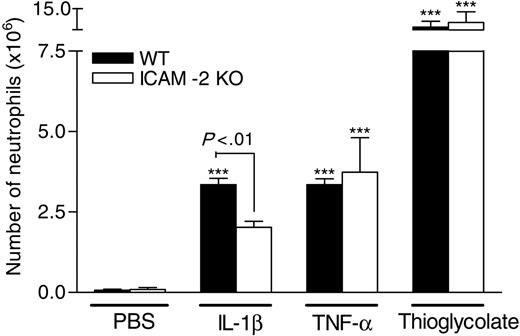
Because we have previously found that PECAM-1–mediated neutrophil transmigration is stimulus specific,7,23 we next investigated whether the same phenomenon applied to ICAM-2. For this purpose, neutrophil migration into mouse peritoneal cavities was investigated in response to IL-1β (10 ng/1 mL per cavity), TNF-α (100 ng/1mL per cavity), and thioglycolate (4% solution in 1 mL PBS) in WT and ICAM-2–deficient mice. As shown in Figure 2, all 3 stimuli when injected intraperitoneally induced a significant increase in neutrophil migration in WT mice over a 4-hour test period as compared with mice injected with intraperitoneal PBS. As previously found (Figure 1), the response induced by IL-1β was suppressed in ICAM-2 KO animals; however, the responses induced by TNF-α or thioglycolate were unaltered in ICAM-2–deficient mice, indicating their independence of ICAM-2.
Effect of anti–ICAM-2 mAb on IL-1β–elicited neutrophil migration into the peritoneal cavity of WT and ICAM-2–deficient mice. Animals were treated intraperitoneally with PBS or IL-1β (10 ng per cavity) 4 hours prior to peritoneal lavage. In additional groups of animals, mice were pretreated intravenously with anti–ICAM-2 mAb 3C4 (3 mg/kg) or isotype-matched control antibody (3 mg/kg) 15 minutes before the intraperitoneal administration of IL-1β. The figure shows the number of neutrophils that transmigrated into cavities. The data represent mean ± SEM from 3 to 8 mice per group. ***P < .001 versus responses obtained from saline-injected animals. Additional statistical comparisons are indicated by lines.
Effect of anti–ICAM-2 mAb on IL-1β–elicited neutrophil migration into the peritoneal cavity of WT and ICAM-2–deficient mice. Animals were treated intraperitoneally with PBS or IL-1β (10 ng per cavity) 4 hours prior to peritoneal lavage. In additional groups of animals, mice were pretreated intravenously with anti–ICAM-2 mAb 3C4 (3 mg/kg) or isotype-matched control antibody (3 mg/kg) 15 minutes before the intraperitoneal administration of IL-1β. The figure shows the number of neutrophils that transmigrated into cavities. The data represent mean ± SEM from 3 to 8 mice per group. ***P < .001 versus responses obtained from saline-injected animals. Additional statistical comparisons are indicated by lines.
Migration of neutrophils into the peritoneal cavity of WT and ICAM-2–deficient mice in response to different stimuli. WT and ICAM-2 KO mice were treated intraperitoneally with PBS (1 mL), IL-1β (10 ng/mL per cavity), TNF-α (100 ng/mL per cavity), or thioglycolate (4% solution in 1 mL per cavity). Four hours later, the peritoneal cavity was opened via a midline incision and the cavity lavaged with PBS (containing 0.25% BSA and 2 mM EDTA). Results represent the number of neutrophils that migrated into cavities. The data are presented as mean ± SEM of 3 to 7 animals. ***P < .001 versus responses obtained from saline-injected animals. In addition, significant differences between the 2 strains of mice are indicated by lines.
Migration of neutrophils into the peritoneal cavity of WT and ICAM-2–deficient mice in response to different stimuli. WT and ICAM-2 KO mice were treated intraperitoneally with PBS (1 mL), IL-1β (10 ng/mL per cavity), TNF-α (100 ng/mL per cavity), or thioglycolate (4% solution in 1 mL per cavity). Four hours later, the peritoneal cavity was opened via a midline incision and the cavity lavaged with PBS (containing 0.25% BSA and 2 mM EDTA). Results represent the number of neutrophils that migrated into cavities. The data are presented as mean ± SEM of 3 to 7 animals. ***P < .001 versus responses obtained from saline-injected animals. In addition, significant differences between the 2 strains of mice are indicated by lines.
ICAM-2 mediates leukocyte migration in vivo at the level of transmigration
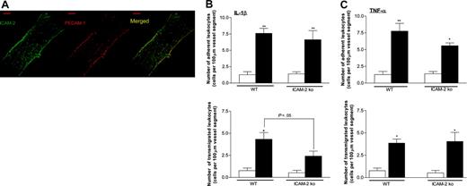
To determine the precise stage in the process of leukocyte emigration at which ICAM-2 was involved, in the following series of experiments the role of ICAM-2 in leukocyte emigration through cremasteric venules was investigated, as directly observed by intravital microscopy. The thin and translucent nature of the murine cremaster muscle also makes this model suitable for analysis of vascular expression of adhesion molecules by immunofluorescent labeling and confocal microscopy. As can be seen in Figure 3A, in cremasteric venules, ICAM-2 is largely expressed at endothelial cell junctions where it appears to colocalize with PECAM-1.
Real-time observation of leukocyte responses by intravital microscopy indicated that IL-1β (50 ng/400 μL per mouse; 4-hour test period), when administered intrascrotally in WT mice, had no significant effect on leukocyte rolling flux (data not shown) but induced a significant increase in leukocyte firm adhesion and transmigration (Figure 3B) as compared with mice injected intrascrotally with saline. In ICAM-2–deficient mice, leukocyte transmigration, but not firm adhesion, was significantly reduced (43.6% inhibition, P < .05) as compared with WT control animals. Importantly, as found in the peritonitis model, leukocyte transmigration through TNF-α (300 ng/400 μL per mouse; 4-hour test period)–stimulated cremasteric venules was not significantly different between WT and ICAM-2–deficient mice (Figure 3C), yet again indicating that ICAM-2 mediates leukocyte transmigration in a stimulus-specific manner.
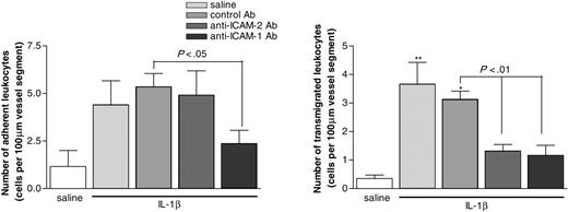
The selective role of ICAM-2 in mediating leukocyte transmigration, but not adhesion, was also indicated in studies in which IL-1β–induced leukocyte responses were quantified in WT mice treated with the anti–ICAM-2 mAb 3C4 (Figure 4). Of interest, in the same series of experiments, an anti–ICAM-1 mAb (YN-1; 3 mg/kg intravenously), but not a control mAb, suppressed both IL-1β–induced leukocyte adhesion and transmigration (Figure 4).
IL-1β– and TNF-α–induced leukocyte responses in mouse cremasteric venules of WT controls and ICAM-2–deficient mice as observed by intravital microscopy. (A) The expression profiles of ICAM-2 and PECAM-1 in representative cremasteric venules of WT mice. Unstimulated cremaster muscles were immunostained and observed for the expressions of ICAM-2 and PECAM-1, as detailed in “Materials and methods.” The figure also shows merged images captured from the 2 channels used. Each image is representative of that obtained from 3 to 5 vessels per tissue (n = 3 mice per group). Bar = 20 μm. (B-C) Mice (WT controls or ICAM-2 KO mice) treated with intrascrotal saline (400 μL per cavity, open bars), IL-1β (50 ng per mouse, closed bars), or TNF-α (300 ng per mouse, closed bars); 4 hours later, leukocyte responses were quantified as detailed in “Materials and methods.” The data represent mean ± SEM of 3 to 7 animals. *P < .05 and **P < .01 versus saline-treated levels. In addition, significant differences between the 2 strains of mice are indicated by lines.
IL-1β– and TNF-α–induced leukocyte responses in mouse cremasteric venules of WT controls and ICAM-2–deficient mice as observed by intravital microscopy. (A) The expression profiles of ICAM-2 and PECAM-1 in representative cremasteric venules of WT mice. Unstimulated cremaster muscles were immunostained and observed for the expressions of ICAM-2 and PECAM-1, as detailed in “Materials and methods.” The figure also shows merged images captured from the 2 channels used. Each image is representative of that obtained from 3 to 5 vessels per tissue (n = 3 mice per group). Bar = 20 μm. (B-C) Mice (WT controls or ICAM-2 KO mice) treated with intrascrotal saline (400 μL per cavity, open bars), IL-1β (50 ng per mouse, closed bars), or TNF-α (300 ng per mouse, closed bars); 4 hours later, leukocyte responses were quantified as detailed in “Materials and methods.” The data represent mean ± SEM of 3 to 7 animals. *P < .05 and **P < .01 versus saline-treated levels. In addition, significant differences between the 2 strains of mice are indicated by lines.
Blockade of ICAM-2 inhibits neutrophil migration in PECAM-1 KO mice
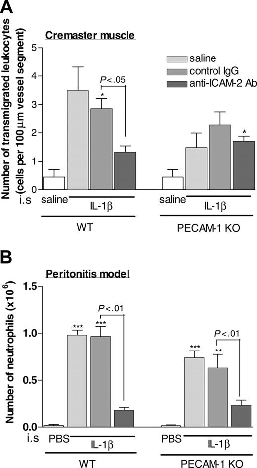
Because the results so far highlighted numerous similarities between ICAM-2 and PECAM-1, in a final series of experiments we sought to investigate the potential additive/cooperative roles of these molecules. For this purpose, the effect of mAb 3C4 was investigated on neutrophil migration in both WT and PECAM-1–deficient mice as induced by IL-1β in the cremaster muscle and peritonitis models (50 ng per mouse intrascrotally or 10 ng per mouse intraperitoneally, respectively, using a 4-hour test period in both). In both models, as previously found (Figures 1 and 4), anti–ICAM-2 mAb suppressed leukocyte transmigration, and PECAM-1–deficient mice exhibited reduced levels of transmigration,7,22 indicating ICAM-2 and PECAM-1 dependency, respectively. However, treatment of PECAM-1 KO mice with the anti–ICAM-2 mAb did not result in a greater level of inhibition of neutrophil transmigration through IL-1β–stimulated cremasteric venules than that seen in untreated PECAM-1 KO mice or PECAM-1 KO mice treated with a control mAb (Figure 5). In contrast, using the peritonitis model, the anti–ICAM-2 mAb induced a significant suppression of neutrophil migration induced by IL-1β in PECAM-1–deficient mice. Collectively, these data suggest that ICAM-2 may mediate PECAM-1–independent neutrophil transmigration in the peritonitis model but not in the cremaster muscle model.
Effect of anti–ICAM-2 mAb (3C4) and anti–ICAM-1 mAb (YN-1) on leukocyte responses in IL-1β–stimulated murine cremasteric venules as observed by intravital microscopy. Animals were treated with saline or IL-1β (50 ng per animal) intrascrotally 4 hours before the surgical preparation. In additional groups of mice, animals were pretreated with intravenous mAbs 3C4 or YN-1 or an isotype-matched control mAb, all at the dose of 3 mg/kg, 15 minutes before the intrascrotal injection of IL-1β. The data represent mean ± SEM from 2 to 8 mice per group. *P < .05 and **P < .01 versus responses obtained from saline-injected animals. Additional statistical comparisons are indicated by lines.
Effect of anti–ICAM-2 mAb (3C4) and anti–ICAM-1 mAb (YN-1) on leukocyte responses in IL-1β–stimulated murine cremasteric venules as observed by intravital microscopy. Animals were treated with saline or IL-1β (50 ng per animal) intrascrotally 4 hours before the surgical preparation. In additional groups of mice, animals were pretreated with intravenous mAbs 3C4 or YN-1 or an isotype-matched control mAb, all at the dose of 3 mg/kg, 15 minutes before the intrascrotal injection of IL-1β. The data represent mean ± SEM from 2 to 8 mice per group. *P < .05 and **P < .01 versus responses obtained from saline-injected animals. Additional statistical comparisons are indicated by lines.
Discussion
In vitro studies have implicated ICAM-2 in leukocyte transendothelial cell migration, but to date there exists no direct in vivo evidence to support this notion. The aim of the present study was to address this issue by the use of both an anti–ICAM-2 mAb and ICAM-2–deficient mice. Collectively, the findings provide direct evidence for the involvement of ICAM-2 in neutrophil transmigration in vivo, an effect that appears to be stimulus specific. Furthermore, by investigating the effect of a blocking anti–ICAM-2 mAb in PECAM-1–deficient mice, we report for the first time that ICAM-2 can mediate PECAM-1–independent leukocyte transmigration.
Historically, the differential patterns of expression of ICAM-1 and ICAM-2 on endothelial cells and the differences in their inducibility by inflammatory mediators has suggested specialized roles for these molecules in the context of leukocyte trafficking. Specifically, because ICAM-1 is expressed at low to moderate levels on resting endothelial cells and its expression is up-regulated by inflammatory cytokines, ICAM-1 has been implicated in regulation of leukocyte migration under inflammatory conditions, and indeed there is now much evidence to support this.17,18,25-27 In contrast, the relatively high constitutive expression of ICAM-2 on endothelial cells (especially on high endothelial venules) and the inability of inflammatory mediators to further enhance this expression led for a long time to the conception that ICAM-2 mediates steady-state recirculation of lymphocytes. Recent in vivo data using both blocking mAbs and ICAM-2–deficient mice has, however, shown that this is not the case.16,19 Indeed, increasing in vitro data indicating a role for ICAM-2 in lymphocyte, monocyte, eosinophil, and neutrophil transmigration through cultured endothelial cells17-19,25-27 suggests a role for ICAM-2 in the regulation of leukocyte migration in inflammation. In vivo evidence to support this possibility is, however, limited.
Effect of anti–ICAM-2 mAb on IL-1β–stimulated leukocyte transmigration in the cremaster muscle and peritoneal cavity of WT and PECAM-1–deficient mice. Wild-type or PECAM-1–deficient mice were injected with intrascrotal (A) or intraperitoneal (B) saline/PBS or IL-1β (50 ng per mouse and 10 ng per cavity, respectively) 4 hours before quantification. In additional groups of mice, IL-1β–injected mice were also pretreated with an isotype-matched control antibody or the anti–ICAM-2 mAb 3C4 (both at 3 mg/kg intravenously) 15 minutes prior to administration of IL-1β. The data represent mean ± SEM from 2 to 9 mice per group in panel A and 3 to 7 mice per group in panel B. *P < .05, **P < .01, and ***P < .001 versus responses obtained from saline-injected animals. Additional statistical comparisons are indicated by lines.
Effect of anti–ICAM-2 mAb on IL-1β–stimulated leukocyte transmigration in the cremaster muscle and peritoneal cavity of WT and PECAM-1–deficient mice. Wild-type or PECAM-1–deficient mice were injected with intrascrotal (A) or intraperitoneal (B) saline/PBS or IL-1β (50 ng per mouse and 10 ng per cavity, respectively) 4 hours before quantification. In additional groups of mice, IL-1β–injected mice were also pretreated with an isotype-matched control antibody or the anti–ICAM-2 mAb 3C4 (both at 3 mg/kg intravenously) 15 minutes prior to administration of IL-1β. The data represent mean ± SEM from 2 to 9 mice per group in panel A and 3 to 7 mice per group in panel B. *P < .05, **P < .01, and ***P < .001 versus responses obtained from saline-injected animals. Additional statistical comparisons are indicated by lines.
The first indications for the involvement of ICAM-2 in leukocyte migration in vivo came from Gerwin et al,19 who using an allergic model of asthma found a delayed and prolonged eosinophil accumulation in the lungs of ICAM-2–deficient mice in response to allergen challenge.19 More recently, Hobden20 reported on the ability of an anti–ICAM-2 mAb to inhibit leukocyte infiltration during ocular infection in ICAM-1–deficient mice.20 The findings of the present study extend these observations in that by using both an anti–ICAM-2 blocking mAb and ICAM-2–deficient mice a direct role for ICAM-2 in mediating neutrophil transmigration in vivo is demonstrated. Specifically, WT mice treated with an anti–ICAM-2 mAb and ICAM-2 KO mice exhibited significantly reduced (about 50% inhibition) levels of neutrophil accumulation into the peritoneal cavity of mice injected with IL-1β. To examine the precise stage in the process of leukocyte migration that is regulated by ICAM-2, IL-1β–induced leukocyte responses in mouse cremasteric venules of WT and ICAM-2 KO mice were investigated, as observed by intravital microscopy. In ICAM-2–deficient animals, while adhesion appeared normal, transmigration was significantly reduced (43%). Similar results were obtained in WT mice treated with an anti–ICAM-2 mAb in that leukocyte transmigration but not firm adhesion was significantly inhibited (64%). In contrast, as expected, an anti–ICAM-1 mAb suppressed both IL-1β–induced leukocyte firm adhesion and transmigration. Indeed, because the anti–ICAM-1 mAb appeared to inhibit leukocyte adhesion and transmigration to the same extent, the findings suggest that in the present model the predominant role of ICAM-1 is in establishing stable leukocyte adhesion while ICAM-2 mediates transmigration (ie, the molecules could be working in series). Hence, while previous studies have suggested that ICAM-2 mediates leukocyte migration through cultured endothelial cells,18,25-27 the present findings provide the first direct evidence for such a role for ICAM-2 in vivo.
Although clear evidence was obtained for a role for ICAM-2 in leukocyte transmigration, the present study did not investigate the relative contribution of leukocyte versus endothelial cell ICAM-2 in this response. Because there is now evidence to suggest that ICAM-2 can exhibit homophilic interaction,15 both leukocyte and endothelial cell ICAM-2 may potentially contribute to ICAM-2–mediated leukocyte transmigration. However, because ICAM-2 can interact with the leukocyte integrins LFA-1 and Mac-1,28,29 it would seem highly probable that ICAM-2–dependent leukocyte transmigration in vivo is mediated via interaction of endothelial cell ICAM-2 with its leukocyte integrin ligands. In support of this, using chimeric mice as developed by bone marrow transfer and in vitro transmigration assays using ICAM-2–deficient endothelial cells, the findings of Gerwin et al suggest that ICAM-2 expressed on endothelial cells mediates eosinophil transmigration.19 In agreement with these findings, ICAM-2 in cremasteric venules was noted to be expressed at endothelial cell junctions, as observed by immunofluorescent staining and confocal microscopy.
The role of ICAM-2 in leukocyte transmigration appeared to be stimulus specific in that, in both the peritonitis model and the cremaster muscle model, while IL-1β–induced transmigration was suppressed by ICAM-2 blockade/deletion, neutrophil migration elicited by TNF-α (and thioglycolate in the peritonitis model) was normal in ICAM-2–deficient animals. The reason for this stimulus-specific activation of the ICAM-2–dependent pathway is currently unclear but may be governed at multiple levels (eg, the target cell being activated [leukocyte versus endothelial cell] and the type of inflammatory receptor and hence signaling pathways activated). Of interest, the detected stimulus-specific profile of ICAM-2–dependent transmigration is directly in line with the observations we have previously made with respect to PECAM-1–dependent transmigration.7,23 Hence, as found in ICAM-2–deficient mice, in PECAM-1–deficient animals, neutrophil transmigration elicited by IL-1β, but not TNF-α or thioglycolate, was suppressed. The molecular basis of these observations is at present unclear.
With respect to PECAM-1, because leukocyte transmigration is highly dependent on the ability of PECAM-1–PECAM-1 interaction to activate leukocyte integrins,22 it is hypothesized that stimuli that can directly stimulate leukocytes and hence activate their integrins can induce leukocyte transmigration in a PECAM-1–independent manner (ie, the need for PECAM-1–mediated integrin activation is bypassed). However, at present there is no evidence to suggest that ICAM-2–mediated transmigration is regulated through integrin activation and indeed, in contrast to PECAM-1 and in the context of leukocyte–endothelial cell interaction, the principal ligands for ICAM-2 appear to be integrins.28-30 Furthermore, because it seems that JAM-A–dependent transmigration also exhibits a similar stimulus-specific profile (ie, IL-1β–induced leukocyte transmigration is JAM-A dependent, but leukocyte transmigration elicited by chemoattractants such as LTB4 and PAF is JAM-A independent [A.W., Christoph Reichel, Andrej Khandoga, Monica Corada, Elisabetta Dejana, Fritz Krombach, and S. N., manuscript in preparation]), a more generalized hypothesis needs to be considered. At present, collectively the available data suggest that stimuli that activate endothelial cells (eg, IL-1β) recruit adhesion pathways involving endothelial cell junctional molecules (eg, PECAM-1, ICAM-2, and JAM-A), whereas stimuli that can directly stimulate leukocytes (eg, TNF-α, FMLP, LTB4) appear to bypass the need for such molecules. While the underlying mechanism associated with these findings is currently unknown, the findings further highlight the existence of stimulus-specific adhesion pathways that mediate leukocyte transmigration. This may account for some apparent contentious and/or inconsistent reports with respect to the potential involvement of specific adhesion molecules in the regulation of leukocyte migration in different inflammatory models in vivo.
Although there is now increasing in vitro and in vivo evidence illustrating the involvement of individual endothelial cell junctional molecules in the process of leukocyte transendothelial cell migration, very few studies have addressed the potential additive/synergistic effects of multiple molecules. One such study is by Schenkel and colleagues in which an anti–PECAM-1 mAb was found to act in an additive manner with a CD99 blocker to inhibit monocyte transendothelial cell migration in vitro.31 Because in numerous inflammatory models PECAM-1 blockade/deletion results in partial suppression of leukocyte transmigration, in a final series of experiments we aimed to investigate the possibility that ICAM-2 may mediate PECAM-1–independent leukocyte transmigration. For this purpose the effect of the anti–ICAM-2 mAb, 3C4, on leukocyte transmigration in WT and PECAM-1–deficient mice was directly compared using both the cremaster muscle and peritonitis models. In line with data discussed above, pretreatment of WT mice with mAb 3C4 resulted in a significant inhibition of neutrophil transmigration in response to IL-1β in both models. However, in PECAM-1–deficient mice, although a significant suppression of IL-1β–induced leukocyte transmigration was noted in both in vivo models (in agreement with previous data),22 the anti–ICAM-2 mAb, 3C4, appeared to have no additional inhibitory effects in the cremaster muscle but significantly inhibited migration in the peritonitis model. This may suggest that the PECAM-1–independent neutrophil transmigration detected in the cremaster muscle model is also ICAM-2 independent. In contrast, the PECAM-1–independent response in the peritonitis model may be mediated by ICAM-2, indicating for the first time an in vivo inflammatory scenario in which ICAM-2 and PECAM-1 can mediate leukocyte transmigration in an additive manner. Because the blockade of leukocyte transmigration in PECAM-1–deficient mice has previously been shown to occur at the level of the perivascular basement membrane,6,7 it is potentially possible that the additive effects of ICAM-2 blockade and PECAM-1 deletion may be due to inhibition of leukocyte transmigration at different steps (ie, migration through endothelial cell junctions and the venular basement membrane, respectively). In line with this, it is of course also possible that in the peritonitis model ICAM-2 may mediate leukocyte firm adhesion as opposed to/in addition to blockade of leukocyte transmigration, though we do not have any evidence at present to support such a notion. Interestingly, because in both inflammatory models mice treated with the anti–ICAM-2 mAb exhibited the same level of leukocyte transmigration in WT and PECAM-1 KO mice, the data can potentially indicate that PECAM-1 has no role in ICAM-2–independent leukocyte transmigration. Studies involving genetic deletion of both PECAM-1– and ICAM-2–dependent pathways are planned to further address such issues.
In summary, the findings of the present study provide direct evidence supporting a role for ICAM-2 in neutrophil transmigration in vivo. Furthermore, the role of ICAM-2 in this response appears to be stimulus specific, and ICAM-2 may act in an additive manner with PECAM-1 to mediate leukocyte transmigration. While these results raise many additional questions related to the process of leukocyte transmigration, the findings do make an important contribution to the field of leukocyte trafficking by identifying ICAM-2 as a significant and nonredundant regulator of this response in vivo.
Prepublished online as Blood First Edition Paper, February 9, 2006; DOI 10.1182/blood-2005-11-4683.
Supported by the British Heart Foundation (M.-T.H., K.Y.L., C.S., D.O.H., S.N.), Overseas Research Student award (M.-T.H.), European Commission Framework 6 (MAIN network of excellence, FP6-502935) (A.W.), and Aventis Pharma Deutschland (N.G.).
M.-T.H. designed and performed experiments, analyzed data, and helped with the writing of the paper; K.Y.L. designed and performed experiments and contributed to data analysis, interpretation, and the writing of the paper; C.S. and A.W. performed experiments; N.G. provided a key reagent; D.O.H. contributed to design of research, data analysis and interpretation, and the writing of the paper; S.N. initiated the study, designed and supervised the research, contributed to data analysis and interpretation, and wrote the paper.
M.-T.H. and K.Y.L. contributed equally to this work.
An Inside Blood analysis of this article appears at the front of this issue.
The publication costs of this article were defrayed in part by page charge payment. Therefore, and solely to indicate this fact, this article is hereby marked “advertisement” in accordance with 18 U.S.C. section 1734.


This feature is available to Subscribers Only
Sign In or Create an Account Close Modal