Abstract
Non-Hodgkin lymphomas are among the most common primary tumors occurring in the ocular adnexa. Herein, we present a 14-year single-institution experience in 62 patients with primary ocular adnexal lymphomas (OALs). Association with Chlamydia psittaci infection is examined in 57 tumor specimens.
Extranodal marginal zone lymphoma (EMZL) was the most frequent histologic subtype (89%). The majority of patients with EMZL (84%) presented with stage E-extranodal (IE), however only 16% had an advanced stage. All stage IE patients were treated with local radiotherapy, whereas patients with disseminated disease received systemic therapy with or without local irradiation. All but 1 patient with EMZL achieved complete remission (CR). During a median follow-up of 52 months (range, 3-153 months), the estimated 5-year overall survival (OS) and freedom from progression (FFP) were 96% and 79%, respectively. During the follow-up, 22% of patients relapsed, mainly in extranodal sites, and 4% transformed to diffuse large B-cell lymphoma. None of the patients exhibited local orbital failure in the radiation field. None of the OAL specimens harbored C psittaci DNA.
Our study demonstrates that EMZLs, accounting for the majority of primary OALs, are characterized by an indolent natural history with frequent, continuous extranodal relapses. In South Florida, OALs are not associated with C psittaci infections.
Introduction
Ocular adnexal lymphomas (OALs) represent a small fraction of all systemic non-Hodgkin lymphomas (NHLs; 6% of all primary extranodal NHLs,1 7% of nongastric marginal zone lymphomas2 ). However, they are among the most common tumors occurring in the ocular adnexa (26%).3 These lymphomas may involve any part of the eye socket, including orbit, conjunctiva, lids, and the lacrimal gland or sac.4-7 OALs can be an initial presentation of NHL, either solitary or systemic, or may represent dissemination of previously diagnosed lymphoma. OALs may present with symptoms of painless proptosis and/or diplopia due to an orbital mass, conjunctival salmon patches, or ptosis from eyelid involvement, and these tumors may spread locally or disseminate systemically.4
Nearly all OALs are of B-cell phenotype, the majority being the extranodal marginal zone lymphomas (EMZLs) of mucosa-associated lymphoid tissue (MALT lymphoma).4,5,8-10 EMZLs account for between 35% and 90% of OALs, whereas follicular and diffuse large-cell lymphomas each account for approximately 10%.4,5,8-10 This is in marked contrast to the overall incidence of EMZLs, which account for only 7% to 8% of all NHLs, being less common than the diffuse large-cell and follicular lymphomas.11-13 Although several studies attempted to address the clinical and pathologic aspects of OALs, interpretation of these studies is confounded by several factors: (a) inclusion of both primary and systemic NHL secondarily involving ocular adnexa5,7,10 ; (b) inclusion of reactive lymphoid hyperplasia cases and improper histologic classification of lymphoproliferative lesions of the ocular adnexa in studies performed before the recognition of EMZL as a distinct pathologic entity and its incorporation into the Revised European-American Classification of Lymphoid Neoplasms (REAL) and the World Health Organization (WHO) classifications4,6,7 ; (c) small cohorts of patients and/or relatively short follow-up.6,10,14
Furthermore, the etiology of OALs and the reason for the predominance of EMZL in this anatomic location are unknown. EMZL arises in lymphoid tissue acquired through chronic antigenic stimulation because of persistent inflammation or infection,15 as exemplified by Helicobacter pylori infection in patients with gastric lymphoma,16 Borrelia burgdorferi and Campylobacter jejuni infections in patients with skin17,18 and intestinal19 MALT lymphomas, respectively, and association of the salivary and thyroid gland MALT lymphomas with Sjögren syndrome20 and Hashimoto thyroiditis.21 However, the majority of patients with OALs do not report any preceding chronic inflammatory or infection processes in the affected tissues. Interestingly, Ferreri et al22 recently reported the presence of Chlamydia psittaci DNA in 80% of the analyzed Italian ocular adnexa EMZL cases, thus raising the possibility that this is the etiologic pathogen of these tumors. Furthermore, the same group of investigators demonstrated complete or partial response to doxycycline treatment in 4 of 9 patients with C psittaci–positive EMZL of the ocular adnexa.23 However, these findings need to be confirmed in independent studies.
Herein we present a 14-year single-institution experience in a large cohort of OAL patients with a long follow-up. Clinicopathologic characteristics, response to therapy, systemic dissemination, and patients' survival are analyzed. Furthermore, association with C psittaci infection is examined.
Patients, materials, and methods
Patients and pathologic specimens
Between 1991 and 2004, 87 patients with primary presentation of NHL in ocular adnexa were seen at the University of Miami, Sylvester Comprehensive Cancer Center. Patients with history of extraocular lymphoma were excluded. Twenty-five of the 87 cases were seen for consultation only and did not have complete lymphoma work-up, histologic specimens for pathologic review, or clinical follow-up data. The remaining 62 cases were evaluated, treated, and followed up at our institution and form the basis of this report. The study was approved by the University of Miami Institutional Review Board. Informed consent was provided according to the Declaration of Helsinki. Patients' charts and laboratory database were reviewed and information on patient age, sex, medical history, clinical complaints at presentation, clinical findings, anatomic location of the lesion, laboratory data, radiologic investigations, stage, type, and extent of therapy, disease course, disease-free survival, freedom from progression survival, re-treatment therapy on recurrence or progression, overall survival, and evidence of disease at the last follow-up were retrieved. Patients with only bilateral ocular adnexal disease were considered to have stage E-extranodal (IE) disease along the convention of the published studies.4,7,10,24 The anatomic localization of OAL was defined as orbital, conjunctival, lacrimal gland, and eyelid, based on ophthalmologic examination and computed tomography (CT) or magnetic resonance imaging (MRI) of the orbits performed in all of the patients. The staging procedures were not standardized and included full physical examination; hematologic and chemical survey with or without lactate dehydrogenase (LDH); CT of the neck, chest, abdomen, and pelvis; bone marrow biopsy and aspiration; sonography of the salivary glands; and gastroscopy.
All pathologic specimens were re-examined and classified according to the WHO classification (G.E.B.) on the basis of the morphologic features observed on routinely prepared hematoxylin and eosin–stained slides of formalin-fixed, paraffin-embedded tissues along with immunophenotypic and genotypic results, without the knowledge of clinical information. Flow cytometry immunophenotyping was performed in most cases and Southern blot analysis or polymerase chain reaction (PCR) for immunoglobulin heavy-chain gene rearrangement were performed in 44 specimens, as previously reported.25,26 The diagnosis of lymphoma was confirmed in all of the patients, however, in 3 cases, the specimens were too small and no flow cytometry data were available to differentiate small lymphocytic lymphoma versus EMZL.
Detection of C psittaci DNA
The DNA was extracted from a total of 59 fresh or paraffin-embedded tumors, which included 49 specimens of EMZL of ocular adnexa, 8 specimens of non-EMZL of ocular adnexa (including several specimens from patients seen for consultation only and thus not included in the cohort that formed the basis of clinical studies in this report), and 2 specimens from patients with reactive ocular lymphoid hyperplasia. From fresh tumors, the DNA was extracted using a commercially available kit as described by the manufacturer (QIAamp DNA mini kit; Qiagen, Valencia, CA). For DNA extraction from paraffin wax–embedded tissue, we have used the protocol reported by Bonin et al.27 The process of paraffin wax embedment of biopsy tissues usually results in extensive degradation of nucleic acids, leading to an average fragment length of DNA of 300 to 400 bp. Therefore, to ascertain integrity and estimate the quality of DNA extracted from fresh specimens, 4 and/or 8 μL of the template DNA was used to amplify a 724–base pair (bp) fragment of intronic β-globin (βG; gene symbol HBB) DNA, spanning exons 2 and 3 (GenBank accession no. V00499) with PCR primers (forward HBB primer, 5′-AGGAAGGGGAGAAGTAAC; and the reverse primer, 5′-AATCCAGCC TTATCCCAA, at a final concentration of 0.5 μM) using a previously reported protocol.28 To ascertain the integrity of DNA extracted from paraffin-embedded tissues, same amounts of DNA were used to amplify a 287-bp fragment of apolipoprotein E (APOE; forward primer, 5′-AAGGAGTTGAAGGCCTACAAAT; and a reverse primer, 5′-GGCCTGGTACACTGCCAG, at a final concentration of 0.5 μM), as previously reported.27 Successful amplification of this fragment would suggest that the extracted DNA is of an expected quality sufficient for amplification of short DNA segments.
DNA samples positive for either βG or ApoE amplicons were used for touchdown enzyme time-release PCR (TETR-PCR) for C psittaci with CPS 100 (5′-CCCAAGGTGAGGCTGATGAC) and CPS 101 (5′-CAAACCGTCCTAAGACAGTTA) primers at a final concentration of 0.6 μM, amplifying a 111-bp amplicon.22,29 Briefly, 4 to 8 μL of template DNA was added to a PCR tube containing 17 to 21 μL of PCR mixture with primers, respectively, and amplified on a GeneAmp PCR System 9700 (Applied Biosystems, Foster City, CA) using the TETR-PCR protocol previously reported by Madico et al.29 These primers amplify a highly conserved and specific rRNA region of the C psittaci genome. DNA prepared from the C psittaci strain 6 BC (American Type Culture Collection [ATCC], Manassas, VA), according to the protocol reported by Shaw et al,30 was used as a positive PCR reaction control. TETR-PCR assay was reported to detect C psittaci DNA at bacterial loads lower than 1 infection-forming unit.22,29 Blank reactions filled with 25 μL PCR mixture were always run concomitantly with the DNA from patients' specimens to monitor for possible contamination of PCR reagents and to rule out false-positive results. All of the reactions were repeated twice. PCR products were separated by 12%polyacrylamide gel electrophoresis and visualized with ethidium bromide.
Statistical analysis
Overall survival (OS) was calculated from the start of treatment to death from any cause or the last follow-up. Freedom from progression (FFP) was calculated from the start of treatment until disease progression, transformation, relapse, or last follow-up. For patients who remained progression free but died of OAL-unrelated causes, the follow-up was censored at the time of death. OS and FFP were estimated by the method of Kaplan-Meier.31 Comparisons were assessed for significance using the log-rank test.32
Results
A total of 62 patients (42 women and 20 men, female-male ratio of 2:1) with primary presentation of NHL in ocular adnexa were treated and followed up at the University of Miami, Sylvester Comprehensive Cancer Center between 1991 and 2004. Clinical characteristics of the patients at the diagnosis and the pathologic diagnoses are summarized in Table 1. The OALs were anatomically distributed as follows: orbit in 33 patients (53%), conjunctiva in 16 patients (26%), lacrimal gland in 12 patients (19%), and lacrimal sac in 1 patient. None of the patients had synchronous involvement of more than 1 ocular anatomic site. However, bilateral involvement was observed in 6 patients (10%). The most common presenting symptoms were mass/swelling in 29 cases (46%), eye redness in 14 (22%), and epiphora in 10 (16%), whereas pain (3%), proptosis (4%), ptosis (9%), and diplopia (3%) were present in a smaller number of patients. None of the patients had extraocular complaints or B symptoms. The duration of the symptoms at presentation ranged from 2 months to more than 2 years with a median of 8 months. None of the patients had a preceding history of Sjögren syndrome, Mickulickz syndrome, or Wegener granulomatosis. Table 1 shows the frequency of the various WHO histologic subtypes of NHL presenting as OAL in our patients. EMZL was the most frequent histologic subtype, accounting for 89% of the cases (55 patients). All of the EMZL cases that were examined by flow cytometry studies expressed the CD19+, CD20+, CD10–, CD5–, and CD23– immunophenotype. A monoclonal immunoglobulin rearrangement band was detected in all of the 44 analyzed EMZL specimens. In 3 additional cases, the biopsy specimens were too small and flow cytometry studies were not performed to enable differentiation between EMZL and small lymphocytic lymphomas (SLLs).
Clinical features of patients with primary ocular adnexal lymphoma
Characteristic . | All . | EMZL . | Non-EMZL . |
|---|---|---|---|
| No. of patients | 62 | 55 | 7 |
| Age, y | |||
| Median | 65 | 66 | 59 |
| Range | 30-92 | 30-92 | 49-91 |
| Sex, no. male/no. female | 42/20 | 35/20 | 7/0 |
| Involved sites, no. | |||
| Orbital | 33 | 26 | 7 |
| Conjunctivae | 16 | 16 | 0 |
| Lacrimal apparatus | 13 | 13 | 0 |
| Stage, no. | |||
| I | 53 | 46 | 7 |
| II | 2 | 2 | 0 |
| III | 0 | 0 | 0 |
| IV | 7 | 7 | 0 |
| Histologic category, no. | |||
| EMZL | 55 | 55 | NA |
| SLL | 2 | NA | 2 |
| DLBCL | 2 | NA | 2 |
| Undetermined | 3 | NA | 3 |
EMZL indicates extranodal marginal zone lymphoma; SLL, small lymphocytic lymphoma; DLBCL, diffuse large B-cell lymphoma; and NA, not applicable.
The initial staging procedure revealed that 46 (84%) patients with EMZL presented with stage IE disease, 2 presented with stage II disease (cervical lymphadenopathy), and 7 patients presented with stage IV disease (bone marrow involvement in 5 patients and asymptomatic skin and palate involvement in 1 patient each). In patients with systemically disseminated EMZL, the ocular adnexas primarily involved were lacrimal apparatus in 4 patients, orbit in 4 patients, and conjunctiva in 1 patient.All of the patients with non-EMZL or unclassified lymphomas presented with stage IE disease.
Radiation treatment techniques varied according to the specific orbital involvement for each patient. In general, patients with lacrimal gland tumors were treated using a wedge pair x-ray technique. Eyes with tumor confined anteriorly to the conjunctiva were usually approached with a direct electron beam and the use of an internal conformer with a corneal shield that was placed beneath the eyelids daily for treatment. Tumors with both anterior and retrobulbar components were treated by a mixed direct photon and electron beam with the electron energy and relative beam weighting being chosen to treat the anterior extent of lymphoma and the orbital contents back to the conus within the 80% isodose line. All of the patients with stage IE unclassified or non-EMZL received solitary orbital radiation therapy with a dose of 36 and 45 Gy in the 2 patients with diffuse large B-cell lymphoma (DLBCL), respectively, and 36 Gy in patients with small lymphocytic and unclassified lymphomas. All of these patients achieved complete remission (CR). During a mean follow-up of 32 months (range, 12 to 75 months) 2 patients relapsed: a 74-year-old patient with DLBCL treated with 36 Gy irradiation to the eye presented 19 months later with histologically confirmed relapse in right knee and brain, which led to patient's death; a 90-year-old patient with SLL treated with orbital irradiation presented 16 months later with histologically confirmed relapse in inguinal lymph node.
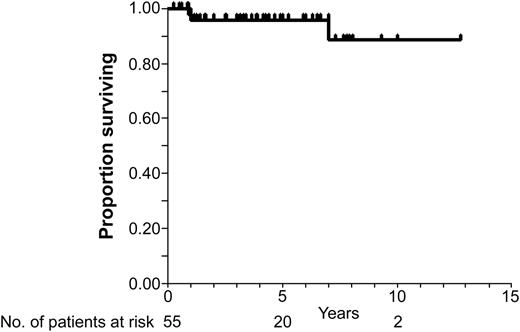
OS for 55 patients with extranodal marginal zone lymphoma of ocular adnexa. Kaplan-Meier estimates of 5- and 7-year survival were 96% (95% CI: 90%-100%) and 89% (95% CI: 74%-100%).
OS for 55 patients with extranodal marginal zone lymphoma of ocular adnexa. Kaplan-Meier estimates of 5- and 7-year survival were 96% (95% CI: 90%-100%) and 89% (95% CI: 74%-100%).
In the patients with stage IE EMZL, the radiation dose ranged from 30 Gy to 45 Gy (in 2 patients); the majority of the patients received either 30 or 36 Gy. Patients with ocular adnexal EMZL presenting at stages II and IV were treated with the following regimens: chlorambucil and prednisone with orbital irradiation (4 patients, 24-30 Gy); rituximab, cyclophosphamide, vincristine, prednisone (R-CVP) alone (1 patient) or in combination with radiation (2 patients, 24 and 30 Gy); and rituximab alone (1 patient) or with radiation (1 patient, 30 Gy). All but one EMZL patient achieved CR or CR unconfirmed (CRu) (1 patient with stage IV disease demonstrated residual retroperitoneal lymphadenopathy, which decreased by more than 75% from its original pretreatment size). The single patient that did not achieve CR was an 87-year-old patient with stage IV disease that achieved local orbital control with irradiation (30 Gy) but progressed systemically on chlorambucil and prednisone treatment. Rebiopsy at the time of progression (6 months after diagnosis) demonstrated transformation to DLBCL.
The orbital radiation therapy was generally well tolerated, with mild acute effects such as dryness or excessive tearing, loss of eyelashes/eyebrow, and periorbital erythema and edema. Cataract development was documented in 10% of irradiated eyes.
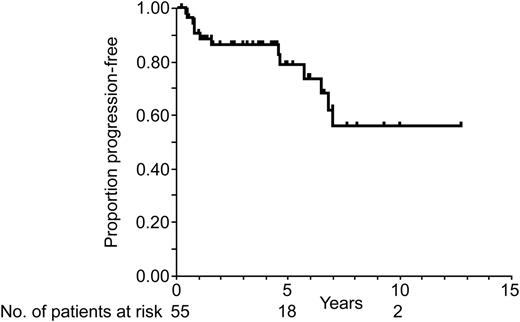
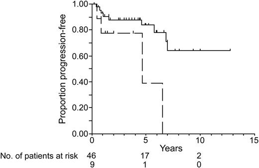
OS and FFP of ocular adnexal patients with EMZL are presented in Figures 1 and 2. Fifty-two patients were alive after a median follow-up of 52 months (range, 3 to 153 months). Kaplan-Meier estimates of 5- and 7-year OS were 96% (95% confidence interval [CI]: 90%-100%), and 89% (95% CI: 74%-100%), respectively. Median survival time was not attained. No difference in OS was observed between patients with conjunctival, orbital, and lacrimal gland EMZL (not shown). Progressive disease, either relapse in 12 patients or progression with transformation to DLBCL in 1 patient, was observed after a median follow-up of 19 months (range, 5 to 84 months), whereas 42 patients remained progression free after a median follow-up of 49 months (range, 3 to 153 months). Kaplan-Meier estimates for the proportion of FFP patients at 1, 5, and 10 years after starting treatment were 91% (95% CI: 83%-98%), 79% (95% CI: 65%-92%), and 56% (95% CI: 34%-77%), respectively. The median time to progression was not attained. None of the relapses occurred in the irradiation field, thus demonstrating local control in all of the irradiated patients. Three of the relapses occurred in patients who originally presented with stage IV disease, whereas 9 relapses were observed in patients with stage IE at presentation. FFP was significantly shorter for patients who presented with stage II-IV disease compared with patients who presented with stage IE disease (P = .016; Figure 3). However, even in the patients with initial stage IE disease, the relapses occurred continuously with estimates of the cumulative relapse/progression of 7% at 1 year, 16% at 5 years, and 36% at 10 years. Only one of the relapsed patients had bilateral OAL at the time of original presentation. The relapse was disseminated in 4 patients but localized to 1 anatomic site in 8 patients. Noticeably, extranodal sites were commonly involved at the time of relapse, including lung and contralateral orbit (each in 2 patients), as well as breast, kidney, muscle, epidural space, and nasal and oral mucosa (each in 1 patient). In all of the cases, the relapse was confirmed by repeated biopsy. In one patient that achieved initial response, biopsy performed at the time of relapse demonstrated transformation to DLBCL. The patient was treated with autologous bone marrow transplantation and is alive at the last follow-up, 12 years after original diagnosis.
FFP for 55 patients with extranodal marginal zone lymphoma of ocular adnexa. Kaplan-Meier estimates for the proportion of progression-free patients 1, 5, and 10 years after starting treatment are 91% (95% CI: 83%-98%), 79% (95% CI: 65%-92%), and 56% (95% CI: 34%-77%), respectively.
FFP for 55 patients with extranodal marginal zone lymphoma of ocular adnexa. Kaplan-Meier estimates for the proportion of progression-free patients 1, 5, and 10 years after starting treatment are 91% (95% CI: 83%-98%), 79% (95% CI: 65%-92%), and 56% (95% CI: 34%-77%), respectively.
FFP according to the initial presentation stage of patients with extranodal marginal zone lymphoma of ocular adnexa. The solid line indicates stage IE patients; the dotted line, stage II-IV patients, P = .016.
FFP according to the initial presentation stage of patients with extranodal marginal zone lymphoma of ocular adnexa. The solid line indicates stage IE patients; the dotted line, stage II-IV patients, P = .016.
During the follow-up period, only 3 patients with EMZL of ocular adnexa had died. One patient with bilateral stage IE disease relapsed systemically 11 months after diagnosis, and, despite treatment with chlorambucil followed by R-CVP, the disease progressed and the patient died 1 year later. Two additional patients at advanced age died in CR from unrelated causes.
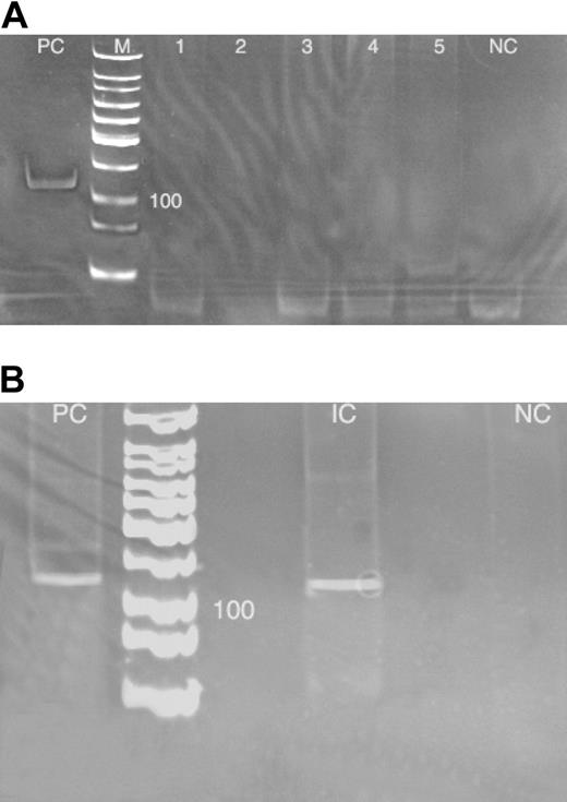
To analyze the recently reported association between C psittaci infection and OALs22 in our patient population, DNA was extracted from 49 specimens of EMZL of ocular adnexa, 8 specimens of non-EMZL of ocular adnexa, and 2 specimens from patients with reactive ocular lymphoid hyperplasia (some of these patients were seen in our center for consultation only, and thus were not part of the clinical follow-up study, as is described in “Patients, materials, and methods”). HBB was successfully amplified from 46 specimens of EMZL of ocular adnexa, 7 specimens of non-EMZL of ocular adnexa, and the 2 specimens with reactive ocular lymphoid hyperplasia. APOE was amplified from an additional 3 specimens of EMZL of ocular adnexa and 1 specimen with non-EMZL of ocular adnexa. C psittaci DNA was not detected in any of the DNA samples positive for either βG or ApoE amplicons, whereas control reactions using DNA prepared from C psittaci strain 6 BC yielded amplicon of expected size (Figure 4A). Furthermore, C psittaci amplicon of expected size was amplified from DNA extracted from a previously reported C psittaci–positive Italian case of EMZL of ocular adnexa, generously provided by Drs Ferreri and Ponzoni (Ferreri et al22 ) (Figure 4B).
Discussion
Primary OAL is a distinct clinical entity in which natural history, etiology, and pathogenesis are not well characterized. Although several large series on OALs have been published recently in ophthalmologic literature,4,5,7-10,14,33,34 they mainly focus on clinical presentation, pathologic aspects, and therapy, whereas the extent of dissemination at the time of diagnosis, relapse pattern, and the natural history are less emphasized. Since the majority of patients with OALs initially present to ophthalmologists, the current subdivision of OALs to primary and secondary adnexal lymphomas, based on limited eye involvement and evidence of disease dissemination or previous history of lymphoma, is not practical, since only thorough patient staging can discriminate between them. However, the frequency of systemic dissemination at the time of diagnosis in patients who present with primary ocular complaints is largely unknown. Herein, we present our experience with a large group of primary OAL patients, defined based on the absence of prior history of lymphoma or extraorbital complaints at presentation, which were treated and followed up for an average of 52 months at a single institution. Furthermore, we examine the recently reported association of OALs with C psittaci in South Florida patients.
Touchdown enzyme time-release PCR for C psittaci in ocular adnexal EMZL tumor specimens. (A) South Florida ocular adnexal EMZL tumor specimens. PC indicates positive control (C psittaci strain 6 BC; ATCC); M, molecular ladder; lanes 1-5, 5 negative ocular adnexal EMZLs; and NC, negative control (PCR mixture with primers, but without DNA input). (B) Reanalysis of an Italian case of a C psittaci–positive ocular adnexal EMZL tumor specimen. PC indicates positive control (see panel A description for details); IC, Italian C psittaci–positive case; and NC, negative control (see panel A description for details).
Touchdown enzyme time-release PCR for C psittaci in ocular adnexal EMZL tumor specimens. (A) South Florida ocular adnexal EMZL tumor specimens. PC indicates positive control (C psittaci strain 6 BC; ATCC); M, molecular ladder; lanes 1-5, 5 negative ocular adnexal EMZLs; and NC, negative control (PCR mixture with primers, but without DNA input). (B) Reanalysis of an Italian case of a C psittaci–positive ocular adnexal EMZL tumor specimen. PC indicates positive control (see panel A description for details); IC, Italian C psittaci–positive case; and NC, negative control (see panel A description for details).
Our study demonstrates several points. EMZL accounts for a majority of primary OALs (89%), which are localized in 84% but not all of the cases. Only thorough staging, including bone marrow examination, reveals disease dissemination in up to 16% (95% CI: 8%-29%) of patients that do not exhibit any extraorbital symptoms. This observation is similar to the recently reported 25% incidence of ocular adnexal EMZL dissemination at the time of diagnosis.24 In our series the lymphoma most commonly originated in the orbit, followed by lacrimal apparatus and conjunctivae, similar to previous reports.4,8,34 However, we did not observe cases with eyelid involvement. The latter observation may result from our definition of primary OALs, since eyelid involvement is frequently associated with dissemination of orbital or systemic lymphomas.7,34
In our cohort of patients, we achieved an excellent control of localized EMZL of ocular adnexa with irradiation, in concordance to previous reports.14,33 There was no difference in local lymphoma control between patients treated with 30 Gy or 36 Gy or higher doses, thus suggesting that the 30-Gy dose may be sufficient for localized disease control. These findings corroborate the recent observations.14,24 Le at al14 reported 100% local control in 31 cases of EMZL of the orbit treated with 30 to 40 Gy, whereas Fung et al24 reported 100% 5-year actuarial local control rate in patients with EMZL of ocular adnexa treated with radiation doses of 30 Gy or higher but only 86% control rate for lower than 30 Gy. Furthermore, our findings may help to narrow the optimal irradiation dose for these patients to 30 Gy. Despite the excellent local control, extraorbital or contralateral relapses occurred in an estimated 16% of patients within 5 years. Some of the relapses occurred after a prolonged follow-up period and no plateau was observed on FFP survival curves, thus suggesting that patients with stage I EMZL of ocular adnexa face a continuous risk of distant relapse that increases from an estimated cumulative progression rate of 16% at 5 years to 36% at 10 years. The high rate of systemic relapse for stage I ocular adnexal EMZL stands in strong contrast to the results reported for localized MALT lymphoma of the stomach, which demonstrate a very low risk of distant recurrence,35,36 but is concordant to the high risk of systemic relapse observed in other nongastric EMZLs.2 Noticeably, similar to other EMZLs, the relapses were frequently extranodal.2 This observation suggests that EMZLs of ocular adnexa express specific homing receptors or adhesion molecules that control extranodal dissemination. Despite the disease dissemination, the relapses are usually well controlled with chemotherapy, since in only 1 patient (2%) with EMZL of ocular adnexa was the death attributed to lymphoma. Furthermore, EMZL of ocular adnexa may transform to DLBCL in 4% of the patients. Although our study clearly demonstrates the indolent natural history of EMZL of ocular adnexa, characterized by continuous risk of systemic dissemination and transformation, the precise incidence of these events might be incorrectly estimated by the inherent weakness of any retrospective study.
The most intriguing finding of this study was the observation that in contrast to Italian cases of OALs,22 EMZLs of ocular adnexa in South Florida are not associated with C psittaci infection. MALT lymphomas of the stomach, thyroid, and salivary glands are commonly associated with chronic inflammations, such as H pylori gastritis, Hashimoto thyroiditis, and Sjögren syndrome, respectively,8,15,16,34 which may predispose to lymphoma development. A tentative pathogenesis of MALT lymphomas might be recruitment to the mucosal regions that normally do not contain lymphocytes, of B and T lymphocytes, as part of the inflammatory or immune response. Recruited lymphocytes undergo chronic antigenic stimulation and clonal selection, which may lead to antigen independence and development of MALT lymphomas. The pathogenesis of OALs and the immune stimuli predisposing to this malignancy are largely unknown. A single case associating Chlamydia trachomatis–induced adult inclusion conjunctivitis with conjunctival EMZL was reported.37 Recently, Ferreri et al22 examined whether Chlamydia psittaci, trachomatis, and pneumoniae infections may be associated with OALs in Italian patients. C psittaci DNA was detected in 80% of 40 patients with OALs but only in 12% of patients with reactive lymphoid hyperplasia, whereas the DNA of other Chlamydia species was not detected. Furthermore, antichlamydial antibiotic therapy, even at the time of relapse, led to objective response (> 50%) in 44% of the reported patients.23 In contrast, in the present study, which used identical methodology, none of the 57 OAL specimens harbored C psittaci DNA. The reason for this marked discrepancy is unclear.
Previous epidemiologic studies analyzing association between hepatitis C and NHL demonstrated marked geographic differences, as summarized by Morgensztern et al.38 For example, hepatitis C infection is commonly associated with lymphoma in Italy but not in South Florida. Similarly, B burgdorferi has been associated with cutaneous extranodal marginal zone lymphomas in several European countries but not in North American cases.17,18,39-41 It is thus possible that there is a regional variation in EMZL etiology and association with infection pathogens. Noticeably, history of chronic conjunctivitis was reported in 35% of Italian cases22 but in none of the cases analyzed herein, thus suggesting different etiology of OALs on the 2 continents. Whether in the case of OALs the genetic and phenotypic differences between C psittaci strains present in Europe and South Florida might account for the differences observed in ours and Ferreri et al's22 studies is currently unknown. Additional studies evaluating the association between C psittaci and EMZL of ocular adnexa in other geographic regions of the world and North America as well as a further search for alternative pathogens in the regions without such association are needed.
Prepublished online as Blood First Edition Paper, September 15, 2005; DOI 10.1182/blood-2005-06-2332.
Supported by RO1 CA109335 from the United States Public Health Service, National Institutes of Health (I.S.L.); and the Dwoskin Family Foundation (I.S.L.).
M.F.R. performed the research, analyzed the data, and participated in preparation of the manuscript; G.E.B. performed the research and data analysis; F.D., P.R., S.R.D., and A.M. performed the research; K.A.F. provided reagents and helped with research performance and data analysis; G.R.W. analyzed the data; and I.S.L. designed the research, performed and supervised the research, analyzed the data, and wrote the manuscript.
The publication costs of this article were defrayed in part by page charge payment. Therefore, and solely to indicate this fact, this article is hereby marked “advertisement” in accordance with 18 U.S.C. section 1734.


This feature is available to Subscribers Only
Sign In or Create an Account Close Modal