Abstract
Studies on CD4+CD25+ regulatory T cells (Tregs) with transgenic T-cell receptors indicate that Tregs may receive continuous antigen (Ag) stimulation in the periphery. However, the consequence of this Ag encounter and its relevance to physiologic polyclonal Treg function are not established. In autoimmune prostatitis (EAP) of the day-3 thymectomized (d3tx) mice, male Tregs suppressed EAP 3 times better than Tregs from female mice or male mice without prostates. Importantly, the superior EAP-suppressing function was acquired after a 6-day exposure to prostate Ag in the periphery, unaffected by sex hormones. Thus, a brief exposure of physiologic prostate Ag capacitates peripheral polyclonal Tregs to suppress EAP. In striking contrast, autoimmune ovarian disease (AOD) was suppressed equally by male and female Tregs. We now provide evidence that the ovarian Ag develops at birth, 14 days earlier than prostate Ag, and that male Tregs respond to neonatal ovarian Ag in the Treg recipients to gain AOD-suppressing capacity. When d3tx female recipients were deprived of ovarian Ag in the neonatal period, AOD was suppressed by female but not by male Tregs, whereas dacryoadenitis was suppressed by both. We conclude that the physiologic autoAg quickly and continuously enhances disease-specific polyclonal Treg function to maintain self-tolerance.
Introduction
The naturally arising CD4+ T cells that express the interleukin 2 receptor α chain (CD25; regulatory T cells [Tregs]) efficiently suppress spontaneous autoimmune disease, including those in severe combined immunodeficient mice targeted by the naturally occurring CD25- pathogenic T cells1-3 and the disease of the day 3 thymectomized (d3tx) mice.4,5 The T-cell receptor (TCR) repertoire of peripheral Tregs showed more bias toward self antigen (Ag) recognition than the TCR of CD4+CD25- T cells.6 In vivo, TCR transgenic Tregs proliferate and expand in the periphery in response to their cognate Ag presented by dendritic cells (DCs).7-10 Recently, Setoguchi et al11 have shown that a small proportion of polyclonal Tregs in normal mice continuously undergo interleukin 2 (IL-2)-dependent proliferation in the lymph node (LN), presumably in response to the physiologically expressed self Ag. However, the importance of endogenous Ag stimulation on Treg function with respect to autoimmune disease suppression and physiologic tolerance remains unresolved.
Previous studies that compared Tregs from Ag+ versus Ag- donors in suppressing the Ag-relevant organ-specific autoimmune disease yielded controversial results. In murine autoimmune prostatitis (EAP) induced by d3tx, prostate Ag was found to be important for Treg generation.12,13 Thus spleen cells from mice without prostates, including female or neonatally orchiectomized (NOr) males, were less effective in preventing EAP. Similarly autoimmune thyroiditis in rats, induced by adult thymectomy and γ irradiation, was suppressed by the peripheral CD4+ T cells from donors with thyroid Ag but not by those from Ag- donors.14 In contrast, our study on autoimmune ovarian disease (AOD) in d3tx mice indicated that suppression was independent of ovarian Ag in the Treg donors; thus, splenic T cells from males, or females with neonatal ovariectomy (NOv), suppressed AOD with a dose response similar to that of T cells from untreated female donors.15
The design of these earlier studies had several unavoidable pitfalls. First, they were conducted before the discovery of CD25 as a selective and functional Treg marker. Because the total CD4+ population contains both Treg and CD4+CD25- effector T cells, the studies would have addressed the influence of the relative ratio of 2 T-cell populations with opposing functions rather than the influence of Tregs per se. Second, autoimmune disease suppression may also have been conferred by Ag-inducible suppressor T cells, such as Th2 cells, Th3 cells, and IL-10-producing Tr1 cells that are present within the CD4+ population.16,17 Third, because the nature of the cognate Ag relevant to the autoimmune disease was not yet defined, the previous studies did not investigate the temporal relationship between self Ag expression and the gain or loss of Treg function. Finally, most of the earlier studies did not titer the cell dose required for suppression, providing little quantitative data.
In this study, we have investigated in parallel EAP and AOD suppression in d3tx (C57BL/6 × A/J) F1 (B6AF1) mice by CD25+ Tregs from male versus female donors. The effect of Ag on Treg function was determined by the expression pattern of disease-associated cognate Ag. Because EAP-relevant Ag was unknown, we first identified and cloned 2 important prostate-specific Ags targeted by serum autoantibodies (autoAbs) from d3tx mice. Based on these prostate Ag and the AOD-associated oocyte Ag, which is known as “maternal Ag that embryo requires” (MATER),18 we have obtained strong evidence that exposure to self Ag critically influences Treg function in the suppression of both autoimmune diseases. Importantly, the study resolved the controversy surrounding the earlier studies on the suppression of AOD versus the other autoimmune diseases.
Materials and methods
Mice and surgery
Female C57BL/6 and male A/J mice, obtained from the National Cancer Institute (Frederick, MD), were mated to generate B6AF1 progeny for all experiments. The mice were housed in a specific pathogen-free facility and manipulated according to Animal Care and Use Committee guidelines of the University of Virginia. The d3tx was performed by the suction techniques on 3-day-old mice anesthetized by hypothermia as described.5 NOr was performed on mice within 3 days after birth, with dissecting microscopy and hypothermia as general anesthetics. A small suprapubic incision was made, the testis removed, and the wound closed with the use of the tissue adhesive, Nexaband Liquid-VPL (Henry Schein Veterinary Supplies, Topsfield, MA). Bilateral NOv was performed on mice at day of birth through a posterior midline incision. Ovarian and prostate implantation was performed by placing age-matched organs under the renal capsule through a posterior incision as described.5 To induce prostate Ag expression, a 60-day release 5α-dihydrotestosterone (DHT) pellet (Innovative Research of America, Sarasota, FL) was implanted subcutaneously.
Histopathologic grading
Bouin-fixed tissues were embedded in paraffin and 5-μm thick sections were stained with hematoxylin and eosin. The pathology was semi-quantitated blindly. Histopathology of AOD is graded from mild with focal oophoritis (grade 1) to severe and diffuse inflammation (grade 4). Intermediate grades 2 and 3 had incremental monocyte inflammation. Loss of oocytes, which led to loss of ovarian function, was also graded from 1 to 4. Total AOD score was computed as: oophoritis grade + (oocyte loss grade × 2). Histopathology of EAP was graded as 1 (focal monocytic submucosal inflammation) to 4 (severe EAP with diffuse and heavy inflammation and loss of mucosal architecture) to intermediate grades of 2 or 3 (with inflammatory cells invasion of mucosa). Histopathology of dacryoadenitis was graded similarly to EAP.
Cell purification
The LN cells were first enriched for T cells using T-cell enrichment columns (R&D Systems, Minneapolis, MN), incubated with biotin-conjugated anti-CD25 Ab (clone 7D4; BD PharMingen, San Diego, CA), followed by phycoerythrin (PE)-conjugated streptavidin (Rockland Immunochemicals, Gilbertsville, PA), and then anti-PE microbeads (Miltenyi Biotec, Auburn, CA). The labeled cells were separated using auto-magnetically activated cell sorting (auto-MACS; Miltenyi Biotec). The purity of Tregs typically ranged from 90% to 95% as determined by flow cytometry. To suppress d3tx disease, known numbers of Tregs in 50 μL HBSS (Hank balanced salt solution; Cambrex, Walkersville, MD) were injected intraperitoneally into d3tx pups between day 3 and 7.
Immunoprecipitation and peptide sequencing
Fresh mouse anterior prostate lobes were homogenized in 50 mM phosphate buffer containing 0.3 M sucrose, 1 mM PMSF, and 1 mM EDTA (pH 8.0). The suspension was centrifuged at 10 000g for 20 minutes at 4°C to remove nuclei and unbroken cells. The protein concentration was adjusted to 1 mg/mL and the supernatant was subsequently incubated with protein A agarose beads (Bio-Rad, Hercules, CA) coated with serum Ab overnight at 4°C. The protein A complexes were then separated in 10% polyacrylamide gel and stained with Coomassie blue. The stained protein bands were subjected to peptide sequencing at the W. M. Keck Biomedical Mass Spectrometry Laboratory at the University of Virginia as described.19 The peptide data were analyzed by searching nonredundant and expressed sequence tag databases using the Sequest algorithm (available through GenBank; www.ncbi.nlm.nih.gov).
cDNA cloning and RT-PCR
Total RNA was extracted using RNeasy mini kit (Qiagen, Valencia, CA) and reverse-transcribed using superscript III (Invitrogen, Carlsbad, CA) and oligo (dT)12-18. Full-length cDNAs of EAPA1 and EAPA2 were obtained from murine anterior prostate by polymerase chain reaction (PCR) with the following primers: EAPA1, 5′-GGATTCTGGCAGTGCCTTTGGCA-3′, 5′-TGTGCCCAGGTTCTCTGGCTCC-3′; EAPA2, 5′-GTGTAAGACTCTGTGAAAACACT-3′,5′-AGCTTTTGTAAACAAACAGAAC-3′. The DNA sequencing was performed on an Applied Biosystems 377 Prism DNA Sequencer (Foster City, CA), using BigDye terminator chemistry with Taq DNA polymerase (Applied Biosystems). To study the expression pattern of autoAgs, mRNAs from different organs were subjected to reverse transcription-PCR (RT-PCR) with primers specific for EAPA1, EAPA2, MATER, and hypoxanthine guanine phosphoribosyl transferase (HPRT), as internal control. The PCR primer pairs were as follows: EAPA1, 5′-CTGCCCATCCCTTTGACTAA-3′, 5′-TTCTCGGCATGAACCTCTTT-3′; EAPA2, 5′-TCCCCCTGTGATGACCTATGTG-3′, 5′-AACTCGCTGATGATGCCTTCTACT-3′; MATER, 5′-AGGACTGTCTGCATCAAGGAGAT-3′, 5′-AGTGTCGTCAGTTCTCTTCA-3′; HPRT, 5′-GTTGGATACAGGCCAGACTTTGTTG-3′, 5′-GAGGGTAGGCTGGCCTATAGGCT-3′.
Immunostaining
Frozen sections of prostate or ovary from normal B6AF1 mice were fixed in cold acetone and incubated with serum (diluted 1:100), followed by incubation with fluorescein isothiocyanate (FITC)-labeled anti-mouse IgG (diluted 1:300).
ELISA
Prostate antigens (10 μg/mL) in 0.1 M bicarbonate buffer, pH 9.6, were plated (100 μL/well) in 96-well ELISA plates (Corning, Corning, NY), and the wells were dried at 37°C overnight. The plates were rinsed with PBS. All subsequent incubations were carried out for 2 hours at room temperature, and washes between incubations were with PBS with 0.05% Tween 20. The plates were blocked with 3% BSA in PBS and then incubated with sera (diluted to 1:100 in 3% BSA/PBS). Bound antibody was detected with horseradish peroxidase conjugated to goat anit-mouse IgG (1:3000 dilution; Southern Biotech, Birmingham, AL). The reaction was developed with orthophenylenediamine and H2O2 in citrate phosphate buffer (pH 5.0) for 10 minutes and stopped with 5N H2SO4. The plates were read at 490 nM in an ELISA reader (Biotek, Woburn, MA). Data are plotted as OD 490 at 1:100 serum dilution.
Statistical analyses
Statistical differences were determined by the nonparametric Wilcoxon test or nonparametric Mann-Whitney test. Significance was defined as P less than or equal to .05 in all experiments.
Results
Two prostate-specific Ags are targeted by Ab of the d3tx mice and by Ab of mice deficient in the Aire gene
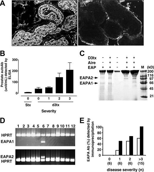
We investigated the influence of Ag on Tregs in autoimmune disease suppression by monitoring the expression pattern of disease-related self Ag. Because the prostate autoAgs in EAP were not yet defined, we used the serum Ab from d3tx mice with EAP to identify and characterize them. Serum Ab of mice with EAP reacted with Ags in the anterior (Figure 1A) and dorsal prostate lobes, and the Ab levels strongly correlated with EAP incidence (Figure S1, available on the Blood website; see the Supplemental Figures link at the top of the online article) and EAP severity (Figure 1B). The serum Ab immunoprecipitated 2 prominent protein bands from the detergent extract of murine anterior prostate lobe with the apparent molecular weights of 68 kDa and 110 kDa (Figure 1C). Trypsin digestion of the 68-kDa protein yielded 6 peptide sequences (underlined in Figure S2), which were determined by mass spectroscopy to have homology with the rat prostate transglutaminase or the dorsal protein 120 (GenBank no. NM022713). Similar analysis of the 110-kDa prostate Ag yielded 15 peptide sequences, of which 12 (underlined in Figure S3) matched the sequence of a putative rat protein of unknown function (GenBank no. BC061995). DNA primers flanking the coding sequence were used to clone the complete cDNA for both proteins from mouse anterior prostate lobe. We named the murine homolog of rat prostate transglutaminase experimental autoimmune prostatitis antigen 1 (EAPA1), and the 110-kDa protein EAPA2 (Figures S2-S3; GenBank nos. AF486627 and AY528666). EAPA1 shared 81% and 53% homology with the rat20 and the human prostate transglutaminases,21 respectively, and had 32% homology to the murine tissue transglutaminase.22 The EAPA2 cDNA encoded a protein with 914 amino acids and a predicted molecular weight of 109 kDa. The expressions of EAPA1 and EAPA2 were prostate-specific, with mRNA transcripts detectable in the prostate but not in other tissues including the thymus (Figure 1D) or the thymic medullary epithelial cells (J. X. S., manuscript submitted). Abs to EAPA2 were detected more frequently than Abs to EAPA1 in d3tx mice with EAP, and the serum titer of EAPA2-precipitating Abs also correlated well with EAP severity (Figure 1E). Both Abs reacted with the recombinant EAPA proteins expressed in Escherichia coli (data not shown).
Identification of prostate autoAgs by autoAb from d3tx male B6AF1 mice with EAP. (A) Cytoplasmic Ag is detected by indirect immunofluorescence in anterior prostate lobe with serum IgG autoAb from d3tx mice with EAP (i; original magnification × 200). The reaction of serum from a d3tx mouse without EAP is shown in panel ii as a negative control (original magnification × 200). Histological studies were done using an Olympus BH2 microscope (Olympus, Melville, NY), and pictures were taken with an Olympus DP12 digital microscope camera. Images were taken with 20×/__ NA (panels __) and 40×/__ NA (panels __) objectives. (B) Correlation between EAP severity and level of serum autoAb to anterior prostate extract detected by enzyme-linked immunosorbent assay (ELISA) as arbitrary units. STx indicates sham thymectomized mice. (C) Two protein bands with molecular weight of 68 kDa and 110 kDa (EAPA1 and EAPA2, respectively) in B6AF1 anterior prostate extract were immunoprecipitated by serum autoAb from d3tx mice and the aire knockout mice with EAP, detected by SDS-PAGE. (D) Prostate-specific expression of EAPA1 and EAPA2 is determined by RT-PCR. Lane 6 contains prostate RNA, whereas the lanes with negative result contain RNA of kidney, stomach, lung, brain, testis, muscle, spleen, liver, heart, pancreas, and thymus. (E) The incidence of serum autoAb that immunoprecipitated EAPA1 (□) and EAPA2 (▪) correlated with EAP severity of d3tx mice, and EAPA2 Ab is detected more frequently.
Identification of prostate autoAgs by autoAb from d3tx male B6AF1 mice with EAP. (A) Cytoplasmic Ag is detected by indirect immunofluorescence in anterior prostate lobe with serum IgG autoAb from d3tx mice with EAP (i; original magnification × 200). The reaction of serum from a d3tx mouse without EAP is shown in panel ii as a negative control (original magnification × 200). Histological studies were done using an Olympus BH2 microscope (Olympus, Melville, NY), and pictures were taken with an Olympus DP12 digital microscope camera. Images were taken with 20×/__ NA (panels __) and 40×/__ NA (panels __) objectives. (B) Correlation between EAP severity and level of serum autoAb to anterior prostate extract detected by enzyme-linked immunosorbent assay (ELISA) as arbitrary units. STx indicates sham thymectomized mice. (C) Two protein bands with molecular weight of 68 kDa and 110 kDa (EAPA1 and EAPA2, respectively) in B6AF1 anterior prostate extract were immunoprecipitated by serum autoAb from d3tx mice and the aire knockout mice with EAP, detected by SDS-PAGE. (D) Prostate-specific expression of EAPA1 and EAPA2 is determined by RT-PCR. Lane 6 contains prostate RNA, whereas the lanes with negative result contain RNA of kidney, stomach, lung, brain, testis, muscle, spleen, liver, heart, pancreas, and thymus. (E) The incidence of serum autoAb that immunoprecipitated EAPA1 (□) and EAPA2 (▪) correlated with EAP severity of d3tx mice, and EAPA2 Ab is detected more frequently.
To generalize the importance of EAPA1 and EAPA2 as autoAgs in prostate autoimmunity, we studied the Aire knockout mice. The aire gene encodes a transcription factor expressed primarily in the medullary epithelial cells of the thymus.23 Deficiency in aire function resulted in spontaneous human and murine polyendocrine autoimmunity.24,25 Mice with targeted disruption of the Aire gene developed a collection of autoimmune diseases and autoAb responses similar to those of the d3tx mice.26 Recently, we discovered severe EAP in 67% (n = 9) of Aire-/- mice of the C57BL/6 background older than 6 weeks of age (Ruan et al, manuscript submitted). All Aire-/- mice with EAP produced Abs to EAPA1 and EAPA2, identified by immunoprecipitation (Figure 1C) and peptide sequence analysis (data not shown). Therefore, the murine EAPA1 and EAPA2 are prostate-specific Ags that are targeted by autoAbs in 2 spontaneous EAP models.
Male Tregs suppress EAP better than female Tregs and the supremacy is eliminated by donor prostate Ag ablation
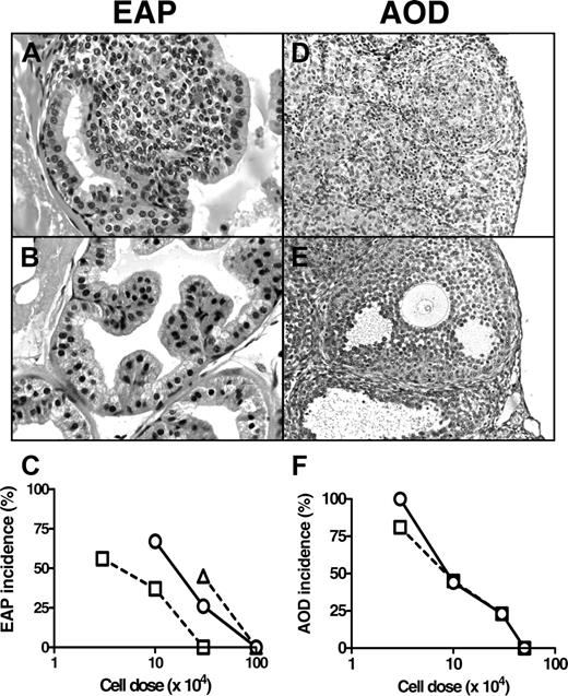
Fifty-three percent of d3tx B6AF1 male mice developed EAP (Table 1; Figure 2A), which affected primarily the anterior and dorsal lobes. To establish the dose response of EAP suppression, we infused Tregs of 90% to 95% purity into d3tx mice on day 5 and examined prostate pathology at 9 weeks. With male Tregs, 1 × 10⁵ cells reduced the incidence of EAP to 37% and 3 × 10⁵ cells completely suppressed disease (Table 1; Figure 2B). Although female Tregs also suppressed EAP, 3 times more cells were required (Figure 2C). For example, 3 × 10⁵ female Tregs reduced the incidence of EAP to 26%, whereas complete suppression required 1 × 10⁶ cells (Table 1). NOr deprived the endogenous testosterone required for prostate development and prostate Ag expression,27 including the expression of EAPA1 and EAPA2 (Figure 3). In parallel, the Tregs from NOr males suppressed EAP less effectively than Tregs from male donors (P = .001), with a dose response approximating that of female Tregs (P = .11; Table 1; Figure 2C).
Incidence and severity of EAP in d3tx B6AF1 mice that received different numbers of Tregs from donors with or without prostates
. | . | . | EAP in d3tx recipients . | . | |
|---|---|---|---|---|---|
| Treg donor . | Treg donor treatment (duration) . | Treg nos. . | Incidence, % (no.) . | Severity* . | |
| None | – | – | 53 (17) | 2.3 ± 0.2 | |
| Male 1 | – | 3 × 10⁴ | 56 (9) | 1.9 ± 0.4 | |
| Male 2 | – | 1 × 10⁵ | 37 (8) | 1.8 ± 0.6 | |
| Male 3 | – | 3 × 10⁵ | 0 (14) | – | |
| Female 1 | – | 3 × 10⁴ | 67 (12) | 2.2 ± 0.3 | |
| Female 2 | – | 1 × 10⁵ | 67 (12) | 2.4 ± 0.3 | |
| Female 3 | – | 3 × 10⁵ | 26 (23) | 1.6 ± 0.2 | |
| Female 4 | – | 1 × 10⁶ | 0 (5) | – | |
| Male 4 | NOr | 3 × 10⁵ | 45 (11) | 1.9 ± 0.5 | |
| Male 5 | NOr | 1 × 10⁶ | 0 (16) | – | |
| Male 6 | NOr-DHT (6 wk) | 3 × 10⁵ | 11 (9) | 0.5 ± 0 | |
| Male 7 | NOr-DHT (2 wk) | 3 × 10⁵ | 6 (16) | 2.0 ± 0 | |
| Male 8 | NOr-DHT (1 wk) | 3 × 10⁵ | 24 (21) | 1.6 ± 0.4 | |
| Male 9 | NOr-ATx-DHT† | 3 × 10⁵ | 0 (20) | – | |
| Female 5 | DHT-prostate‡ | 3 × 10⁵ | 8 (12) | 2.0 ± 0 | |
| Female 6 | DHT‡ | 3 × 10⁵ | 58 (12) | 1.8 ± 0.3 | |
| Female 7 | Prostate‡ | 3 × 10⁵ | 30 (10) | 2.0 ± 0.6 | |
– indicate none.
Mean ± SD of mice with disease
Six-week-old NOr males were thymectomized and received implants of DHT pellets for 2 weeks
Six-week-old females received implants of prostate or DHT pellet or both for 2 weeks
Histopathology of AOD and EAP in d3tx B6AF1 mice and comparison of suppression of the 2 autoimmune diseases by Tregs. (A) Prostatitis with lymphocytic infiltration in a d3tx mouse. (B) Normal anterior prostate of a d3tx mouse suppressed with 3 × 10⁵ male Tregs. (D) Severe oophoritis in a d3tx female mouse, with heavy inflammatory infiltrate and oocyte loss. (E) Normal ovary of a d3tx mouse suppressed by 5 × 10⁵ Tregs from normal female donors. Original magnification × 400 (A-B) and × 200 (D-E); hematoxylin and eosin. Histological studies were done using an Olympus BH2 microscope (Olympus, Melville, NY), and pictures were taken with an Olympus DP12 digital microscope camera. Images were taken with 20×/__ NA (panels __) and 40×/__ NA (panels __) objectives. (C) The cell dose response of EAP suppression by Tregs from male (□), female (○), and NOr male (▵) donors. (F) The cell dose response of AOD suppression by Tregs from male (□) and female (○) donors.
Histopathology of AOD and EAP in d3tx B6AF1 mice and comparison of suppression of the 2 autoimmune diseases by Tregs. (A) Prostatitis with lymphocytic infiltration in a d3tx mouse. (B) Normal anterior prostate of a d3tx mouse suppressed with 3 × 10⁵ male Tregs. (D) Severe oophoritis in a d3tx female mouse, with heavy inflammatory infiltrate and oocyte loss. (E) Normal ovary of a d3tx mouse suppressed by 5 × 10⁵ Tregs from normal female donors. Original magnification × 400 (A-B) and × 200 (D-E); hematoxylin and eosin. Histological studies were done using an Olympus BH2 microscope (Olympus, Melville, NY), and pictures were taken with an Olympus DP12 digital microscope camera. Images were taken with 20×/__ NA (panels __) and 40×/__ NA (panels __) objectives. (C) The cell dose response of EAP suppression by Tregs from male (□), female (○), and NOr male (▵) donors. (F) The cell dose response of AOD suppression by Tregs from male (□) and female (○) donors.
DHT induced EAPA mRNA expression in 4 days and restored the EAP-suppressing capacity of peripheral Tregs of NOr mice in 14 days
To further study the relation of Ag expression and Treg function, we took advantage of the hormonal dependency of prostate development and function. Postpubertal development and Ag expression of the prostate are dependent on testicular androgen, and they do not occur in mice with neonatal orchiectomy (NOr). However, the reintroduction of androgen to NOr mice, in the form of a DHT pellet, rapidly triggers prostate maturation and Ag expression.
The treatment of NOr mice with DHT at 6 weeks of age for 6 weeks restored prostate Ag expression and also the capacity of their Tregs to suppress EAP. Compared with the Tregs from NOr donors, the Tregs from NOr/DHT donors had greater EAP-suppressing activity, and the EAP incidences in the d3tx recipients were 45% and 11%, respectively (Table 1; P = .045). In contrast, the Tregs from NOr mice treated with DHT for 6 weeks suppressed EAP to a comparable extent as the Tregs of normal male donor (P = .220).
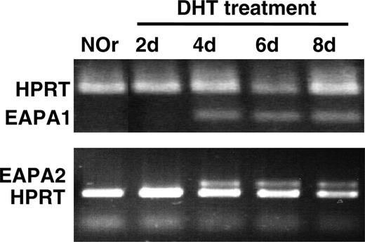
We next used this approach to determine the duration of EAPA exposure required for the gain in EAP-suppressing Treg function. As shown in Table 1, when the NOr male Treg donors were treated with DHT for 2 weeks, their EAP-suppressing capacity was restored to that of untreated males (P = .359), surpassing that of Tregs from NOr male donors (P = .003). In contrast, the Tregs of NOr mice exposed to DHT for 1 week exhibited comparable EAP-suppressing capacity as the Tregs of NOr males (P = .159). To relate the change in EAP-suppressing capacity of Tregs to the duration of their exposure to EAPA, we studied the expression of EAPA mRNA in the anterior prostate lobes of NOr mice at different time intervals following DHT treatment. EAPA1 and EAPA2 transcripts, absent in the NOr males, were detectable 4 days after the DHT implant (Figure 3). Because our study on the ontogeny EAPA (see “The ontogeny of prostate and ovarian Ag”) indicated that EAPA protein expression occurred 4 days after EAPA mRNA expression (Figure 4), we conclude that the Tregs from the prostate Ag- mice can gain EAP-suppressing capacity to reach the level of Tregs of normal males after 6 days of prostate Ag expression.
Because the natural CD4+CD25+ Treg development is dependent on Ag recognition in the thymus,28 we investigated whether the observed effect of Ag on Treg function requires the presence of the thymus. NOr donors were thymectomized at 6 weeks of age and then given implants of DHT for 2 weeks before their Tregs were studied. As shown in Table 1, adult thymectomy (ATx) did not alter the acquisition of Treg function of NOr mice by DHT treatment. We conclude that the education of Treg by prostate Ag occurs mainly in the peripheral lymphoid tissues.
Based on EAP suppression by female Tregs, modulation of testosterone by DHT and NOr is not responsible for the altered Treg function
DHT was required for our investigation on EAP-suppressing Treg function in the NOr male mice. Because testosterone is known to affect T-cell responses,29 it was necessary to rule out a direct effect of DHT on Treg function. This was investigated in EAP suppression by the Tregs from female mice. Tregs from DHT-treated female donors were somewhat less effective in suppressing EAP than Tregs from untreated female mice, but the difference was not statistically significant (P = .064; Table 1). However, when the DHT-treated female donors were simultaneously engrafted with normal adult anterior prostate, their Tregs gained EAP-suppressing function that matched those of normal male Tregs in 2 weeks (Table 1; P = .289). An additional control showed that Treg suppression of EAP was not enhanced in female donors with prostate implant alone (Table 1). The result indicates that the DHT effect on EAP-suppressing Treg function is mediated through promotion of prostate Ag expression.
Detection of EAPA1 and EAPA2 mRNA. Both the EAPA1 mRNA and EAPA2 mRNA are detected in the anterior prostate of NOr B6AF1 mice 4 days after implantation of DHT pellets. HPRT served as control.
Detection of EAPA1 and EAPA2 mRNA. Both the EAPA1 mRNA and EAPA2 mRNA are detected in the anterior prostate of NOr B6AF1 mice 4 days after implantation of DHT pellets. HPRT served as control.
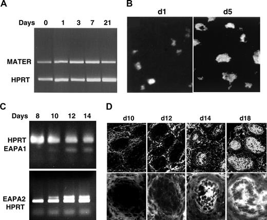
The ontogeny of prostate Ag and oocyte Ag expression in B6AF1 mice. The mRNA of the ovarian MATER Ag is detected from birth by RT-PCR (A), and the MATER Ag is detected in ovarian oocytes in 1-day-old ovary by immunofluorescence with serum autoAb from d3tx mice with AOD (original magnification × 400). (B) The RNAs of prostate EAPA1 and EAPA2 are detectable in prostates at and after 10 days of age by RT-PCR. (C) The EAPA1 or EAPA2 Ag is detected in the prostate at and after 14 days of age by indirect immunofluorescence with serum autoAb from d3tx male mice with EAP (D; original magnification × 200 for top panels and × 400 for bottom panels). Panels A-B are reproduced from Alard et al,5 with permission of the Journal of Immunology (Copyright 2001. The American Association of Immunologists, Inc).
The ontogeny of prostate Ag and oocyte Ag expression in B6AF1 mice. The mRNA of the ovarian MATER Ag is detected from birth by RT-PCR (A), and the MATER Ag is detected in ovarian oocytes in 1-day-old ovary by immunofluorescence with serum autoAb from d3tx mice with AOD (original magnification × 400). (B) The RNAs of prostate EAPA1 and EAPA2 are detectable in prostates at and after 10 days of age by RT-PCR. (C) The EAPA1 or EAPA2 Ag is detected in the prostate at and after 14 days of age by indirect immunofluorescence with serum autoAb from d3tx male mice with EAP (D; original magnification × 200 for top panels and × 400 for bottom panels). Panels A-B are reproduced from Alard et al,5 with permission of the Journal of Immunology (Copyright 2001. The American Association of Immunologists, Inc).
Two additional experiments were conducted to investigate a direct effect of NOr on Treg function. In the in vitro assay, the response of CD4+CD25- T cells to CD3 monoclonal Ab was suppressed to a similar extent by the Tregs from normal male, normal female, and NOr male mice (data not shown). Moreover, dacryoadenitis, which occurred in 47% of d3tx male mice (data not shown), was suppressed to comparable levels by male Tregs (0% in 11 mice), female Tregs (13% in 23 mice; P = .167), or Tregs from NOr males (9% in 11 mice; P = .268) at 3 × 10⁵ Tregs per recipient. Thus, NOr alone and DHT treatment alone had no detectable effect on Treg function; these findings further strengthen the conclusion that prostate Ag exposure modulates EAP-suppressing function of male and female Tregs.
Male and female Tregs suppress AOD equally
Following d3tx, 92% of 8- to 9-week-old female B6AF1 mice developed AOD (Table 2; Figure 2D). When infused with female Tregs, the incidence and severity of AOD were partially but significantly reduced at the Treg dose of 1 × 10⁵ and 3 × 10⁵ cells, and complete disease suppression was achieved at 5 × 10⁵ cells (Table 2; Figure 2E). In striking contrast to the finding in EAP suppression, the dose responses of male and female Tregs in AOD suppression were essentially identical (Table 2; Figure 2F). Additionally, NOv had no effect on the AOD-suppressing capacity of female mice (P = .123; Table 2).
Incidence and severity of AOD in d3tx B6AF1 mice that received different numbers of Tregs from donors with or without ovaries
. | . | . | AOD in d3tx recipients . | . | |
|---|---|---|---|---|---|
| Treg donor . | Treg donor treatment . | Treg nos. . | Incidence,% (no.) . | Severity* . | |
| None | – | – | 92 (25) | 8.0 ± 0.8 | |
| Female 1 | – | 3 × 10⁴ | 100 (9) | 4.0 ± 1.3 | |
| Female 2 | – | 1 × 10⁵ | 44 (9) | 3.7 ± 1.8 | |
| Female 3 | – | 3 × 10⁵ | 23 (13) | 2.3 ± 0.6 | |
| Female 4 | – | 5 × 10⁵ | 0 (32) | – | |
| Male 1 | – | 3 × 10⁴ | 81 (16) | 5.6 ± 1.0 | |
| Male 2 | – | 1 × 10⁵ | 45 (11) | 5.4 ± 1.7 | |
| Male 3 | – | 3 × 10⁵ | 23 (13) | 1.5 ± 0.3 | |
| Male 4 | – | 5 × 10⁵ | 0 (15) | – | |
| Female 5 | NOv | 3 × 10⁵ | 55 (11) | 1.0 ± 0.1 | |
| Female 6 | NOv | 5 × 10⁵ | 0 (8) | – | |
NOv indicates neonatal ovariectomy; –, none.
Mean ± SD of mice with disease
The results on AOD suppression do not support the influence of endogenous Ag on autoimmune disease suppression by organ-specific Tregs. However, we were equally impressed by the rapidity of the prostate Ag to enhance EAP-suppressing function of Tregs. To reconcile this dichotomy, we considered the possibility that the ovarian Ag in the neonatal d3tx female recipients might rapidly educate the incoming male Tregs and enable them to suppress AOD efficiently. To investigate this, we first compared the ontogeny of expression of the ovarian MATER Ag and the prostatic EAPA.
The ontogeny of prostate and ovarian Ag
The murine ovarian MATER Ag is detectable very early in oocyte development though it does not manifest its physiologic function until after fertilization in the 2-cell embryo.18 MATER is the major oocyte Ag targeted by Abs of d3tx mice with AOD.30 As we have reported previously,5 the mRNA of the ovarian MATER was detectable from birth (Figure 4A) and the oocytes of 1-day-old ovaries reacted positively with the MATER Ab (Figure 4B). Moreover, neonatal ovaries were targeted by pathogenic T cells adoptively transferred from d3tx donors with AOD.5 In contrast to the ovarian MATER, the mRNAs of both EAPA1 and EAPA2 were first detectable in the prostate on day 10 and the EAPA proteins 4 days later, at 2 weeks (Figure 4C-D). The ovarian Ag expression therefore precedes prostate Ag expression by at least 2 weeks. When the male Tregs are infused into the female d3tx mice on day 5, they should immediately encounter the ovarian MATER Ag, whereas the female Tregs in the neonatal male d3tx recipients would not encounter prostate Ag for the next 9 days.
Late ontogeny of ovarian Ag caused differential suppression by male and female Tregs
To directly determine whether the early ontogeny of ovarian Ag influences the function of Tregs that are transferred into the neonatal d3tx mice, we repeated an experiment described in our recent study,31 because the results are so germane to the present investigation. The experiment was designed to delay ovarian Ag expression in female recipient for 3 weeks to simulate the ontogeny of prostate Ag expression in the male. We ablated the ovarian Ag by ovariectomy at birth. We then thymectomized the mice at day 3 and transferred 3 × 10⁵ Tregs from normal male or female donors at day 5. At 3 weeks of age, we engrafted the mice with age-matched syngeneic ovaries to serve as a source of endogenous Ag to stimulate pathogenic T-cell response, and as targets of AOD. We evaluated the pathology of the ovarian graft at 7 to 8 weeks.
In control recipients that were devoid of neonatal ovarian Ag for 3 weeks and did not receive any Tregs, 63% of the ovarian grafts were found to develop AOD with intermediate severity (Table 3). In similar recipients, the male Tregs did not suppress AOD and the recipients developed the same frequency of ovarian disease as the control mice (63%). In contrast to the male Tregs, the female Tregs were able to suppress AOD in the d3tx female recipients devoid of neonatal ovaries and reduced the incidence of AOD to 8% (n = 12; P = .001). The sex difference in AOD suppression was disease-specific because dacryoadenitis that occurred in the same cohort of mice was suppressed equally by male and female Tregs (Table 3).
Incidence and severity of AOD in the ovarian grafts of NOv-d3tx B6AF1 mice that received 3 × 10⁵ Tregs
. | AOD in ovarian graft . | . | Dacryoadenitis . | . | ||
|---|---|---|---|---|---|---|
| Treg donor . | Incidence, % (n) . | Severity* . | Incidence, % (n) . | Severity* . | ||
| None | 63 (11) | 2.0 ± 0.2 | 72 (11) | 1.5 ± 0.2 | ||
| Female | 8 (12) | 1.5 ± 0 | 0 (12) | – | ||
| Male | 63 (11) | 1.9 ± 0.2 | 0 (11) | – | ||
The female recipients were ovariectomized at the day of birth, thymectomized at day 3, received implants of an age-matched syngeneic ovary at week 3, and studied at week 7 to 8. They also received the Tregs at day 5.
Mean ± SD of mice with disease
The experimental results, which are completely consistent with our earlier study, provided 3 interpretations that are pertinent to the present study. First, ovarian Ag expressed in Treg recipients in the neonatal weeks can modify AOD-suppressing function of the incoming male Tregs. Second, the effect of neonatal ovarian Ag stimulation on the acquisition of AOD-suppressing Treg function is disease-specific. Third, compared to male Tregs, the capacity of female Tregs to suppress AOD was not diminished even when the Tregs did not encounter ovarian Ag for 16 days; therefore, the female Tregs are more able to retain their capacity to suppress AOD than male Tregs in the absence of ovarian Ag.
Discussion
This study has provided compelling evidence that the functional acquisition by polyclonal Tregs to suppress an autoimmune disease is a process that critically depends on the presence of endogenous autoAg. We showed that EAP and AOD in the d3tx mice were suppressed by fewer numbers of Tregs from Ag+ donors than the Treg from Ag- donors. Thus male Tregs surpassed female Tregs in suppressing EAP, whereas the reverse is true for the suppression of AOD. In addition, we showed that the disease-suppressing function of Tregs from Ag- donors was rapidly enhanced by a short exposure to the Ag, and the effect was Ag-specific and extrathymic. The peripheral Tregs of NOr male mice, and the Tregs of normal female mice, when exposed to prostate Ag induced by DHT, gained full EAP-suppressing function within 6 days. Equally striking was the finding that AOD-suppressing function was acquired by male Tregs in response to the neonatal ovarian Ag of the d3tx female recipients. The extent to which the recipient Ag affected donor Treg function in the d3tx model depended on the ontogeny of expression of the self Ag. Ovarian Ag expression preceded prostate Ag expression, and accordingly, the neonatal ovarian Ag had more of an impact on the AOD-suppressing function of the incoming male Tregs than that of prostate Ag on incoming female cells. Indeed, the capacity of male Tregs to suppress AOD was strictly dictated by the neonatal expression of ovarian Ag in the d3tx female recipients. We have not, however, ruled out the possibility that the recipient prostate Ag of late ontogeny (after day 14) might also contribute to the ability of female Tregs to suppress EAP.
Studies on the Ag dependency in polyclonal Treg function began before CD25 was identified to be a useful Treg marker. However, although the result of studies on EAP and autoimmune thyroiditis supports the influence of Ag on disease suppression by total T cells, this was not observed in the study on AOD. We have now uncovered the explanation for the contradictory result. We have found that the intrinsic supremacy of the female Tregs to suppress AOD was masked in our experiment by the ability of male Tregs to acquire AOD-suppressing function when they were exposed to the ovarian Ag in the neonatal female d3tx recipients. The male Tregs, stimulated by the recipient ovarian Ag, gained AOD-suppressing efficacy, leading to the false impression that they had equal intrinsic capacity to suppress AOD as the Tregs from female mice. Ironically, the finding that prompted the false impression also provided one of the strongest pieces of evidence supporting Ag requirement in Treg function.
The studies on AOD, EAP, and thyroiditis suppression by polyclonal Tregs collectively support the importance of polyclonal Ag-specific Tregs in the maintenance of physiologic tolerance. The studies on AOD further indicated that self Ag plays a critical role in the functional acquisition by polyclonal Tregs as well as in the process of autoimmune disease prevention. The participation of endogenous Ag in autoimmune disease prevention was documented in a recent study that tracked the polyclonal Treg during AOD suppression.31 The study, based on the paradigm that Ag-specific Treg response should occur in the regional LNs, made the following points. First, whereas the polyclonal Thy1.1+ Tregs proliferated and were distributed diffusely, the Tregs retrieved from the ovary-draining LNs exhibited far greater AOD-suppressing function than those from nondraining LNs or the spleen. Thus, the Tregs that accumulated exclusively in ovary-draining LNs were 5 times more effective in suppressing AOD than the total LN cells from normal female donors. Second, the regional LN was the unique location where profound, persistent, and reversible suppression of host T-cell response and AOD occurred. Third, similar changes were observed in the suppression of EAP and dacryoadenitis by polyclonal Tregs. These results support the regional LN as the location where normal tissue Ag continuously stimulates polyclonal Tregs to maintain physiologic tolerance.
It has been documented that Tregs with transgenic TCRs proliferate in vivo in response to their cognate Ag7-10 ; thus, clonal expansion of Ag-specific Tregs is a likely explanation for enhancement of Treg function in response to physiologic Ag. There is, however, evidence that Ag exposure also changes the intrinsic Treg function. Compared with male Tregs, which were unable to suppress AOD in neonatal females devoid of ovaries, female Tregs were found to retain full capacity to suppress AOD in the same ovarian Ag- recipients. Indeed, a similar discrepancy in the retention of male Treg function was observed in EAP suppression; thus, the male Tregs retained their capacity to suppress EAP in neonatal male d3tx recipients that were normally devoid of prostate Ag for 9 days after Treg transfer. The difference in the retention of Treg function is unknown and it could be determined by the higher frequency of memory Tregs in the Ag+ donors. Studies have shown that Tregs previously stimulated by Ag in vitro had a greater capacity to regulate in an Ag-specific manner,32 and subpopulations of Tregs with phenotype and function of memory and naive Tregs have been identified.33,34
In conclusion, based on the study of 2 sex-specific autoimmune diseases, we demonstrated that the polyclonal Tregs from normal mice can respond quickly and gain regulatory function in response to physiologically expressed self Ag, and that Tregs that have encountered Ag retain superior regulatory capacity. Our study has focused on the response of polyclonal Tregs and physiologic self Ag and is dependent on autoimmune disease suppression as the experimental end point. The results are therefore physiologically relevant and strongly support the prevailing concept of constant peripheral Treg stimulation by self Ag as an important physiologic tolerance mechanism.
Prepublished online as Blood First Edition Paper, October 13, 2005; DOI 10.1182/blood-2005-08-3088.
Supported by the National Institutes of Health grants AI 41236, AI 51420, and AR 45222 (K.S.K.T). Y.Y.S., K.O. and E.T.S. contributed equally to this study.
The online version of the article contains a data supplement.
An Inside Blood analysis of this article appears at the front of this issue.
The publication costs of this article were defrayed in part by page charge payment. Therefore, and solely to indicate this fact, this article is hereby marked “advertisement” in accordance with 18 U.S.C. section 1734.
We thank Dr Osamu Taguchi for helpful suggestions, Sharon Mangawang and Yuefang Sun for expert surgery, the Cancer Center Research Histology Core of the University of Virginia for histology support, the Biomolecular Core of the University of Virginia for peptide sequence analysis, and the Paul Mellon Prostate Institute at University of Virginia for financial support.


This feature is available to Subscribers Only
Sign In or Create an Account Close Modal