The roles of aberrant expression of constitutively active ALK chimeric proteins in the pathogenesis of anaplastic large-cell lymphoma (ALCL) have been well defined; nevertheless, the notion that ALK is a molecular target for the therapeutic modulation of ALK+ ALCL has not been validated thus far. Select fused pyrrolocarbazole (FP)–derived small molecules with ALK inhibitory activity were used as pharmacologic tools to evaluate whether functional ALK is essential for the proliferation and survival of ALK+ ALCL cells in culture. These compounds inhibited interleukin 3 (IL-3)–independent proliferation of BaF3/NPM-ALK cells in an ALK inhibition-dependent manner and significantly blocked colony formation in agar of mouse embryonic fibroblast (MEF) cells harboring NPM-ALK. Inhibition of NPM-ALK phosphorylation in the ALK+ ALCL-derived cell lines resulted in significant inhibition of cell proliferation and induction of apoptotic-cell death, while having marginal effects on the proliferation and survival of K562, an ALK- leukemia cell line. ALK inhibition resulted in cell-cycle G1 arrest and inactivation of ERK1/2, STAT3, and AKT signaling pathways. Potent and selective ALK inhibitors may have therapeutic application for ALK+ ALCL and possibly other solid and hematologic tumors in which ALK activation is implicated in their pathogenesis.
Introduction
Chromosomal translocations occur frequently in a select group of human cancers, including most lymphomas, leukemias, and sarcomas. Individual translocations have shown a high degree of specificity for particular cancer types and the presence of a particular translocation often correlates well with clinical behavior and outcome for specific types of cancer.1 Consequently, therapies directed at molecular targets dysregulated by tumor-specific genetic aberrations will potentially provide more effective and less toxic therapies than conventional chemotherapy.1,2 Anaplastic large-cell lymphomas (ALCLs) comprise a group of non-Hodgkin lymphomas (NHLs) that are usually of T-cell origin and often present with extranodal disease, especially the skin, and are characterized by the expression of the CD30/Ki-1 antigen. Roughly 2500 to 3000 new cases of ALCLs are diagnosed in the United States each year and 50% to 60% of these ALCLs are associated with a specific t(2;5) (p23;q35) chromosome translocation.4,5 The genes altered in the t(2;5) translocation contain the N-terminal portion of nucleophosmin (NPM) gene, a nuclear phosphoprotein, fused to the catalytic domain of anaplastic lymphoma kinase (ALK) gene. ALK is a cell-membrane–spanning receptor tyrosine kinase and a member of the insulin receptor superfamily. Although the precise physiologic function and regulation of ALK have not been well defined, the NPM-ALK fusion gene encodes for an 80-kDa NPM-ALK chimeric oncoprotein with constitutively active ALK tyrosine kinase activity, which plays a key role in lymphomagenesis by the aberrant phosphorylation of multiple intracellular substrates downstream of NPM-ALK.4,5 Subsequently, other fusion partners of ALK were also reported in ALCL, and dysregulated expression and constitutive activation of the ALK protein was demonstrated in approximately 60% to 70% of ALCLs, termed ALK+ lymphomas.4,6-8 Preclinical experimental data have demonstrated that the aberrant expression of constitutively active ALK is directly implicated in the pathogenesis of ALCL and that ALK down-regulation or inhibition of ALK-mediated pathways can markedly impair the growth of ALK+ lymphoma cells.9-15 Currently there is no optimal therapeutic regimen for ALK+ ALCL. Doxorubicin-based combination chemotherapy has limited effectiveness, resulting in a substantial number of patients with ALK+ ALCL with a poor outcome, either failing to enter remission or relapsing within a few months from the start of treatment.3 Thus, optimal and more effective therapeutic approaches are needed for these patients. The mechanisms by which deregulation of the ALK tyrosine kinase is linked with subset of ALCL are essentially similar to those involved in the development of more than 90% of chronic myeloid leukemia (CML) due to the dysregulated activity of the ABL tyrosine kinase. The clinical efficacy and safety demonstrated by imatinib mesylate (Gleevec), a small-molecule ABL inhibitor, for ABL+ CML,16 suggests that the inhibition of ALK with a specific and potent small-molecule ALK inhibitor would potentially offer a more effective and less toxic therapy for patients with ALK+ lymphomas. Due to the high expression level and relatively long half-life of NPM-ALK in ALCL cells, specific ablation of ALK expression in these cells with either siRNA- or ribozyme-based approaches has not been successfully achieved to provide solid proof-of-concept validation that inhibition of ALK is sufficient to attenuate the growth and proliferation of ALK+ ALCL cells.17 Currently no small-molecule inhibitors of ALK have been described for the treatment of ALK+ ALCL.
In this study, several cell-permeable, fused-pyrrolocarbazole (FP)–derived kinase inhibitors varying in their potency to ALK were used as pharmacologic tools to evaluate whether ALK activity is essential for the proliferation and survival of ALK+ ALCL cells in culture. Our data strongly suggest that ALK inhibition is sufficient to attenuate the growth and survival of ALK+ ALCL cells and provide an experimental basis for targeting ALK as a therapeutic strategy in ALK+ lymphomas.
Materials and methods
Compounds, cell lines, antibodies, and transfection
The synthesis of CEP compounds, CEP-11988, CEP-14083, and CEP-14153, was described in a recently issued US patent (no. US 2005-0143442A1).18
The ALK+ ALCL-derived cell lines, Karpas-299, SR-786, Sup-M2, and Sudhl-1, were purchased from DSMZ (Heidelberg, Germany) and cultured in RPMI with 10% to 15% fetal bovine serum (FBS). Characterization of these ALCL cell lines has been described previously.19 The BaF3 cells were kindly provided by Dr Stephan Morris of St Jude Children Research Hospital (Memphis, TN) and cultured in RPMI with 10% FBS and 5 to 10 μg/mL murine IL-3 (catalogue no. 403-ML; R&D Systems, Minneapolis, MN). K562 cells were purchased from American Type Culture Collection (Manassas, VA). The mouse embryonic fibroblast (MEF)–Tet-Off-NPM-ALK cell line was kindly provided by Dr Giorgio Inghirami of New York University School of Medicine (New York, NY).
The rabbit phospho-NPM-ALK(Y664) (catalogue no. 3341), ALK antibody (catalogue no. 3342), and the STAT3 and phospho-STAT3 antibodies (catalogue nos. 9132 and 9138) were purchased from Cell Signaling Technology (Beverly, MA), and the mouse ALK antibody (catalogue no. 35-4300) was obtained from Zymed (Seattle, WA). ERK1/2 and phospho-ERK1/2 antibodies (catalogue nos. SC-94 and SC-7383), and AKT and phospho-AKT antibodies (catalogue nos. SC-8312 and SC-7985-R) were purchased from Santa Cruz Biotechnology (Santa Cruz, CA).
The cDNA of NPM-ALK N-terminal region (1-216 amino acid [aa]) was isolated by reverse transcription-polymerase chain reaction (RT-PCR) with a one-step Cells-to-cDNA TMII kit (catalogue no. 1722; Ambion, Austin, TX) using SR-786 ALCL cells according to the manufacturer's protocol. The cDNAs were subcloned into a pCMV-Tag2B vector (Stratagene, La Jolla, CA) to generate pCMV-Tag2B/NPM-ALK (1-216 aa). The remaining cDNAs of NPM-ALK C-terminal region (217-680 aa) were isolated from a human pan cDNA library and subcloned into pCMV-Tag2B/NPM-ALK (1-216 aa) to complete the pCMV-Tag2B/NPM-ALK (1-680 aa) construct. BaF3/NPM-ALK cell lines were generated by transfecting BaF3 cells with pCMV-Tag2B/NPM-ALK (1-680 aa) and the transfectants were then selected with G418 for 2 to 3 weeks in RPMI without IL-3.
Expression of GST-ALK and recombinant ALK kinase assay
Human ALK cDNA encoding the cytoplasmic domain (1058-1620 aa) was PCR amplified from a pan cDNA library (pool of cDNAs from 21 tissues) purchased from Panomics (Redwood City, CA) and subcloned into a pFastBac expression vector (Invitrogen, Carlsbad, CA) engineered to encode a chimera with an NH2-terminal glutathione-S-transferase (GST) moiety. The GST-ALK (1058-1620 aa) protein was expressed in Sf21 insect cells and purified by standard glutathione affinity chromatography as described previously.20
An in vitro recombinant ALK kinase assay was performed using a modification of the enzyme-linked immunosorbent assay (ELISA) described for trkA.21 Briefly, the 96-well microtiter plate was coated with 10 μg/mL substrate (recombinant human PLC-γ/GST). The kinase reaction mixture consisting of 20 mM HEPES, pH 7.2, 1 μM ATP, 5 mM MnCl2, 0.1% BSA, 2.5% DMSO, and test compound (various concentrations) was added to the plate. Recombinant GST-ALK (30 ng/mL) was added and the reaction was allowed to proceed for 15 minutes at 37°C. Detection of the phosphorylated product was performed by adding Eu-N1–labeled PT66 antibody (catalogue no. AD0041; Perkin Elmer, Wellesley, MA). Incubation at 37°C then proceeded for 1 hour, followed by addition of enhancement solution (catalogue no. 1244-105, Perkin Elmer). Fluorescence was then measured using the time-resolved fluorescence protocol on the EnVision 2100 (or 2102) multilabel plate reader (Perkin Elmer). Inhibition data were analyzed using ActivityBase (IDBS, Guildford, UK) and median inhibitory concentration (IC50) curves were generated using XLFit 3.0.5 (IDBS).
Phospho-ALK ELISA and immunoblot analysis
To measure ALK tyrosine phosphorylation in an ELISA, fluoronunc plates (catalogue no. 437796; Nalge Nunc, Rochester, NY) were precoated with goat anti–mouse IgG and incubated with the capture mouse ALK antibody (Zymed) diluted 1:1000 in Superblock (Pierce, Rockford, IL). Following blocking, cell lysates were incubated overnight at 4°C. Plates were incubated with the detecting antibody, anti–phospho-ALK (Y664; Cell Signaling Technology), diluted at 1:2000, followed by incubation with goat anti–rabbit IgG alkaline phosphatase to amplify the detection. Wells were exposed to the fluorogenic substrate 4-MeUP and the signal quantified using a CytoFluor (series 4000) Fluorescence Multi-Well Plate Reader (Applied Biosystems, Foster City, CA).
Immunoblotting of phospho-NPM-ALK and total NPM-ALK, phospho-AKT and AKT, phospho-ERK1/2 and ERK1/2, and phospho-STAT3 and STAT3 was carried out according to the protocols provided by the antibody suppliers. In brief, after treatment, cells were lysed in Frak lysis buffer (10 mM Tris, pH 7.5, 1% Triton X-100, 50 mM sodium chloride, 20 mM sodium fluoride, 2 mM sodium pyrophosphate, 0.1% BSA, plus freshly prepared 1 mM activated sodium vanadate, 1 mM DTT, and 1 mM PMSF, protease inhibitors cocktail III [1:100 dilution]). After brief sonication, the lysates were cleared by centrifugation, mixed with sample buffer and subjected to sodium dodecyl sulfate-polyacrylamide gel electrophoresis (SDS-PAGE). Following transfer to membranes, the membranes were blotted with individual primary and secondary antibodies and washed in TBS/0.2% Tween, and protein bands visualized with enhanced chemiluminescence. For immunoblotting phospho-AKT and AKT, the cells were lysed in sample buffer instead and the samples were then analyzed as described.
MTS assay, caspase activation assay, and flow cytometry
Living cells were measured with the celltiter 96 nonradioactive cell proliferation assay kit (MTS kit) (catalogue no. G5430; Promega, Madison, WI). In brief, the cells were seeded on 96-well plates and 48 hours after compound treatment, equal volume of reagents from the kit were added to the culture medium. The plates were measured with a plate reader and calculated based on the standard curve.
Caspase 3/7 activity was measured with an Apo-one homogenous caspase 3/7 assay kit (catalogue no. G7791, Promega). Briefly, the cells seeded on 96-well plates were treated with compounds for 24 hours. The reaction reagents from the kit were added to the culture medium, and after incubation, the plates were measured with a plate reader for the relative caspase 3/7 activity.
Cell cycle analysis was carried out by flow cytometry on cells treated with compounds for 24 hours, subsequently fixed with ethanol, and stained with propidium iodide according to the procedure of DNA-prep Coulter reagent kit (catalogue no. PN4238055-B; Beckman-Coulter, Miami, FL). Cellular DNA content was analyzed using an EPICS XL flow cytometer (Beckman-Coulter), and the percentage of cells in G1, S, and G2/M were calculated with a Multicycle program (Phoenix Flow Systems, San Diego, CA).
Results
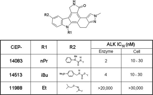
High-throughput screening efforts have identified several fused pyrrolocarbazoles as ALK inhibitors. The inhibitory effects of these small molecules against recombinant enzyme have been confirmed in biochemical and functional cellular assays. As shown in Figure 1, CEP-14083 and CEP-14513 displayed potent ALK inhibitory activity in both in vitro enzymatic assay (IC50 < 5 nM) and cell-based assays of ALK tyrosine phosphorylation (IC50 < 30 nM). In contrast, CEP-11988, a structurally similar compound, displayed relatively weak activity against ALK (IC50 > 20 μM in the enzymatic assay and > 30 μM in the cell-based ALK tyrosine phosphorylation assay; Figure 1). With the exception of ALK inhibition, all 3 compounds have similar kinase inhibition profiles based on the limited number of protein kinases tested. For example, all 3 compounds are weak inhibitors or inactive against CDK1, CDK2, CDK5, c-Met, and IKK, but are potent inhibitors of VEGFR2, Tie2, and DLK (data not shown).
The enzymatic and cellular ALK inhibitory activity of FP kinase inhibitors. The IC50 values from in vitro enzymatic and cellular assays were calculated as described in “Materials and methods.” nPr indicates nPropyl; iBu, isobutyl; and E+, ethyl.
The enzymatic and cellular ALK inhibitory activity of FP kinase inhibitors. The IC50 values from in vitro enzymatic and cellular assays were calculated as described in “Materials and methods.” nPr indicates nPropyl; iBu, isobutyl; and E+, ethyl.
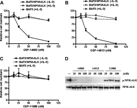
Given the potent enzyme and cellular ALK inhibitory activities of CEP-14083 and CEP-14513, we reasoned that these compounds at certain concentration ranges may effectively inhibit ALK-dependent biologic activities in ALK+ cells with minimal cellular cytotoxicity toward ALK- cells. To test this hypothesis, we first determined whether these compounds attenuated ALK-mediated biologic activity in ALK-activated cell lines without exerting significant cytotoxicity toward ALK- cells. For this purpose, 2 ALK-dependent cell lines were used, the BaF3/NPM-ALK cell line and MEF-Tet-Off-NPM-ALK cell line. BaF3, a murine lymphoid cell line, requires IL-3 for proliferation and survival.22 Overexpression of NPM-ALK in BaF3 cells converted cells from IL-3–dependent to IL-3–independent growth10 (Figure 2). The BaF3/NPM-ALK cell line is an excellent system to evaluate ALK inhibition-dependent cytotoxicity of inhibitors as indicated by the impairment of Ba/F3 NPM-ALK cell growth without alterations in the growth of the parental Ba/F3 cells. Treatment of BaF3/NPM-ALK cells with CEP-14083 or CEP-14513 at concentrations ranging from 10 to 100 nM led to significant inhibition of cell proliferation and survival, correlating with the degree of inhibition of ALK tyrosine phosphorylation. Neither compound affected the total NPM-ALK protein level. Neither compound attenuated the proliferation and survival of parental BaF3 and BaF3/NPM-ALK cells in the presence of IL-3 (Figure 2A,B,D). In contrast, CEP-11988 did not inhibit ALK tyrosine phosphorylation in BaF3/NPM-ALK cells at concentrations ranging from 10 to 100 nM and had marginal effects on the proliferation and survival of BaF3/NPM-ALK cells (Figure 2C,D).
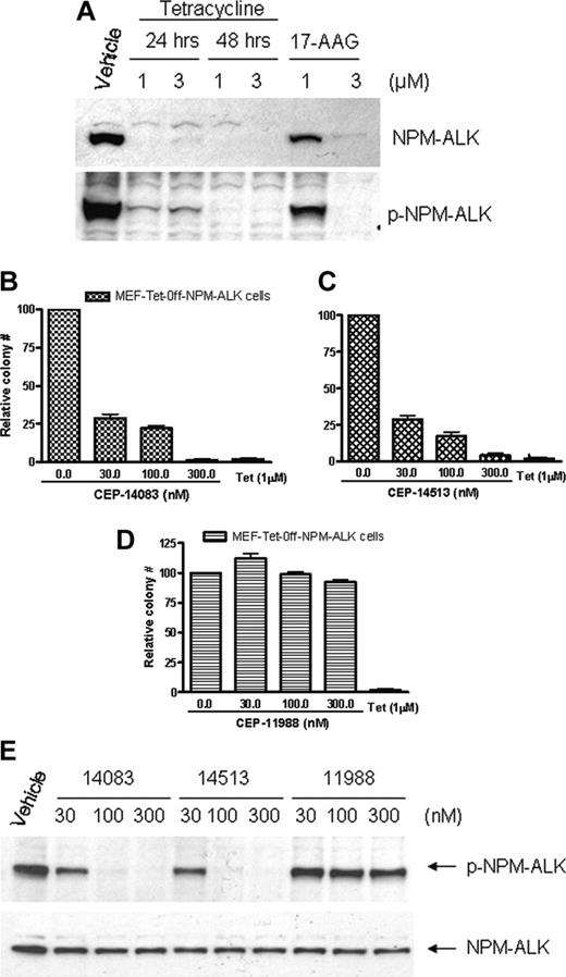
In MEF-Tet-Off-NPM-ALK cells, NPM-ALK expression is controlled by a promoter negatively regulated by tetracycline.23 Treatment of these cells with tetracycline led to down-regulation of NPM-ALK protein levels within 24 to 48 hours (Figure 3A). These transformed cells exhibit NPM-ALK–dependent colony formation in soft agar as demonstrated by the inhibition of colony formation following tetracycline treatment (Figure 2B-D). Treatment of MEF-Tet-Off-NPM-ALK cells with CEP-14083 and CEP-14513 at concentrations of 30 to 300 nM led to dose-related inhibition of NPM-ALK tyrosine phosphorylation and inhibition of colony formation of MEF-tet-off-NPM-ALK cells in agar (Figure 2B-C,E). In contrast, CEP-11988 did not inhibit ALK tyrosine phosphorylation in MEF cells in the same concentration range and exhibited marginal effects on the colony formation of MEF-tet-off-NPM-ALK cells (Figure 2D-E).
ALK inhibition-dependent attenuation of BaF3/NPM-ALK cell proliferation. (A-C) The BaF3 and BaF3/NPM-ALK cell lines were seeded on 96-well plates and treated with CEP compounds at indicated concentrations for 48 hours with or without IL-3. Viable cells were detected with MTS assay. The values presented are the average of relative cell numbers from 2 independent experiments with standard error. (D) For immunoblotting, the BaF3/NPM-ALK cells were treated with CEP compounds at indicated concentrations for 2 hours and lysed, and the lysates were subjected to SDS-PAGE. After transfer, membranes were blotted with either phospho-ALK or total ALK antibody.
ALK inhibition-dependent attenuation of BaF3/NPM-ALK cell proliferation. (A-C) The BaF3 and BaF3/NPM-ALK cell lines were seeded on 96-well plates and treated with CEP compounds at indicated concentrations for 48 hours with or without IL-3. Viable cells were detected with MTS assay. The values presented are the average of relative cell numbers from 2 independent experiments with standard error. (D) For immunoblotting, the BaF3/NPM-ALK cells were treated with CEP compounds at indicated concentrations for 2 hours and lysed, and the lysates were subjected to SDS-PAGE. After transfer, membranes were blotted with either phospho-ALK or total ALK antibody.
ALK inhibition-dependent attenuation of MEF-Tet-off-NPM-ALK cell colony formation in soft agar. (A) The MEF-Tet-off-NPM-ALK cells were treated with indicated concentrations of tetracycline for 24 or 48 hours or 17-AAG for 24 hours. The cells were lysed and the lysates were subjected to SDS-PAGE. After transfer, the members were blotted with either phospho-ALK or total ALK antibody. (B-D) The MEF-Tet-off-NPM-ALK cells were treated with indicated concentrations of CEP compounds or 1 μM tetracycline for 24 hours. The cells were diluted with 0.65% agar in culture medium with or without compounds or tetracycline and seeded on 35-mm3 culture dishes at density of 2 × 10³/dish. The plates were cultured until individual colonies were formed. The values presented are the average of relative colony numbers from 2 independent assays with standard error. (E) For immunoblotting, the MEF-Tet-Off-NPM-ALK cells were treated with CEP compounds at indicated concentrations for 2 hours and lysed, and the lysates were subjected to SDS-PAGE. After transfer, the membranes were blotted with either phospho-ALK or total ALK antibody.
ALK inhibition-dependent attenuation of MEF-Tet-off-NPM-ALK cell colony formation in soft agar. (A) The MEF-Tet-off-NPM-ALK cells were treated with indicated concentrations of tetracycline for 24 or 48 hours or 17-AAG for 24 hours. The cells were lysed and the lysates were subjected to SDS-PAGE. After transfer, the members were blotted with either phospho-ALK or total ALK antibody. (B-D) The MEF-Tet-off-NPM-ALK cells were treated with indicated concentrations of CEP compounds or 1 μM tetracycline for 24 hours. The cells were diluted with 0.65% agar in culture medium with or without compounds or tetracycline and seeded on 35-mm3 culture dishes at density of 2 × 10³/dish. The plates were cultured until individual colonies were formed. The values presented are the average of relative colony numbers from 2 independent assays with standard error. (E) For immunoblotting, the MEF-Tet-Off-NPM-ALK cells were treated with CEP compounds at indicated concentrations for 2 hours and lysed, and the lysates were subjected to SDS-PAGE. After transfer, the membranes were blotted with either phospho-ALK or total ALK antibody.
These data strongly indicate that at concentrations of 300 nM or less, CEP-14083 or CEP-14513 can effectively inhibit NPM-ALK tyrosine phosphorylation and ALK-dependent biologic activities (cell proliferation in BaF3/NPM-ALK cells and colony formation in MEF-Tet-Off-NPM-ALK cells) in cells, without exerting significant cytotoxicity toward ALK- cells. Therefore, these compounds, as well as CEP-11988 (lacking ALK inhibitory activity), were used as pharmacologic tools to address whether functional ALK is essential for the growth and survival of ALK+ ALCL cells.
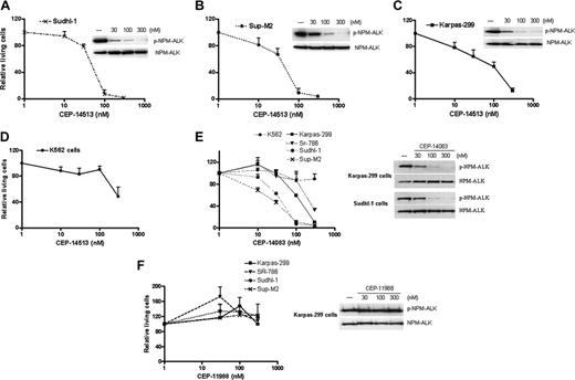
The effects of CEP-14083 and CEP-14513 on the proliferation of ALK+ ALCL cells and ALK- cells in culture were measured by MTS assay. As shown in Figure 4, treatment of ALK+ ALCL cells, including Karpas-299, Sudhl-1, SR-786, and Sup-M2 cells, with CEP-14083 and CEP-14513 at concentrations ranging from 10 to 300 nM significantly decreased the proliferation of these ALK+ ALCL cell lines in culture, with the magnitude of inhibition correlating with the inhibition of ALK tyrosine phosphorylation (Figure 4A-C,E). In contrast, at the same concentrations, CEP-14083 or CEP-14513 had marginal inhibitory effects on K562, a T cell-derived leukemia cell line lacking ALK expression (Figure 4D-E). Treatment with CEP-11988 did not inhibit ALK tyrosine phosphorylation in ALK+ ALCL cells and had no effects on the proliferation and survival of ALK+ ALCL cells in culture over the same concentration range (Figure 4F). Treatment with CEP-14083 or CEP-14513 led to inhibition of colony formation in agar of ALK+ ALCL cells (Figure 5A) and a significant increase in caspase 3/7 activities in ALK+ ALCL cells, while inducing minimal caspase activation in K562 cells (Figure 5B). These data validated the assumption that ALK activity is essential for the proliferation and survival of ALK+ ALCL cells.
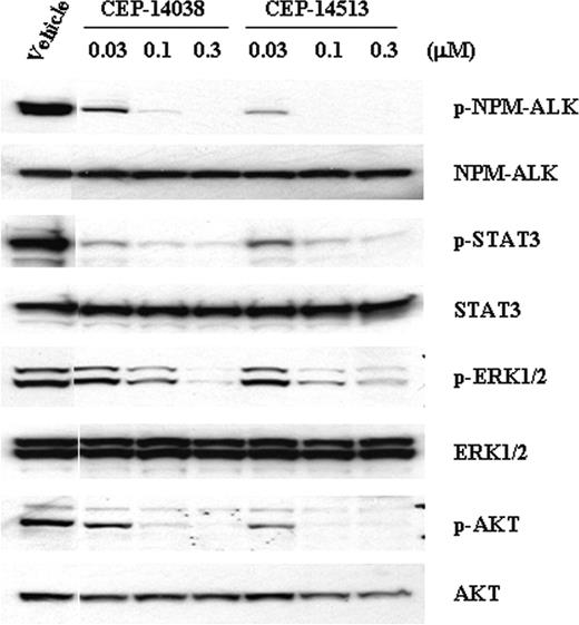
The mechanisms for ALK-mediated cell proliferation and survival have not been well defined; however, several signaling pathways including the MAP kinase pathway (ERK1 and 2), the PI3K/AKT pathway, and the JAK/STAT3 pathway, have been implicated in mediating downstream biologic effects of ALK.10-11,13-15 To characterize the effects of inhibition of ALK activity on these signaling pathways, the phosphorylation (activation) status of these pathways in ALCL cells treated with CEP-14083 and CEP-14513 was examined. As shown in Figure 6, AKT, ERK1/2, and STAT3 were readily phosphorylated (activated) in serum-starved Sup-M2 cells, correlating with the degree of ALK tyrosine phosphorylation in these cells. Following inhibition of ALK tyrosine phosphorylation by exposure to CEP-14083 or CEP-14513, ERK1/2, STAT3, and AKT phosphorylation/activation were significantly decreased in these cells (Figure 6). Neither CEP-14083 nor CEP-14513 inhibited AKT and ERK phosphorylation/activation directly in ALK- cell lines at concentrations up to 300 nM (data not shown). These results suggest that all 3 signaling pathways are downstream targets mediating ALK signaling and that modulation of these pathways by ALK inhibitors could contribute to the growth inhibition and induction of apoptosis in ALK+ cells.
The effects of ALK inhibition on cell-cycle progression in ALK+ ALCL cells were also examined. Treatment of CEP-14513 or CEP-14083 led to cell-cycle G1 arrest, followed by accumulation of sub-G1 population (an indication of apoptosis) in ALK+ ALCL cells, including Sudhl-1 and Karpas-299 cells (Figure 7 and data not shown). The G1 arrest observed following ALK inhibition correlated with the inhibition of PI3K/AKT and STAT3 activation in these cells.
Discussion
Functional ALK is essential for the proliferation and survival of ALCL cells
The results of these studies indicate the essential role of functional ALK in the proliferation and survival of ALK+ ALCL cells and validated the notion that ALK is a promising therapeutic target for treatment of ALK+ ALCL. To the best of our knowledge, this is the first direct demonstration that inhibition of ALK activation is sufficient to attenuate the growth and survival of ALK+ ALCL cells. Previously, Turturro et al demonstrated that down-regulation of NPM-ALK with 17-allylamino-17-demethoxygeldanamycin (17-AAG), an ansamycin-derived HSP90 inhibitor, led to increased apoptosis and reduced AKT/PKB activity in ALK+ ALCL cells.9 Treatment with 17-AAG led to the degradation of multiple protein kinases, including AKT/PKB and Raf, important for cell proliferation and survival.24 Consequently, the specific role of NPM-ALK degradation contributing to increased apoptosis in ALK+ ALCL cells could not be defined unequivocally in those studies. Hubinger et al recently described the use of a hammerhead ribozyme-mediated approach for the cleavage of the NPM-ALK transcript in ALK+ ALCL cell lines. Although in vitro assays demonstrated essentially complete degradation of NPM-ALK mRNA, repeated transfections of the ribozyme did not produce a significant reduction in levels of endogenous NPM-ALK protein in Karpas-299 cells.17 In this report, using small-molecule kinase inhibitors with potent ALK inhibitory activity, we were able to completely abolish ALK activity in ALK+ ALCL cells at nanomolar concentration ranges. Our data clearly demonstrated that ALK activity is indispensable for the proliferation and survival of ALK+ ALCL cells.
ALK activity is essential for the proliferation of ALK+ ALCL cells in culture.ALK+ ALCL cell lines (Karpas-299, SR-786, Sup-M2, and Sudhl-1) and ALK- cells (K562) were seeded on 96-well plates and treated with compounds at indicated concentrations for 48 hours. Viable cells were detected with MTS assay. The values presented are the average of relative cell numbers from 2 independent experiments with standard error. The effects of compounds on ALK tyrosine phosphorylation in ALCL cells are shown, in which the ALCL cells were treated with compounds at indicated concentrations for 2 hours and phospho- and total NPM-ALK were detected by immunoblotting as described in “Materials and methods.”
ALK activity is essential for the proliferation of ALK+ ALCL cells in culture.ALK+ ALCL cell lines (Karpas-299, SR-786, Sup-M2, and Sudhl-1) and ALK- cells (K562) were seeded on 96-well plates and treated with compounds at indicated concentrations for 48 hours. Viable cells were detected with MTS assay. The values presented are the average of relative cell numbers from 2 independent experiments with standard error. The effects of compounds on ALK tyrosine phosphorylation in ALCL cells are shown, in which the ALCL cells were treated with compounds at indicated concentrations for 2 hours and phospho- and total NPM-ALK were detected by immunoblotting as described in “Materials and methods.”
The unfavorable physical properties of the FP ALK inhibitors used in the in vitro studies described precluded their use for in vivo proof-of-concept studies. However, using siRNA approaches, Inghirami et al recently demonstrated that functional ALK is essential for the proliferation and survival of ALK+ ALCL tumor xenografts in mice.40
ALK is a potential molecular target for ALK+ ALCL and solid tumors
Our data indicate that ALK is a potential molecular target for the therapeutic modulation of ALK+ ALCL. Similar to BCR-ABL translocation associated with more than 90% of CML, the roles of aberrant expression of NPM-ALK or other ALK chimeric oncoproteins in the pathogenesis of ALCL have been well defined. Studies by Kuefer et al reported that mice given transplants of bone marrow from donor mice infected with retroviral constructs containing the human NPM-ALK cDNA developed lymphomas with 4 to 6 months.25 Subsequently, it was reported that the NPM-ALK transgenic mice spontaneously develop T-cell lymphomas with a relatively short latency period.26,27 The lack of ALK expression in adult human normal tissues with the exception of neuronal and glial cells4 and the observation that ALK knock-out mice are viable without gross or histologic defects (S. W. Morris, personal oral communication, December 2004) suggest that systemic ALK inhibition should be relatively well tolerated without significant side effects. In agreement with this hypothesis, the FP ALK inhibitors used here had minimal effects on the proliferation and survival of ALK- cell lines evaluated at the same concentrations that effectively inhibit ALK tyrosine phosphorylation and ALK-dependent biologic activities in ALK+ tumor cells
In addition to ALK+ lymphomas, ALK may also be implicated in the pathogenesis of other solid and hematologic tumors.28 Constitutively activated chimeric ALKs, including TPM3-ALK, TPM4-ALK, CTLC-ALK, CARS-ALK, and RANBP2-ALK, have been demonstrated in approximately 60% of inflammatory myofibroblastic tumors (IMTs), a slow-growing sarcoma that mainly affects children and young adults with about 250 new incidences each year in the United States. Although most IMTs can be cured by surgical resection of the tumor, cases of malignant, invasive, and metastatic IMTs are usually fatal.4,29 Therefore, an ALK inhibitor could have some clinical benefit for at least some patients with IMTs.
ALK inhibition leads to inhibition of colony formation and enhanced caspase activity in ALK+ ALCL cells. (A) ALK+ ALCL cells were treated with indicated concentrations of CEP compounds for 24 hours. The cells were diluted with 0.65% agar in culture medium with or without compounds and seeded on 35-mm3 culture dishes at density of 2 × 10³/dish. The plates were cultured until individual colonies were formed. The values presented are the average of relative colony numbers from 2 independent assays with standard error. (B) ALK+ ALCL cells (Karpas-299 and Sudhl-1) or ALK- cells (K562) were seeded on 96-well plates and treated with CEP compounds at indicated concentrations for 24 hours. Caspase 3/7 activity was quantitated with apo-one homogenous caspase 3/7 assay kit. The values presented are the average of relative caspase 3/7 activities from 3 individual samples with standard error.
ALK inhibition leads to inhibition of colony formation and enhanced caspase activity in ALK+ ALCL cells. (A) ALK+ ALCL cells were treated with indicated concentrations of CEP compounds for 24 hours. The cells were diluted with 0.65% agar in culture medium with or without compounds and seeded on 35-mm3 culture dishes at density of 2 × 10³/dish. The plates were cultured until individual colonies were formed. The values presented are the average of relative colony numbers from 2 independent assays with standard error. (B) ALK+ ALCL cells (Karpas-299 and Sudhl-1) or ALK- cells (K562) were seeded on 96-well plates and treated with CEP compounds at indicated concentrations for 24 hours. Caspase 3/7 activity was quantitated with apo-one homogenous caspase 3/7 assay kit. The values presented are the average of relative caspase 3/7 activities from 3 individual samples with standard error.
ALK inhibition leads to the attenuation of STAT3, ERK1/2, and AKT activation. Sup-M2 cells were serum-starved for about 24 hours by culturing in RPMI/0.1% FBS. The cells were then treated with indicated concentrations of compounds for 2 hours. Cells were lysed and the tyrosine phosphorylated and total NPM-ALK, P-AKT and total AKT, P-ERKs and total ERKs, and P-STAT3 and total STAT3, were detected by immunoblotting as described in “Materials and methods.”
ALK inhibition leads to the attenuation of STAT3, ERK1/2, and AKT activation. Sup-M2 cells were serum-starved for about 24 hours by culturing in RPMI/0.1% FBS. The cells were then treated with indicated concentrations of compounds for 2 hours. Cells were lysed and the tyrosine phosphorylated and total NPM-ALK, P-AKT and total AKT, P-ERKs and total ERKs, and P-STAT3 and total STAT3, were detected by immunoblotting as described in “Materials and methods.”
ALK inhibition leads to cell-cycle G1 arrest of ALK+ ALCL cells. Sudhl-1 cells were treated with CEP compounds at indicated concentrations for 24 hours. Cells were collected, fixed, and stained with propidium iodide. The DNA contents were analyzed by flow cytometry. Representative histograms from 2 independent experiments are shown.
ALK inhibition leads to cell-cycle G1 arrest of ALK+ ALCL cells. Sudhl-1 cells were treated with CEP compounds at indicated concentrations for 24 hours. Cells were collected, fixed, and stained with propidium iodide. The DNA contents were analyzed by flow cytometry. Representative histograms from 2 independent experiments are shown.
The fact that the most abundant expression of ALK occurs in the neonatal brain suggests a possible role for ALK in brain development.4,5 Recently, pleiotrophin and midkine, secreted heparin-binding polypeptide growth factors highly expressed in the developing nervous system, were identified as putative ALK ligands.30,31 Preclinical studies have suggested that pleiotrophin/midkine contribute to tumor growth, invasion, and metastasis of a variety of solid tumors.31-34 Both pleiotrophin and ALK were found to be overexpressed in human glioblastomas,31 and depletion of ALK transcripts by ribozymes reduced tumor growth of glioblastoma xenografts in athymic nude mice and prolonged survival of the animals.35 In particular, a 15-kDa COOH-terminal truncation of pleiotrophin was shown to promote glioblastoma proliferation through ALK.31 These data implicate ALK signaling as a rate-limiting factor in the growth of glioblastomas and suggest the potential utility of therapeutic targeting of ALK in this patient population, with about 17 000 new cases in the United States each year.
The expression of ALK receptor protein or fusion proteins (or both) has also been reported in neuroblastomas,36 diffuse large B-cell lymphoma,37,38 malignant peripheral nerve sheath tumor, and leiomyosarcoma.39 It has been reported that cell lines established from solid tumors of ectodermal origin, including melanoma and breast carcinoma, exhibited ALK receptor mRNA expression.28 Further studies should elucidate the role, if any, of either full-length or fusion forms of ALK in the pathogenesis or progression of these tumors.
Prepublished online as Blood First Edition Paper, October 27, 2005; DOI 10.1182/blood-2005-08-3254.
The publication costs of this article were defrayed in part by page charge payment. Therefore, and solely to indicate this fact, this article is hereby marked “advertisement” in accordance with 18 U.S.C. section 1734.
We thank Dr Stephan W. Morris of St Jude Children's Research Hospital for providing the BaF3 cells and Dr Giorgio Inghirami of New York University Medical School for providing the MEF-Tet-Off-NPM-ALK cell line.



This feature is available to Subscribers Only
Sign In or Create an Account Close Modal