The iron requirements of the erythroid compartment modulate the expression of hepcidin in the liver, which in turn alters intestinal iron absorption and iron release from the reticuloendothelial system. We have taken advantage of an inherited anemia of the mouse (hemoglobin deficit, or hbd) to gain insights into the factors regulating hepcidin expression. hbd mice showed a significant anemia but, surprisingly, their iron absorption was not increased as it was in wild-type animals made anemic to a similar degree by dietary iron depletion. In wild-type mice hepatic hepcidin levels were decreased but in hbd animals a significant and unexpected increase was observed. The level of absorption was appropriate for the expression of hepcidin in each case, but in hbd mice did not reflect the degree of anemia. However, this apparent inappropriate regulation of hepcidin correlated with increased transferrin saturation and levels of diferric transferrin in the plasma, which in turn resulted from the reduced capacity of hbd animals to effectively use transferrin-bound iron. These data strengthen the proposal that diferric transferrin is a key indicator of body iron requirements.
Introduction
Hemoglobin deficit (hbd) is a strain of mouse with a mild to moderate anemia that was first described by Scheufler in 1969.1 The mutation in these animals arose spontaneously and is inherited in an autosomal recessive manner.1,2 The hbd gene has been mapped to chromosome 193 but has yet to be identified. Affected animals are characterized by a microcytic, hypochromic anemia, reticulocytosis, elevated free erythrocyte protoporphyrin, and raised plasma iron.1,2,4 The anemia is most severe in newborn animals but persists throughout adult life.1,2
The early demonstration that the hbd anemia failed to respond to parenteral iron suggested that the basic defect led to a reduction in iron uptake by immature erythroid cells.1,2 The observations that the plasma iron concentration and free erythrocyte protoporphyrin levels were elevated in these mice supported this proposal.2,4 Garrick et al5 found that while hbd reticulocytes were able to take up diferric transferrin normally, their capacity to extract iron from this transferrin was significantly impaired. Molecules involved in transferrin-bound iron uptake include transferrin receptors 1 and 2 (Tfr1, Tfr2)6,7 and divalent metal transporter 1 (Dmt1),8 but the genes encoding these proteins are not located on chromosome 19, suggesting that a novel molecule is involved. Interestingly, although uptake of transferrin-bound iron is a property shared by all nucleated cells in the body, bone marrow transplantation studies suggest that expression of the hbd defect is restricted to hematopoietic cells and leads to a late block in erythroid differentiation.4
Regardless of the basic defect underlying the hbd anemia, these mice provide an ideal model for investigating the relationship between disturbed erythroid iron supply and iron absorption. The erythroid marrow requires a large amount of iron for hemoglobin synthesis and thus represents the major sink for iron in the body.9 Under normal circumstances, the vast majority of this iron is derived from the macrophages of the reticuloendothelial system following breakdown of hemoglobin from senescent erythrocytes. However, if iron supply to the marrow is insufficient to meet the erythroid demand, then intestinal iron absorption is stimulated.9 Enhanced erythropoiesis is a potent stimulus for iron absorption, and recent studies have suggested that this effect is mediated by the liver-derived regulatory peptide hepcidin.10,11 In individuals with iron deficiency anemia, or in clinical situations where erythropoiesis is markedly ineffective (in β-thalassemia, for example), iron absorption is strongly stimulated and hepcidin expression is reduced.9,12,13 Hepcidin is synthesized in the liver14 and appears to act on intestinal enterocytes to repress iron absorption,15,16 but it is not known how erythroid iron requirements are relayed to the liver to modulate hepcidin expression.
In this study, we have used hbd mice as a model to study the relationship between systemic markers of iron homeostasis and intestinal iron absorption. Despite their anemia, hbd animals do not show elevated iron absorption, nor do they show a decrease in hepcidin expression. However, the hbd anemia differs from iron deficiency anemia in that transferrin saturation is elevated due to the inability of the mice to efficiently use transferrin-bound iron. The increased circulating level of diferric transferrin correlates well with the inappropriately high hepcidin expression and provides further evidence that the level of diferric transferrin in the plasma is an important means of communicating body iron requirements to the regulatory system that modulates intestinal iron absorption.
Materials and methods
Animals, diets, and tissue collection
Hbd mice were originally obtained from the Jackson Laboratories (Bar Harbor, ME), and a breeding colony was established in Brisbane, Australia. The hfe knockout mice used were on a C57BL/6J background and were a generous gift from Professor William Sly (St Louis University, MO).17 Control C57BL/6J mice were obtained from the Animal Resource Centre (Perth, Western Australia). Male mice were used for all studies and animals were investigated when they were 10 to 12 weeks old. Mice were maintained on either a semisynthetic iron-deficient diet (3 mg/kg wet weight) or the equivalent control diet (159 mg/kg wet weight).18 Prior to analysis, animals were anesthetized (44 mg/kg ketamine and 8 mg/kg xylazine) and blood was withdrawn from the abdominal aorta for hematologic analysis and for measurement of plasma iron indices. Duodenal enterocytes were isolated as previously described19 and snap-frozen in liquid nitrogen, as was liver tissue. In some studies, 4-week-old C57BL/6J mice were treated with Freund complete adjuvant and processed as previously described.20 All experiments described in this study were approved by the Queensland Institute of Medical Research Animal Ethics Committee.
Assessment of hematologic parameters and hepatic iron levels
Hemoglobin, hematocrit, and red blood cell (RBC) count were measured with a Cell-Dyn 1600 automated blood analyzer (Abbott Laboratories, Brisbane, Australia). Reticulocyte counts were determined by examining blood smears of cells stained with new methylene blue as described previously.21 Plasma iron and transferrin saturation were measured using an Iron and Iron Binding Capacity Kit (Sigma-Aldrich, Sydney, Australia). Levels of diferric transferrin were determined using urea polyacrylamide gel electrophoresis as previously described.11 Western blotting was used to determine total transferrin levels in the same samples and was carried out as previously described22 using a polyclonal antibody to human transferrin (1 in 1000 dilution; Silenus Laboratories, Hawthorne, Australia) that crossreacts with the mouse protein. To measure hepatic iron concentration, a small amount of liver from each animal was dried overnight at 110°C, and tissue nonheme iron content determined colorimetrically as previously described.23
Evaluation of intestinal iron absorption
Intestinal iron absorption measurements were performed by whole-body counting after oral administration of 59FeCl3 as previously described.24
RNA extraction and ribonuclease protection assay
Total RNA was extracted from tissue samples using TRIZOL reagent (Invitrogen, Melbourne, Australia) as per the manufacturer's instructions. Ribonuclease protection assays (RPAs) were performed as previously described22 using 5 μg total RNA. The housekeeping gene acidic ribosomal protein P0 (Arbp) was used for normalization. The riboprobes used corresponded to the following cDNA sequences (the data presented in parentheses indicate official gene symbol, section of cDNA, and GenBank accession number): Dcytb (Cybrd1, nt213-452, AF354666), Dmt1 (Slc11a2, nt1473-1747, L33415), ferroportin 1 (Slc40a1, nt1156-1328, AF231120), acidic ribosomal phosphoprotein P0 (Arbp, nt81-400, X15267), hemojuvelin (Hfe2, nt790-1049, NM_027126), hepcidin (HAMP, nt1-225, AF297664), hfe (Hfe, nt455-687, U66849), Tfr2 (Trfr2, nt501-787, AF222895), orosomucoid 2 (Orm2, nt241-500, M27009) and serum amyloid A-1 (Saa1, nt361-580, NM_009117).
Western blot analysis
Western blotting on liver samples (100 μg total protein per lane) was carried out as previously described.22,24 A rabbit polyclonal antibody to Tfr2 (affinity purified; 0.5 μg/mL) was a generous gift from Dr Nathan Subramaniam (Queensland Institute of Medical Research, Brisbane, Australia).25 Actin was used as a loading control and was detected using a rabbit polyclonal antiactin antibody (1 in 1000 dilution; Sigma-Aldrich).
Statistical analysis
All experimental groups contained 3 to 11 mice and values are expressed as means ± SEM. Statistical differences between means were calculated by analysis of variance followed by Tukey post-hoc testing using SPSS software (SPSS Australasia, Sydney, Australia).
Results
Anemia and iron status of hbd mice
In order to confirm the anemic phenotype of hbd mice, we measured various hematologic parameters in these animals and compared them with wild-type mice. On a control diet, hbd mice had significantly lower hemoglobin concentrations, hematocrits, and mean corpuscular volumes than their wild-type counterparts (Figure 1A-C), and a mild reticulocytosis (data not shown). These data confirmed that the animals carried a hypochromic, microcytic anemia. The differences between hbd and wild-type mice were retained when the animals were fed an iron-deficient diet, although all values were reduced.
Despite the anemia in hbd mice, the hepatic iron concentration in these animals was not significantly different from wild-type mice, regardless of whether they were maintained on an iron-sufficient or iron-deficient diet (Figure 1D). All animals on an iron-deficient diet had reduced hepatic iron stores. While anemia in a wild-type mouse would normally be associated with decreased transferrin saturation, this was not the case in hbd mice. In fact, transferrin saturation was increased in these animals (Figure 2A). This reached statistical significance in the animals maintained on a control diet, and was increased, but not significantly so (at the 5% level), in animals on an iron-deficient diet. The increased transferrin saturation is, however, consistent with the inability of immature erythroid cells from hbd mice to effectively remove diferric transferrin from the plasma. To examine this directly, we used urea gel electrophoresis to determine the relative level of diferric transferrin in the plasma (Figure 2B-C) and found it to be significantly increased in hbd mice. There were no differences in total iron-binding capacity (TIBC; data not shown, or in total transferrin as measured by Western blot; Figure 2B) between hbd and wild-type mice.
Hematologic parameters and hepatic iron levels in hbd and wild-type mice. Hemoglobin concentrations (A), hematocrits (B), mean corpuscular volumes (C), and hepatic iron concentrations (D) were measured in 10- to 12-week-old wild-type (C57BL/6J;  ) and hbd (▪) mice as described in “Assessment of hematologic parameters and hepatic iron levels.” Some animals were maintained on a diet of normal iron content (“Control Diet”) while others had been on an iron-deficient diet (“Deficient Diet”) from the time of weaning. The data represent the mean ± SEM (n = 5-10; *P < .05; **P < .01 for hbd mice relative to the wild-type group for each diet).
Hematologic parameters and hepatic iron levels in hbd and wild-type mice. Hemoglobin concentrations (A), hematocrits (B), mean corpuscular volumes (C), and hepatic iron concentrations (D) were measured in 10- to 12-week-old wild-type (C57BL/6J;  ) and hbd (▪) mice as described in “Assessment of hematologic parameters and hepatic iron levels.” Some animals were maintained on a diet of normal iron content (“Control Diet”) while others had been on an iron-deficient diet (“Deficient Diet”) from the time of weaning. The data represent the mean ± SEM (n = 5-10; *P < .05; **P < .01 for hbd mice relative to the wild-type group for each diet).
Transferrin saturation and diferric transferrin levels are increased in hbd mice. (A) Plasma transferrin saturation was measured in 10- to 12-week-old wild-type (C57BL/6J;  ) and hbd (▪) mice on either a control or an iron-deficient diet (n = 7-11; **P < .01 relative to wild-type mice). (B) To determine the levels of diferric transferrin in wild-type and hbd mice on a control diet, rivanol extracts derived from the equivalent of 60 nL serum were subjected to urea polyacrylamide gel electrophoresis followed by Western blotting with an antibody to transferrin (top panel). Total transferrin levels determined by analysis of the same samples by sodium dodecyl sulfate–polyacrylamide gel electrophoresis (SDS-PAGE) followed by Western blotting demonstrated that there was no difference between C57BL/6J mice and hbd mice (bottom panel). Total transferrin was used as a loading control for the diferric transferrin gels because an independent measure of serum transferrin levels (total iron-binding capacity) showed no differences between the strains (data not shown). Four replicates are shown for each group. (C) Densitometric analysis of the gels shown in panel B. The graph shows the relative diferric transferrin levels expressed as a proportion of the control level of diferric transferrin in C57BL/6J mice. The data represent the mean ± SEM (n = 4; **P < .01).
Transferrin saturation and diferric transferrin levels are increased in hbd mice. (A) Plasma transferrin saturation was measured in 10- to 12-week-old wild-type (C57BL/6J;  ) and hbd (▪) mice on either a control or an iron-deficient diet (n = 7-11; **P < .01 relative to wild-type mice). (B) To determine the levels of diferric transferrin in wild-type and hbd mice on a control diet, rivanol extracts derived from the equivalent of 60 nL serum were subjected to urea polyacrylamide gel electrophoresis followed by Western blotting with an antibody to transferrin (top panel). Total transferrin levels determined by analysis of the same samples by sodium dodecyl sulfate–polyacrylamide gel electrophoresis (SDS-PAGE) followed by Western blotting demonstrated that there was no difference between C57BL/6J mice and hbd mice (bottom panel). Total transferrin was used as a loading control for the diferric transferrin gels because an independent measure of serum transferrin levels (total iron-binding capacity) showed no differences between the strains (data not shown). Four replicates are shown for each group. (C) Densitometric analysis of the gels shown in panel B. The graph shows the relative diferric transferrin levels expressed as a proportion of the control level of diferric transferrin in C57BL/6J mice. The data represent the mean ± SEM (n = 4; **P < .01).
Absorption and the expression of intestinal iron transport genes
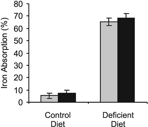
Intestinal iron absorption is enhanced in many forms of anemia, but this was not the case in hbd mice, where absorption was similar to that of wild-type mice regardless of their diet (Figure 3). Nevertheless, hbd animals were able to increase their absorption as effectively as wild-type animals when they were placed on an iron-deficient diet.
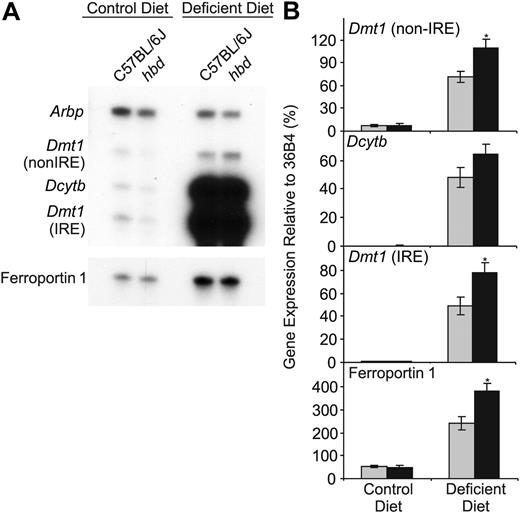
Analysis of the expression of several iron transport genes in the small intestine supported the whole-body iron absorption studies. Levels of Dcytb, Dmt1 (both the iron-responsive element [IRE] and the non-IRE forms), and ferroportin 1 mRNA were low in animals maintained on an iron-sufficient diet, as was iron absorption, and there were no significant differences between wild-type and hbd mice (Figure 4). However, the expression of each of these genes was significantly enhanced when the animals were placed on an iron-deficient diet, consistent with the observed increase in iron absorption.
Iron absorption is normal in hbd mice. The absorption of iron in wild-type ( ) and hbd (▪) mice on either a control or iron-deficient diet was determined following the oral administration of 59FeCl3. Whole-body counting for 59Fe was carried out 1 hour and 5 days after the dose, and absorption is expressed as the percentage of the initial dose retained by the animals after 5 days. The data represent the mean ± SEM (n = 3-7).
Iron absorption is normal in hbd mice. The absorption of iron in wild-type ( ) and hbd (▪) mice on either a control or iron-deficient diet was determined following the oral administration of 59FeCl3. Whole-body counting for 59Fe was carried out 1 hour and 5 days after the dose, and absorption is expressed as the percentage of the initial dose retained by the animals after 5 days. The data represent the mean ± SEM (n = 3-7).
Expression of iron regulatory genes in the liver
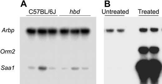
The importance of the liver in the regulation of body iron homeostasis has been recognized in recent years,26,27 so we undertook an examination of the expression of several of the key hepatic regulatory molecules (Figure 5). Expression of the antimicrobial peptide hepcidin was significantly enhanced in hbd mice compared with their wild-type counterparts, a somewhat surprising finding because increased hepcidin levels are usually associated with iron-loading rather than anemia.28 This effect was only seen in animals on an iron-sufficient diet. To ensure that the observed increase in hepcidin expression was not due to inflammation, we examined the hepatic expression of serum amyloid A-1 (Saa1) and orosomucoid-2 (Orm2) in hbd and control mice. The hepatic expression of Saa1 is increased during acute inflammation, and hepatic Orm2 is increased during both acute and chronic inflammation.29 Neither of these markers of inflammation was increased in the liver of hbd mice; however, strong increases were seen in wild-type mice treated with Freund Complete Adjuvant (Figure 6). When an iron-deficient diet was used, hepcidin expression was barely detectable and no differences between the 2 types of mice could be discerned.
The expression of iron transport genes in the duodenum of hbd and wild-type mice. Duodenal expression of Dmt1 (both the IRE and non-IRE splice variants), Dcytb, and ferroportin 1 mRNA in wild-type ( ) and hbd (▪) mice on either a control or iron-deficient diet was determined by ribonuclease protection assay. The mRNA encoding ribosomal protein Arbp was used as a normalization control. (A) Representative assays for each gene. (B) Band intensities for each gene were measured by densitometry and graphed as a proportion of 36B4 expression. The data represent the mean ± SEM (n = 5-11; *P < .05).
The expression of iron transport genes in the duodenum of hbd and wild-type mice. Duodenal expression of Dmt1 (both the IRE and non-IRE splice variants), Dcytb, and ferroportin 1 mRNA in wild-type ( ) and hbd (▪) mice on either a control or iron-deficient diet was determined by ribonuclease protection assay. The mRNA encoding ribosomal protein Arbp was used as a normalization control. (A) Representative assays for each gene. (B) Band intensities for each gene were measured by densitometry and graphed as a proportion of 36B4 expression. The data represent the mean ± SEM (n = 5-11; *P < .05).
The expression of iron regulatory genes in the liver of hbd and wild-type mice. Hepatic expression of hepcidin, Tfr2, hemojuvelin, and hfe mRNA in wild-type ( ) and hbd (▪) mice on either a control or iron-deficient diet was determined by ribonuclease protection assay. The mRNA encoding ribosomal protein Arbp was used as a normalization control. (A) Representative assays for each gene. (B) Band intensities for each gene were measured by densitometry and graphed as a proportion of 36B4 expression. The data represent the mean ± SEM (n = 5-11; *P < .05; **P < .01).
The expression of iron regulatory genes in the liver of hbd and wild-type mice. Hepatic expression of hepcidin, Tfr2, hemojuvelin, and hfe mRNA in wild-type ( ) and hbd (▪) mice on either a control or iron-deficient diet was determined by ribonuclease protection assay. The mRNA encoding ribosomal protein Arbp was used as a normalization control. (A) Representative assays for each gene. (B) Band intensities for each gene were measured by densitometry and graphed as a proportion of 36B4 expression. The data represent the mean ± SEM (n = 5-11; *P < .05; **P < .01).
The expression of genes induced by inflammation in the liver of hbd and wild-type mice. The hepatic expression of Saa1 (induced in acute inflammation) and Orm2 (induced in both acute and chronic inflammation) mRNA in wild-type and hbd mice on control diet was determined by ribonuclease protection assay. The mRNA encoding ribosomal protein Arbp was used as a normalization control. (A) A representative gel is shown (16 hours of exposure). Note that Orm2 mRNA could not be detected in these animals. (B) RNA extracted from 4-week-old C57BL/6J mice that were either untreated or had been injected with Freund complete adjuvant 16 hours earlier was used to show the induction of the genes during acute inflammation (2 hours of exposure). No evidence of inflammation was observed in any hbd mouse tested (n = 9).
The expression of genes induced by inflammation in the liver of hbd and wild-type mice. The hepatic expression of Saa1 (induced in acute inflammation) and Orm2 (induced in both acute and chronic inflammation) mRNA in wild-type and hbd mice on control diet was determined by ribonuclease protection assay. The mRNA encoding ribosomal protein Arbp was used as a normalization control. (A) A representative gel is shown (16 hours of exposure). Note that Orm2 mRNA could not be detected in these animals. (B) RNA extracted from 4-week-old C57BL/6J mice that were either untreated or had been injected with Freund complete adjuvant 16 hours earlier was used to show the induction of the genes during acute inflammation (2 hours of exposure). No evidence of inflammation was observed in any hbd mouse tested (n = 9).
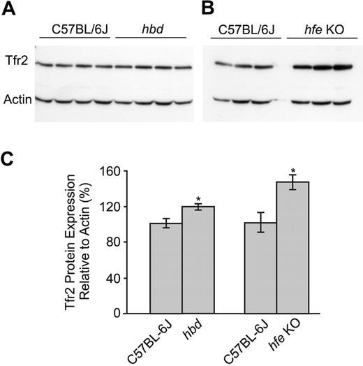
Expression of Tfr2 and hemojuvelin, both of which have been implicated in the regulation of hepcidin,30,31 was identical in hbd and wild-type mice at the RNA level. However, when Tfr2 protein expression was examined, we found a small but significant increase in the hbd animals (Figure 7). Previous in vitro and in vivo studies have suggested that Tfr2 protein is stabilized in the presence of diferric transferrin,32,33 and our finding that Tfr2 protein is increased in hbd animals is consistent with these data. As a positive control, we investigated Tfr2 protein levels in hfe knockout mice, which have an even higher transferrin saturation (85.2 ± 3.8% for hfe knockout mice versus 48.0 ± 2.4% for hbd mice), and found Tfr2 expression to be further increased as previously described.33 The other molecule known to modulate hepcidin levels is hfe,26 and we found hfe mRNA to be slightly, but significantly, increased in hbd mice.
Discussion
Iron deficiency anemia has long been known to be a potent stimulus for intestinal iron absorption since the erythroid compartment has the highest iron requirements of any body tissue.9 How the body signals the intestine to alter iron absorption in response to anemia remains poorly understood, although it is likely to do so by altering the expression of the regulatory peptide hepcidin in the liver. In this study we have taken advantage of an inherited anemia of the mouse to investigate this relationship. Hemoglobin-deficit mice show a significant anemia but, surprisingly, their iron absorption is normal. The level of absorption in hbd mice is consistent, however, with the unexpectedly high expression of hepcidin in these animals. It is unlikely that the increase in hepcidin expression is the result of inflammation in the hbd animals because no increase in the hepatic expression of Saa1 or Orm2, which are increased during acute and chronic inflammation, respectively,29 was observed. This apparent inappropriate regulation of hepcidin correlates with increased transferrin saturation and levels of diferric transferrin in the plasma, which in turn result from the reduced capacity of hbd animals to effectively utilize transferrin-bound iron. Wild-type animals made anemic to a similar degree by dietary iron depletion have much lower transferrin saturations and show appropriately elevated iron absorption. Consistent with these findings, we observed an increase in Tfr2 protein expression in the liver of hbd animals. Tfr2 is a key regulator of hepcidin expression,30 and previous studies have shown that Tfr2 protein stability is increased by diferric transferrin in cell culture;32,33 this is supported by animal models.33 These data strengthen the proposal that diferric transferrin is a key indicator of body iron requirements.34
The expression of Tfr2 protein in the liver of hbd and wild-type mice. (A) The hepatic expression of Tfr2 protein in wild-type and hbd mice on the control diet was determined by Western blotting. Actin was used as a loading control. (B) Protein extracted from the liver of 10-week-old C57BL/6J mice and hfe knockout (hfe KO) mice was used to demonstrate the increase in Tfr2 protein expression that occurs when the serum transferrin saturation is high. (C) Band intensities for each gene were measured by densitometry and normalized to actin expression before being graphed as a percentage of controls. The data represent the mean ± SEM (n = 3-4; *P < .05).
The expression of Tfr2 protein in the liver of hbd and wild-type mice. (A) The hepatic expression of Tfr2 protein in wild-type and hbd mice on the control diet was determined by Western blotting. Actin was used as a loading control. (B) Protein extracted from the liver of 10-week-old C57BL/6J mice and hfe knockout (hfe KO) mice was used to demonstrate the increase in Tfr2 protein expression that occurs when the serum transferrin saturation is high. (C) Band intensities for each gene were measured by densitometry and normalized to actin expression before being graphed as a percentage of controls. The data represent the mean ± SEM (n = 3-4; *P < .05).
It has been known for many decades that the amount of iron absorbed from the diet reflects events at distant sites in the body (eg, the level of storage iron in the liver or the rate of red blood cell production in the erythroid marrow).9 How such sites communicate their iron requirements to the small intestine has been the subject of considerable interest and the necessary “humoral factor” has long been sought, but until recently no satisfactory candidates had been identified. Levels of erythropoietin,35 serum ferritin,36 transferrin saturation,37-39 and several other factors have at various times been considered potential signaling molecules, but it has proved difficult to place any of these into a strong mechanistic framework. Transferrin saturation has been a particularly appealing candidate, as diferric transferrin is the major source of iron for most tissues, and particularly for developing erythroid cells.9,40 Changes in the level of diferric transferrin would thus be expected to reflect the iron requirements of the tissues.
The identification in recent years of hepcidin as a central regulator of iron absorption and iron release from other tissues has played a major role in focusing this field. There is evidence that the effects of all stimuli known to alter absorption are mediated through hepcidin. These stimuli include changes in body iron stores,15,28 the rate of erythropoiesis,10 hypoxia,10 and inflammation.10 Following a stimulus to increase red cell production, for example, hepcidin levels decline10,11 and the repression on iron absorption is decreased to facilitate enhanced dietary iron intake. Hepcidin is synthesized in the liver and has recently been shown to interact with ferroportin 1,41 the major protein responsible for iron efflux from enterocytes,42 so a clear link between the liver and the gut has been established. Changes in storage iron in the liver could alter hepcidin expression directly (although this is far from resolved), but to explain how changes in erythropoiesis alter hepcidin expression, a link between the marrow and the liver must be invoked.
Previous studies from our laboratory indicated that the level of diferric transferrin in the plasma could provide such a signal. When rats were switched from a control diet to an iron-deficient diet we observed a decline in hepcidin expression before changes in hepatic iron levels were apparent.24 However, the decline correlated with changes in transferrin saturation, with levels below approximately 20% associated with reduced hepcidin expression. This agrees very well with earlier studies indicating that iron supply to the developing erythroid cells in the bone marrow becomes limiting at transferrin saturations below 16% in humans.43 In addition, we found that the transient decrease in hepcidin levels following a stimulus to increase erythropoiesis (phenylhydrazine-induced hemolysis) was associated with a transient reduction in the level of circulating diferric transferrin.11 These data support the proposal that transferrin saturation, and more specifically diferric transferrin, plays a major role in directing changes in hepcidin expression,34 although the effects of aberrant erythropoiesis in hbd mice on hepcidin expression via some independent factor could not be formally excluded.
The studies with hbd mice described in this paper support this proposal. Since these mice cannot use transferrin-bound iron efficiently, their transferrin saturation and levels of diferric transferrin were higher than those of wild-type animals. In addition, despite their anemia, hbd mice showed an increase in hepcidin expression relative to controls. Wild-type mice made anemic to a similar degree by dietary iron restriction showed very low transferrin saturations and negligible hepcidin expression as expected. The iron absorption mechanism in hbd mice per se does not appear to be defective, as the animals are able to mount an appropriate absorption response when they were maintained on an iron-deficient diet. In this case both transferrin saturation and hepcidin expression decreased and absorption increased. Our data also suggest that other consequences of the hbd anemia that could influence absorption, such as hypoxia, are less important than diferric transferrin levels. Hypoxia has been shown to lead to a decrease in hepcidin expression,10 so our observed increase in hepcidin in hbd mice is incompatible with a primary overriding hypoxic response. It is of interest that in his landmark paper on factors regulating iron balance, Finch9 concluded that iron supply to the marrow was the single most important factor influencing iron absorption.
A reciprocal relationship has been found between hepatic hepcidin expression and iron absorption.24 In this study, however, we observed a significant increase in hepcidin in hbd mice maintained on a control diet, but no corresponding decrease in iron absorption or the expression of intestinal iron transport genes. This may be because iron absorption was quite low on this diet and the sensitivity of the whole-body iron absorption technique was not sufficient to distinguish any differences between the 2 groups of mice. An alternative explanation is that relatively small changes in hepcidin are not sufficient to bring about changes in absorption, and that hepcidin must reach a certain threshold before it can act to repress intestinal iron transport. In other studies we have performed we have found evidence for this by demonstrating that a low level of hemolysis is unable to stimulate absorption even though hepcidin expression is reduced, whereas more significant hemolysis will decrease hepcidin expression even further and stimulate iron absorption (D.M.F., S.J.W., G.J.A., unpublished observations, April 2004). Nevertheless, these smaller changes in hepcidin expression may be sufficient to modulate iron release from other cells such as macrophages. As noted, when hbd mice are placed on an iron-deficient diet they are able to increase their iron absorption as effectively as wild-type animals, and hepcidin expression declines significantly. This demonstrates that the mice are able to show an appropriate hepcidin/absorption relationship when the stimulus to alter absorption is strong enough.
The mechanism by which changes in transferrin saturation might alter hepcidin expression is poorly understood. Recent studies showing that hepcidin levels are reduced in patients with mutations in HFE,26 Tfr2,30 or hemojuvelin31 suggest that these molecules act upstream of hepcidin and, since all are membrane proteins, that they represent potential sensors of diferric transferrin. This concept is strengthened by the realization that Tfr2 is a transferring-binding protein,7 and HFE interacts with Tfr1,44 the archetypal plasma membrane transferrin receptor with a high affinity for diferric transferrin. We recently proposed a model whereby a competition between HFE and diferric transferrin for binding to Tfr1 would modulate the level of free HFE on the hepatocyte surface, which would in turn initiate a signal transduction process that would ultimately lead to a stimulation of hepcidin expression.34 We similarly envisioned a competition between Tfr1 and Tfr2 for binding to diferric transferrin, with the Tfr2/transferrin complex initiating a signal transduction pathway. While recent data have supported the concept that HFE and diferric transferrin can compete for binding to Tfr1 at the cell surface,45 other aspects of this model remain unresolved. For example, it is not yet clear whether HFE and Tfr2 promote parallel hepcidin activation pathways or are components of the same pathway.
The hbd mouse has provided us with an ideal model system for investigating the relationship between anemia and iron absorption. Our results are consistent with the proposal that erythroid iron demand can signal the liver to alter hepcidin expression through changes in the circulating concentration of diferric transferrin. Since the basic defect in hbd mice is unknown, it remains possible that the abnormality could directly alter hepcidin expression in the liver. However, the demonstration that the hbd phenotype is transplantable with the bone marrow4 makes this unlikely and suggests that an indirect signal, such as diferric transferrin, is required. It is also likely that changes in storage iron exert their effects on hepcidin expression indirectly by influencing diferric transferrin levels, rather than through a direct effect on the hepcidin gene. Indeed, a number of situations have been described recently where the expression of hepcidin can be dissociated from changes in hepatic iron concentration.10,11,24 Further studies on the regulation of hepcidin will clarify the mechanisms involved.
Prepublished online as Blood First Edition Paper, October 20, 2005; DOI 10.1182/blood-2005-07-2614.
Supported in part by grants from the National Health and Medical Research Council of Australia, the National Institute of Diabetes, Digestive Diseases and Kidney (DK57648), and the Human Frontier Science Program (RGY0328/2001-M).
The publication costs of this article were defrayed in part by page charge payment. Therefore, and solely to indicate this fact, this article is hereby marked “advertisement” in accordance with 18 U.S.C. section 1734.


This feature is available to Subscribers Only
Sign In or Create an Account Close Modal