The α4β7 integrin plays a central role in the homing of T cells to the gut. We hypothesized that absence of the β7 subunit would result in a reduction of intestinal graft-versus-host disease (GVHD) and an improvement in overall GVHD morbidity and mortality in recipients of hematopoietic stem cell transplantation (HSCT). Analysis of alloreactive β7-/- T cells showed intact activation, proliferation, cytokine production, and cytotoxicity. However, recipients of β7-/- donor T cells in murine HSCT models experienced less GVHD morbidity and mortality than recipients of wild-type (WT) T cells, associated with a decrease in donor T-cell infiltration of the liver and intestine and with an overall significant decrease in hepatic and intestinal GVHD. In graft-versus-tumor (GVT) experiments, we demonstrated intact or even enhanced GVT activity of β7-/- donor T cells. In conclusion, β7-/- donor T cells caused less GVHD morbidity and mortality than WT donor T cells because of selectively decreased T-cell infiltration of the liver and intestines. Our data suggest that strategies to target the β7 integrin have the clinical potential to alleviate or prevent GVHD while sparing or potentiating GVT activity.
Introduction
Allogeneic hematopoietic stem cell transplantation (HSCT) is a potentially curative therapy for a variety of malignancies and nonmalignant conditions. In addition to the antitumor effect of the conditioning regimen, the graft-versus-tumor (GVT) activity of donor T cells is increasingly recognized as an important component of the overall antitumor effect of allogeneic HSCT. However, it has been well established that alloreactive T cells also play a critical role in the development of acute graft-versus-host disease (GVHD), which remains one of the main complications of allogeneic HSCT. Acute intestinal GVHD is a significant cause of posttransplantation morbidity and mortality. Damage to the gastrointestinal tract increases the circulation of inflammatory stimuli (such as endotoxin), which can amplify acute GVHD in the intestines and other target organs. Thus, reducing or preventing GVHD, especially of the gut, while preserving the beneficial GVT effect of donor T cells would significantly improve overall survival in patients who undergo allogeneic HSCT.
Although virtually any host tissue is a potential source of alloantigens, GVHD develops only in skin, liver, the gastrointestinal tract, and possibly lung and thymus.1 The reason for this target organ specificity remains unclear but may be attributed to organ-specific differences in susceptibility to damage from the conditioning regimen, inflammatory cytokine response, activation of antigen-presenting cells, and activation and infiltration of donor T cells. A fundamental hypothesis regarding the pathobiology of GVHD suggests that the dysregulation of leukocyte trafficking is important for the disease process,2 and recent studies have demonstrated the important role of T-cell homing and its regulation by integrins, chemokine receptors, and ligands in the T-cell immune response and T cell-mediated diseases, including acute GVHD.3-9
T-cell trafficking through the circulation, secondary lymphoid organs, and specific tissues is a multifaceted process requiring precise communication between lymphocytes, endothelial cells, and the extracellular matrix; chemokines, selectins, integrins, and their receptors play crucial roles in these complex interactions. The expression of specific adhesion molecules and chemokine receptors on T cells, in combination with a spatial and temporal expression pattern of the ligands for these receptors, is largely responsible for the tissue tropism of T-cell migration.6,10,11 The migration patterns of T cells are further influenced by inflammatory stimuli, including cytokines and chemokines associated with inflammation.
Circulating T cells, on activation, may undergo altered surface expression of adhesion molecules and receptors, leading to specific tissue tropism.6 The α4β7 integrin, or LPAM-1 (lymphocyte Peyer patch adhesion molecule), is expressed on T cells and acts as an intestinal homing receptor. The integrin exists as a 154-kDa α chain and a 130-kDa β chain joined as a transmembrane heterodimer with a short intracytoplasmic tail.12,13 The α4 subunit also associates with β1 chains, forming an integrin that binds to VCAM-1 and to fibronectin, resulting in cell adhesion and entry to inflamed tissues.14 The β7 integrin subunit also associates with the αE subunit, forming an integrin thought to play a role in lymphocyte homing to and retention in the lamina propria of the gut epithelium.15,16 The αϵβ7 integrin may also play a role in thymocyte adhesion to thymic epithelial cells.17,18
The α4β7 integrin interacts specifically with MAdCAM-1 (mucosal addressin cell adhesion molecule-1) on high endothelial venules in the Peyer patches and intestinal lamina propria,19,20 and it interacts less specifically with VCAM-1 and fibronectin.21 Circulating lymphocytes in spleen and mesenteric lymph nodes (MLNs) have low levels of α4β7 expression, which may be up-regulated on activation.22,23 Up-regulation is highly dependent on passage through the Peyer patches, with the subsequent increase in α4β7 expression leading to lymphocyte infiltration of gut endothelium.3,24
It has been well established that the α4β7 integrin plays a specific role in intestinal homing of lymphocytes; previous studies have shown that interference with MAdCAM-1 or with the α4 integrin subunit can prevent or ameliorate the development of various inflammatory conditions of the gut.3,25,26 Administration of monoclonal antibodies directed against the α4β7 integrin or against the α4 integrin subunit has been shown to resolve colitis in a well-established monkey model of inflammatory bowel disease (IBD).27 Recently, natalizumab, a monoclonal antibody directed against the α4 integrin subunit, was demonstrated to ameliorate IBD in human subjects.28-30 The β7 integrin subunit has been targeted in several experimental disease models, also with promising results; monoclonal antibodies directed against the β7 subunit have been shown to ameliorate colitis and to attenuate intestinal allograft rejection in murine models.15,31 Relatively few studies, however, have addressed the role of integrins in T-cell homing in patients with GVHD.
We previously demonstrated in murine models that α4β7- donor T cells cause less GVHD morbidity and mortality than α4β7+ unselected donor T cells, specifically because of reduced homing of donor T cells to host intestinal mucosa.32 However, we also found that even when using α4β7- selected donor T cells, α4β7 expression will be up-regulated on activation, resulting in the delay, but not the prevention, of intestinal infiltration of T cells.32 In a significant departure from these findings, we sought to investigate whether permanent loss of the β7 subunit of the α4β7 integrin would provide similar or better benefits.
The phenotype of β7-/- mice has been well described: mice are healthy and normal in all respects except for impaired development of gut-associated lymphoid tissue (GALT).20 Although lymphocyte numbers in the Peyer patches and intestine are reduced, lymphocytes are found in all other organs in normal quantities.20,33 In addition, lymphocyte development is not affected in β7-/- mice, suggesting that impaired GALT development is secondary to a homing defect and not to defective lymphocyte maturation.20
In this study we used β7-/- donor T cells in murine HSCT models to study the role of the β7 integrin in the trafficking of donor alloreactive T cells in HSCT recipients with GVHD.
Materials and methods
Cell line and antibodies
P815 (H-2d), from ATCC (Manassas, VA), is a mastocytoma cell line of DBA/2 mouse origin. Cell culture medium contained RPMI, 10% heat-inactivated fetal bovine serum (FBS), 100 U/mL penicillin, 100 mg/mL streptomycin, and 2 mM l-glutamine. P815-TGL (H-2d) was generated by transducing P815 with a retroviral vector containing a fusion reporter gene coding for HSV1-TK (T), enhanced green fluorescent protein (G), and firefly luciferase (L).34
Antimurine CD16/CD32 FcR block (2.4G2) and fluorochrome-labeled antimurine antibodies against CD3 (145-2C11), CD4 (RM4-5), CD8 (53-6.7), CD62L (MEL-14), Ly-9.1 (30C7), α4β7 (DATK32), CD44 (IM7), NK1.1-PE (PK136), T-cell receptor β (TCR-β; H57-597), Thy 1.1, H-2k, and H-2b were obtained from Pharmingen (San Diego, CA).
Mice and HSCT
Female B10.BR (H-2k), C57BL/6 (B6, H-2b), B6D2F1 (H-2b/d), B6.Thy1.1 (H-2b), and B6.β7-/- (H-2b) mice were obtained from The Jackson Laboratory (Bar Harbor, ME). Mice used were between 8 and 12 weeks old. HSCT protocols were approved by the Memorial Sloan-Kettering Cancer Center (MSKCC) Institutional Animal Care and Use Committee and have been described previously.32,35-38 Briefly, BM cells removed from femurs and tibias were T cell-depleted (TCD) with anti-Thy-1.2 and low-TOX-M rabbit complement (Cedarlane Laboratories, Hornby, ON, Canada). Purified splenic T cells were obtained by nylon wool column passage, followed by staining with anti-CD3-FITC antibodies for T-cell purity. Allografts consisted of 5 × 10⁶ TCD wild-type (WT) BM cells with or without 1 × 10⁶ WT or β7-/- T cells. Cells were resuspended in DMEM and injected into lethally irradiated recipients on day 0 after 1300 cGy total body irradiation (cesium Cs 137 [137Cs] source) as a split dose 3 hours apart. Mice were housed in the MSKCC pathogen-free facility in sterilized micro-isolator cages and were given normal chow and autoclaved hyperchlorinated drinking water (pH 3.0).
Tumor induction, assessment of GVHD, and determination of cause of death
P815 cells (1 × 10⁵) were infused on day 0 of HSCT after irradiation. Survival was monitored daily, and mice were individually scored weekly for 5 clinical parameters (weight, posture, activity level, fur ruffling, and skin lesions) on a scale from 0 to 2. A clinical GVHD score was generated by summation of the 5 criteria scores, as described by Cooke et al39 ; mice scoring 5 or greater were killed. All animals, regardless of macroscopic tumor at autopsy, underwent histopathologic examination of liver and spleen, performed by a veterinary pathologist (Dr. Krista La Perle, Cornell University Medical College, New York, NY) for evidence of tumor and to determine cause of death (GVHD vs tumor).
Histopathologic analysis
Small and large bowel, liver, and skin were assessed by experts in a blinded fashion. Formalin-preserved organs were embedded in paraffin, sectioned, stained with hematoxylin/eosin, and scored with a semiquantitative scoring system, as previously described.35 Bowel and liver were scored for 19 to 22 different parameters associated with GVHD, as previously described40 ; skin was evaluated for number of apoptotic cells per millimeter of epidermis, as previously described.41
Lymphocyte isolation from liver and gut
Mice were killed, and small intestine was dissected from the gastric-duodenal junction to the ileocecal junction. Intestines were flushed with 10% HEPES (N-2-hydroxyethylpiperazine-N ′-2-ethanesulfonic acid), 10% FBS, cut into 1-cm-long pieces, and incubated for 1 hour at 37° C with continuous shaking. Intestinal pieces were then vortexed for 15 seconds, and the supernatant was strained and centrifuged at 325 g for 5 minutes. Pellets were resuspended in 40% Percoll (Sigma Aldrich, St Louis, MO), overlaid on 70% Percoll, and centrifuged at 1300 g for 30 minutes. Lymphocytes were recovered from the interface. Livers were homogenized and passed through a 70-μm cell strainer. Pellets were resuspended in 40% Percoll, and lymphocytes were isolated as described.
Organ harvest for T-cell infiltrate analysis
Female B10.BR recipients underwent HSCT, as described. Allografts consisted of 5 × 10⁶ T cell-depleted WT BM cells with mixtures of 2 × 10⁶ WT (B6.Thy1.1) and 2 × 10⁶ β7-/- (Thy1.2) T cells infused into lethally irradiated recipients on day 0. Mice were harvested for GVHD target organs on day 8; lymphocytes were isolated for fluorescence-activated cell sorter (FACS) analysis to determine relative percentages of WT and β7-/- T cells.
Flow cytometry analysis
Lymphocytes were washed in FACS buffer (phosphate-buffered saline [PBS], 0.5% bovine serum albumin [BSA], 0.1% sodium azide). Cells (106) were incubated for 20 minutes at 4°C with anti-CD16/CD32 FcR block and subsequently with fluorochrome-labeled primary antibodies at saturating concentrations for 20 minutes at 4°C. Appropriate isotype controls were used. Cells were resuspended in FACS buffer, and flow cytometry analysis was performed on a FACScalibur (Becton Dickinson, San Jose, CA) with CellQuest software. Data analysis was performed with FlowJo software (Treestar, San Carlos, CA).
Carboxyfluorescein diacetate succinidyl ester labeling
Cells were labeled with CFSE, as described previously.42 Briefly, splenocytes were incubated with CFSE at a concentration of 2.5 μM in PBS at 37°C for 15 minutes, washed with PBS, and infused into sublethally irradiated (750 cGy) B10.BR allogeneic recipients. Splenocytes from recipients were harvested 72 hours later and analyzed by FACS, as described.
Cytotoxicity assay
Target cells were labeled with 100 μCi (3.7 MBq) of 51Cr at 3 × 10⁶ cells/mL for 1 hour at 37°C and plated at 2.5 × 10³ cells/well in 96-well U bottom plates (Costar, Cambridge, MA). Splenocytes from mice 14 days after HSCT with allografts consisting of WT bone marrow and either WT or β7-/- T cells were analyzed by FACS for donor markers and CD3/CD8 purity and were added at various effector-target ratios in a final volume of 200 μL in triplicate and incubated for 4 hours at 37°C. Subsequently, 35 μL supernatant was removed from each well and was counted in a gamma counter (Topcount-Packard, Meriden, CT) to determine experimental release. Spontaneous release was obtained from wells receiving target cells with medium, and total release was obtained from wells receiving 5% Triton X-100. Percentage cytotoxicity was calculated as follows: percentage cytotoxicity = 100 × (experimental release - spontaneous release)/(total release - spontaneous release).
Enzyme-linked immunosorbent assay
Blood was obtained by cardiac puncture from GVHD and control animals and was centrifuged at 13 400 g for 1.5 minutes. Sera were collected and stored at -80°C. Concentration of IFN-γ was determined by ELISA (R&D Systems, Minneapolis, MN) according to the manufacturer's instructions.
Bioluminescent imaging
Animals that received P815-TGL were given intraperitoneal (150 mg/kg) d-Luciferin (Xenogen, Alameda, CA). Ten minutes after injection, mice were anesthetized with isofluorane and placed supine in the Xenogen IVIS bioluminescence imaging system, and recordings were made for 5 minutes. Pseudocolor images showing whole body distribution of bioluminescent signal were superimposed on conventional grayscale photographs.
Statistical analysis
Histopathologic scores and cell counts were compared between groups using the nonparametric unpaired Mann-Whitney U test; the Mantel-Cox log-rank test was used for survival data. The log rank statistic was applied for comparison of survival data between groups. Area under the curve (AUC) was used to summarize the GVHD trajectory of each mouse. Pairwise difference in AUC between groups, using all possible pairwise contrasts, was used to test whether a differential GVHD change occurred between treatment groups. Not all mice were observed for the full length of the study. The primary reason for censoring was death. To account for informative dropouts, AUCs were calculated up to the minimum follow-up time for each pairwise difference in the double sum above. Permutation distribution was used to compute the achieved significance level.
Results
Recipients of β7-/- donor T cells experienced significantly less GVHD morbidity and mortality than recipients of WT donor T cells
We performed GVHD experiments in a well-described major histocompatibility complex (MHC)-mismatched murine allogeneic HSCT model: B6→B10.BR. Lethally irradiated recipients were infused with 5 × 10⁶ WT TCD-BM cells, and GVHD was induced by the addition of 1 × 10⁶ WT or β7-/- donor splenic T cells to the allograft. Recipients of allografts with only WT TCD-BM and without GVHD were used as controls.
At a dose of 1 × 10⁶ cells, B10.BR recipients of β7-/- donor T cells experienced a significant delay and decrease in GVHD mortality compared with recipients of WT T cells (Figure 1A). In addition, clinical GVHD scores in the mice that received β7-/- T cells compared with those receiving WT T cells showed significantly worse GVHD morbidity in the WT group (Figure 1B). From these results we conclude that at lower doses, β7-/- donor T cells have significantly less potential to induce GVHD than WT T cells. However, with escalating T-cell doses, the survival benefit for recipients of β7-/- donor T cells becomes insignificant (Figure 1C) at 3 × 10⁶ T cells, though at a dose of 2 × 10⁶, a small survival benefit (did not reach statistical significance) favoring the β7-/- T cell recipients could still be observed.
Recipients of β7-/- donor T cells have less GVHD mortality and morbidity. (A) Lethally irradiated (1300 cGy) B10.BR recipients underwent transplantation with 5 × 10⁶ WT TCD BM and either 1 × 10⁶ WT (□) or β7-/- (▴) splenic T cells. Survival from 3 combined experiments (n = 30) is depicted as a Kaplan-Meier curve. Statistical analysis: ▴ versus □ (P = < .04). (B) Clinical GVHD score curve is shown for mice (A), again representing 3 combined experiments (n = 30), where □ versus ▴ (P = .023). (C) Lethally irradiated (1300 cGy) B10.BR recipients underwent transplantation with 5 × 10⁶ WT TCD BM only and 2 × 10⁶ WT (□) or β7-/- (▴) splenic T cells or 3 × 10⁶ WT (♦) or β7-/- (+) splenic T cells. Statistical analysis: ▴ versus □ (P = NS), ♦ versus + (P = NS). TCD BM only group (n = 4), 2 × 10⁶ WT or β7-/- T cells group (n = 8), 3 × 10⁶ WT or β7-/- T cells group (n = 5).
Recipients of β7-/- donor T cells have less GVHD mortality and morbidity. (A) Lethally irradiated (1300 cGy) B10.BR recipients underwent transplantation with 5 × 10⁶ WT TCD BM and either 1 × 10⁶ WT (□) or β7-/- (▴) splenic T cells. Survival from 3 combined experiments (n = 30) is depicted as a Kaplan-Meier curve. Statistical analysis: ▴ versus □ (P = < .04). (B) Clinical GVHD score curve is shown for mice (A), again representing 3 combined experiments (n = 30), where □ versus ▴ (P = .023). (C) Lethally irradiated (1300 cGy) B10.BR recipients underwent transplantation with 5 × 10⁶ WT TCD BM only and 2 × 10⁶ WT (□) or β7-/- (▴) splenic T cells or 3 × 10⁶ WT (♦) or β7-/- (+) splenic T cells. Statistical analysis: ▴ versus □ (P = NS), ♦ versus + (P = NS). TCD BM only group (n = 4), 2 × 10⁶ WT or β7-/- T cells group (n = 8), 3 × 10⁶ WT or β7-/- T cells group (n = 5).
Alloreactive β7-//- T cells have intact activation, proliferation, and cytotoxicity
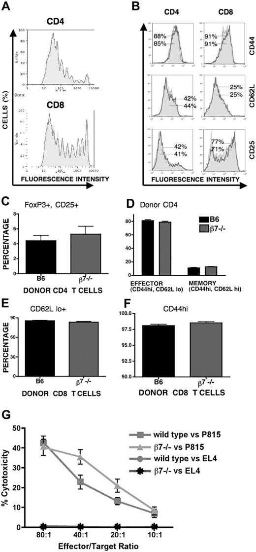
The diminished GVHD activity of alloreactive β7-/- T cells could be attributed to an intrinsic defect in activation, proliferation, or cytotoxicity. To determine the capacity of β7-/- T cells to undergo alloreactive proliferation in vivo, CFSE-labeled T cells were transferred into a sublethally irradiated allogeneic host (B6→B10.BR), and proliferation kinetics were compared with those of CFSE-labeled WT T cells. We found no differences in the percentages of dividing cells and the numbers of divisions between the 2 selected populations of β7-/- and WT T cells (Figure 2A). Additionally, activation of β7-/- T cells, as determined by CD44 and CD25 up-regulation and CD62L down-regulation on fast proliferative T cells,37 was comparable to WT T-cell activation (Figure 2B), indicating that alloreactive β7-/- CD4 and CD8 T cells have intact expression of these activation markers and proliferation kinetics.
We also performed further analyses of the phenotypes and percentages of effector, memory, and regulatory T-cell subsets in spleens on day 11 after HSCT. In the spleens, we observed similar percentages of CD4+CD25+FoxP3+ regulatory T cells (Figure 2C), indicating that differences in the proliferation of suppressor populations do not account for differences in overall morbidity and mortality. Further phenotypic analysis of donor-derived T cells revealed equal percentages of effector (CD44hi CD62Llo) and central memory (CD44hi CD62Lhi) CD4 T cells and effector (CD44hi and CD62Llo) CD8 T cells in the WT and β7-/- T cell groups (Figure 2D-F).
Finally, we assessed the cytolytic capacity of β7-/- T cells against host tumor targets after in vivo stimulation with host antigens (Figure 2G). β7-/- T cells displayed no significant difference in cytolytic activity compared with alloreactive WT T cells. These results indicate that β7 integrin deficiency does not impair the activation, proliferation, or cytotoxic activity of alloreactive T cells.
WT donor T cells infiltrate recipient intestinal mucosa with greater avidity than β7-/- donor T cells
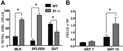
Our previous study with α4β7+ and α4β7- selected donor T cells demonstrated that recipients of α4β7+ donor T cells have higher numbers of infiltrating T cells in their intestinal mucosa than do recipients of α4β7- donor T cells.32 Therefore, we hypothesized that impaired intestinal infiltration of β7-/- donor T cells could explain our improved GVHD morbidity and mortality. To test this hypothesis, we determined the percentages of WT versus β7-/- donor T cells in the spleens, mesenteric lymph nodes, and intestinal mucosa of B10.BR recipients on day 8 after transplantation of equal numbers of WT (Thy1.1) and β7-/- (Thy 1.2) donor T cells (Figure 3A). We found that the intestinal mucosa in recipient B10.BR mice contained significantly higher percentages of WT donor T cells compared with β7-/- donor T cells. In contrast, we found significantly higher percentages of β7-/- donor T cells in recipient spleens and mesenteric lymph nodes. This finding was consistent with results of complete blood counts on days 7 and 14 in lethally irradiated B10.BR mice that underwent transplantation of B10.BR TCD-BM (5 × 10⁶) and splenic T cells (1 × 10⁶) from WT or β7-/- mice (Figure 3B). Mice receiving β7-/- donor T cells consistently showed a higher number of circulating lymphocytes, with the difference reaching statistical significance by day 14. This could be attributed to a defect in the ability of β7-/- donor T cells to traffic to and infiltrate the gut, resulting in more β7-/- lymphocytes remaining in the general circulation and secondary lymphoid organs.
WT and β7-/- T cells do not differ in activation or alloreactive proliferation and have intact cytotoxicity. WT and β7-/- donor T cells were labeled with CFSE and were injected intravenously into sublethally irradiated (750 cGy) B10.BR recipients. Donor splenic T cells from recipient mice were analyzed 72 hours after infusion. (A) Histogram overlay of dividing donor CD4+ and CD8+ CFSE-labeled T cells (shaded area, β7-/-; outline, WT) show nearly identical proliferation kinetics. (B) CD44, CD62L, and CD25 expression on CD4+ and CD8+ donor T cells, with percentage positive T cells expressed as a percentage of fast proliferating T cells (outline = β7-/-; shaded area = WT). (C) Lethally irradiated (1300 cGy) B10.BR recipients underwent transplantation with 5 × 10⁶ WT TCD BM and either 1 × 10⁶ WT or β7-/- splenic T cells, and spleens were analyzed at day 11 after HSCT by flow cytometry. Graph represents the percentage of donor-derived CD4+CD25+FoxP3+ T cells (P = NS). (D) Mice underwent transplantation and harvest as in panel C. Percentage of donor CD4+CD44hiCD62Llo (effector) and CD4+CD44hiCD62Lhi (central memory) populations. (E) Percentage of donor CD8+CD62Llo population. (F) Percentage of donor CD8+CD44hi population. (G) Splenocytes from B6D2F1 HSCT recipients of WT versus β7-/- T cells were analyzed at day 14 after HSCT against syngeneic (EL4) and allogeneic (P815) target cells (n = 5).
WT and β7-/- T cells do not differ in activation or alloreactive proliferation and have intact cytotoxicity. WT and β7-/- donor T cells were labeled with CFSE and were injected intravenously into sublethally irradiated (750 cGy) B10.BR recipients. Donor splenic T cells from recipient mice were analyzed 72 hours after infusion. (A) Histogram overlay of dividing donor CD4+ and CD8+ CFSE-labeled T cells (shaded area, β7-/-; outline, WT) show nearly identical proliferation kinetics. (B) CD44, CD62L, and CD25 expression on CD4+ and CD8+ donor T cells, with percentage positive T cells expressed as a percentage of fast proliferating T cells (outline = β7-/-; shaded area = WT). (C) Lethally irradiated (1300 cGy) B10.BR recipients underwent transplantation with 5 × 10⁶ WT TCD BM and either 1 × 10⁶ WT or β7-/- splenic T cells, and spleens were analyzed at day 11 after HSCT by flow cytometry. Graph represents the percentage of donor-derived CD4+CD25+FoxP3+ T cells (P = NS). (D) Mice underwent transplantation and harvest as in panel C. Percentage of donor CD4+CD44hiCD62Llo (effector) and CD4+CD44hiCD62Lhi (central memory) populations. (E) Percentage of donor CD8+CD62Llo population. (F) Percentage of donor CD8+CD44hi population. (G) Splenocytes from B6D2F1 HSCT recipients of WT versus β7-/- T cells were analyzed at day 14 after HSCT against syngeneic (EL4) and allogeneic (P815) target cells (n = 5).
Recipients of β7-/- T cells have significantly lower numbers of T cells in their intestinal mucosa but significantly higher numbers of circulating T cells. (A) Lethally irradiated (1300 cGy) B10.BR mice underwent transplantation such that each mouse received WT TCD-BM (5 × 10⁶) and WT (Thy1.1) T cells in combination with β7-/- (Thy1.2) T cells (2 × 10⁶ of each type). T cells were analyzed before transfer into recipient mice to ascertain that equivalent percentages of CD4+/CD8+ cells were being given (not shown). Mice were killed at day 8, and T cells were isolated and analyzed, as described in “Materials and methods.” Donor origin of the isolated T cells was determined by multicolor flow cytometry. Statistical analysis is as follows: for mesenteric lymph nodes and spleen, P < .001; for gut, P = .004 (n = 5); experiment repeated 3 times. (B) Lethally irradiated (1300 cGy) B10.BR recipients underwent transplantation with 5 × 10⁶ WT TCD-BM and either 1 × 10⁶ WT or β7-/- splenic T cells. Cardiac puncture to obtain blood for complete blood counts was performed on day 7 and day 14. *P ≤ .05 (n = 8).
Recipients of β7-/- T cells have significantly lower numbers of T cells in their intestinal mucosa but significantly higher numbers of circulating T cells. (A) Lethally irradiated (1300 cGy) B10.BR mice underwent transplantation such that each mouse received WT TCD-BM (5 × 10⁶) and WT (Thy1.1) T cells in combination with β7-/- (Thy1.2) T cells (2 × 10⁶ of each type). T cells were analyzed before transfer into recipient mice to ascertain that equivalent percentages of CD4+/CD8+ cells were being given (not shown). Mice were killed at day 8, and T cells were isolated and analyzed, as described in “Materials and methods.” Donor origin of the isolated T cells was determined by multicolor flow cytometry. Statistical analysis is as follows: for mesenteric lymph nodes and spleen, P < .001; for gut, P = .004 (n = 5); experiment repeated 3 times. (B) Lethally irradiated (1300 cGy) B10.BR recipients underwent transplantation with 5 × 10⁶ WT TCD-BM and either 1 × 10⁶ WT or β7-/- splenic T cells. Cardiac puncture to obtain blood for complete blood counts was performed on day 7 and day 14. *P ≤ .05 (n = 8).
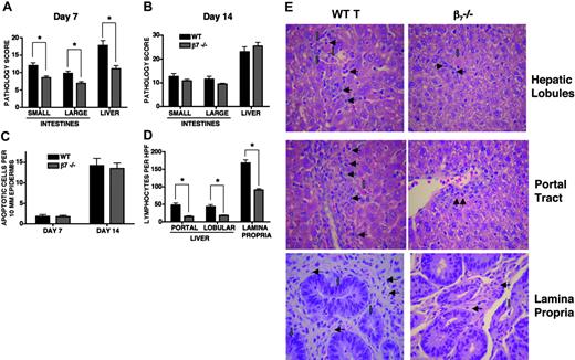
Recipients of β7-/- donor T cells have significantly less gastrointestinal target organ abnormality and no significant difference in skin GVHD. Lethally irradiated (1300 cGy) B10.BR recipients underwent transplantation with 5 × 10⁶ WT TCD BM and either 1 × 10⁶ WT or β7-/- splenic T cells. On day 7 (A) and day 14 (B), mice were killed and gastrointestinal target organs were harvested for evaluation of histopathology, as described in “Materials and methods.” Shown are combined results of pathology scores for 10 mice in each group (*P < .001). (C) Ear and tongue were harvested on days 7 and 14 for evaluation of skin histopathology. (D) Lymphocytes per high-power field (600 ×) from organs on day 7. Shown are results of 5 fields per organ. *P < .001 (n = 10). (E) Representative photomicrographs from day-7 organs. Green arrows show typical apoptotic cells, and black arrows show typical lymphocytes. Original magnification × 600. Images were captured with an Olympus BX 40 microscope (Olympus, Melville, NY) equipped with a 10 ×/0.40 numerical aperture objective lens. Image acquisition was performed with a JVC GC-Qx 5HDU digital camera (JVC, Wayne, NJ).
Recipients of β7-/- donor T cells have significantly less gastrointestinal target organ abnormality and no significant difference in skin GVHD. Lethally irradiated (1300 cGy) B10.BR recipients underwent transplantation with 5 × 10⁶ WT TCD BM and either 1 × 10⁶ WT or β7-/- splenic T cells. On day 7 (A) and day 14 (B), mice were killed and gastrointestinal target organs were harvested for evaluation of histopathology, as described in “Materials and methods.” Shown are combined results of pathology scores for 10 mice in each group (*P < .001). (C) Ear and tongue were harvested on days 7 and 14 for evaluation of skin histopathology. (D) Lymphocytes per high-power field (600 ×) from organs on day 7. Shown are results of 5 fields per organ. *P < .001 (n = 10). (E) Representative photomicrographs from day-7 organs. Green arrows show typical apoptotic cells, and black arrows show typical lymphocytes. Original magnification × 600. Images were captured with an Olympus BX 40 microscope (Olympus, Melville, NY) equipped with a 10 ×/0.40 numerical aperture objective lens. Image acquisition was performed with a JVC GC-Qx 5HDU digital camera (JVC, Wayne, NJ).
Recipients of β7-/- T cells experience significantly less hepatic and intestinal GVHD
To investigate whether recipients of β7-/- T cells had less severe target organ GVHD damage, we analyzed GVHD-associated organ damage in terminal ileum, colon, and liver. We performed semiquantitative histopathologic analysis in a blinded fashion on tissue samples from the target organs.
We found significantly less GVHD in small intestine, large intestine, and liver in recipients of β7-/- T cells when organs were examined at day 7 after transplantation (Figure 4A). When organs were examined at day 14 after transplantation (Figure 4B), no significant difference was found, though a trend was still evident in small and large intestine toward less damage in recipients of β7-/- T cells. Ear and tongue specimens were analyzed to evaluate skin GVHD damage at days 7 and 14 (Figure 4C). No significant difference was found between recipients of WT T cells and recipients of β7-/- T cells on day-7 or -14 samples. Finally, on close histopathologic analysis, we found significantly greater numbers of T cells infiltrating lamina propria, hepatic lobules, and hepatic portal tracts in recipients of WT T cells (Figure 4D-E) on day 7 after transplantation.
Recipients of β7-/- T cells and of WT T cells have similar serum levels of IFNγ
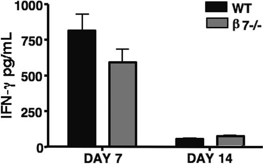
To assess the cytokine response of alloreactive T cells in vivo, we examined serum from mice with GVHD at days 7 and 14 after transplantation (Figure 5). Serum levels of IFNγ were similar in mice that received β7-/- T cells and mice that received WT T cells, suggesting that the difference in morbidity and mortality did not stem from a difference in IFNγ production. In addition, because IFNγ is produced primarily by alloreactive T cells, these data suggest that β7-/- alloreactive T cells have intact IFNγ production in addition to intact alloreactive proliferation, activation, and cytotoxicity (Figure 2).
Recipients of β7-/- donor T cells and of WT T cells generate similar levels of serum IFNγ. Lethally irradiated (1300 cGy) B10.BR recipients underwent transplantation with 5 × 10⁶ WT TCD-BM and either 1 × 10⁶ WT or β7-/- splenic T cells. Serum levels of IFNγ were determined by ELISA at day 7 and 14 after transplantation. Shown are combined results of 2 experiments (n = 9). P = .08 at day 7, and P = .751 at day 14.
Recipients of β7-/- donor T cells and of WT T cells generate similar levels of serum IFNγ. Lethally irradiated (1300 cGy) B10.BR recipients underwent transplantation with 5 × 10⁶ WT TCD-BM and either 1 × 10⁶ WT or β7-/- splenic T cells. Serum levels of IFNγ were determined by ELISA at day 7 and 14 after transplantation. Shown are combined results of 2 experiments (n = 9). P = .08 at day 7, and P = .751 at day 14.
β7-/- T cells have intact GVT activity
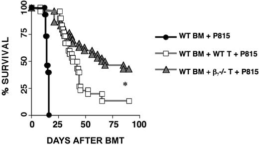
To assess the effects of β7 integrin deficiency on the GVT activity of alloreactive T cells, we performed experiments in a well-characterized GVHD/GVT model: B6→B6D2F1 with the P815 mastocytoma cell line (Figure 6). A low dose of donor T cells (0.5 × 10⁶) was used to decrease GVHD mortality, allowing for better measurement of GVT activity. When possible, dead mice underwent necropsy with histopathologic analysis to determine cause of death (GVHD vs tumor). Results of necropsy and histopathologic analysis for all groups are listed in Table 1. Recipients of β7-/- T cells demonstrated overall improvement in survival over recipients of WT T cells. Our earlier finding that GVHD mortality is decreased in recipients of β7-/- T cells was evident in this transplantation model as well, with fewer recipients of β7-/- T cells dying of GVHD. Surprisingly, recipients of β7-/- T cells not only had intact GVT activity but actually had fewer documented tumor deaths, suggesting improved GVT activity in this group.
Causes of death (GVHD vs tumor) for all recipients who died during the experiment
. | Tumor . | GVHD . | Not analyzed . |
|---|---|---|---|
| BM only | 15 of 15 | 0 of 15 | 0 of 15 |
| P815 + WT T cells | 11 of 30 | 11 of 30 | 4 of 30 |
| P815 + β7-/- T cells | 7 of 30 | 7 of 30 | 3 of 30 |
. | Tumor . | GVHD . | Not analyzed . |
|---|---|---|---|
| BM only | 15 of 15 | 0 of 15 | 0 of 15 |
| P815 + WT T cells | 11 of 30 | 11 of 30 | 4 of 30 |
| P815 + β7-/- T cells | 7 of 30 | 7 of 30 | 3 of 30 |
Values indicate number of mice dead out of total number of mice for the indicated category.
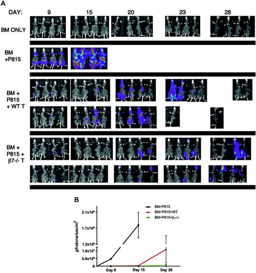
Further support for these findings was obtained using in vivo bioluminescence imaging. Lethally irradiated (1300cGy) B6D2F1 recipients underwent transplantation with 5 × 10⁶ WT TCD BM and either 0.5 × 10⁶ WT or β7-/- splenic T cells. P815 tumor cells (0.5 × 10⁶) that had been transduced with an LTR-HSV1 TK-EGFP-Luc retroviral vector were infused into each mouse at the time of transplantation. Serial imaging (Figure 7) shows that tumor was established in both groups but that recipients of β7-/- donor T cells exhibited significantly delayed tumor growth; in several mice, established tumor even appeared to regress, whereas most recipients of WT donor T cells developed progressively more luminescence and widespread tumor.
GVT activity is preserved in recipients of β7-/- T cells. Lethally irradiated (1100 cGy split dose) B6D2F1 mice underwent transplantation on day 0 with 5 × 10⁶ WT TCD-BM cells with or without the addition of splenic T cells. Recipients were given 1 × 10⁵ P815 murine mastocytoma cells as a separate intravenous injection at the time of transplantation. Survival is depicted as a Kaplan-Meier curve representing mice that received TCD-BM + P815 (•), TCD-BM +β7-/- T cells + P815 (▴), TCD-BM + WT T cells + P815 (□). Causes of death (GVHD vs tumor) for all recipients that died during the course of the experiment are shown in Table 1. Statistical analysis: ▴ versus □ (P = .005). Shown are combined results of 3 experiments (n = 30).
GVT activity is preserved in recipients of β7-/- T cells. Lethally irradiated (1100 cGy split dose) B6D2F1 mice underwent transplantation on day 0 with 5 × 10⁶ WT TCD-BM cells with or without the addition of splenic T cells. Recipients were given 1 × 10⁵ P815 murine mastocytoma cells as a separate intravenous injection at the time of transplantation. Survival is depicted as a Kaplan-Meier curve representing mice that received TCD-BM + P815 (•), TCD-BM +β7-/- T cells + P815 (▴), TCD-BM + WT T cells + P815 (□). Causes of death (GVHD vs tumor) for all recipients that died during the course of the experiment are shown in Table 1. Statistical analysis: ▴ versus □ (P = .005). Shown are combined results of 3 experiments (n = 30).
Discussion
Several recent studies have examined the role of integrins and their respective ligands in the development of acute GVHD. A number of these have confirmed a central role for the α4β7 integrin in lymphocyte homing to gut3,24-26,32 and a pivotal role for Peyer patches and dendritic cells in the Peyer patches in the imprinting of gut-specific tropism on circulating lymphocytes.3,23,24
Tanaka et al25 used GVHD models to demonstrate a moderate improvement in intestinal GVHD in mice treated with anti-α1, -α4, or -β7 antibodies. However, the emphasis of this study was the phenotype of gut-infiltrating lymphocytes during GVHD, and relatively little was assessed in terms of therapeutic benefits of blocking antibodies.25 Li et al26 examined the role of α4 integrin and CD62L in the development of acute graft-versus-host reaction (a-GVHR) and showed that incubating donor splenocytes with anti-α4 and anti-CD62L antibodies resulted in delayed death. However, the potential role of the α4 integrin subunit in other processes, such as stem cell homing to the marrow compartment, was not evaluated. Murai et al3 examined the role of Peyer patches in lymphocyte homing in a-GVHR and reported that the development of a-GVHR could be attenuated through the use of anti-MAdCAM-1 antibodies, emphasizing the key role of the α4β7 integrin-MAdCAM-1 interaction in the imprinting of gut-homing specificity on donor lymphocytes. Yet a limitation of some of these earlier studies is the reliance on a-GVHR models, which use nonirradiated or sublethally irradiated hosts. Our previous study32 used GVHD models to demonstrate that the adoptive transfer of selected α4β7- donor T cells compared with α4β7+ selected donor T cells resulted in a delay of intestinal GVHD and a significant decrease in GVHD morbidity and mortality secondary to reduced donor T-cell infiltration of intestine, but we noted that α4β7--selected alloreactive T cells up-regulate their α4β7 expression on activation and that α4β7--selected alloreactive T cells were, therefore, only delayed in their infiltration of the gut, as reflected by the delayed development of intestinal GVHD.
In this study, using β7-/- donor T cells in murine allogeneic HSCT models, we built on previous findings to assess the role of α4β743,44 in GVHD by permanent inhibition of the β7 integrin in alloreactive T cells. This represents a significant departure from our previous study,32 in which we used α4β7- selection as a method of adoptive cell transfer by preventing re-expression of α4β7 and from previous studies using a-GVHR models, because using lethally irradiated hosts in GVHD models results in an accurate reproduction of clinical GVHD-associated organ damage. Because conditioning regimen toxicity, especially to the intestines, may itself play a role in the development of GVHD,1,45,46 its replication in studies of intestinal GVHD is essential. In addition to previous work with MAdCAM-1 and the α4 integrin subunit, we found that the other significant role of the β7 integrin subunit is (in conjunction with αE) in lymphocyte localization to the gut intraepithelial compartment15,16 and that the β7 integrin subunit is also potentially involved in lymphocyte adhesion and infiltration of target tissues through its less specific interactions with VCAM-1 and fibronectin, all addressed here by the use of β7-/- T cells. From a translational perspective, these studies can serve as a preclinical model for novel drug therapies aimed at inhibition of the β7 integrin (eg, by a neutralizing antibody) that could be used in the HSCT setting to prevent or ameliorate GVHD.
Spatiotemporal analysis of alloreactive T cells and tumor cells by bioluminescence imaging after allogeneic BMT. Lethally irradiated (1300cGy) B6D2F1 recipients underwent transplantation with 5 × 10⁶ WT TCD BM and either 0.5 × 10⁶ WT or β7-/- splenic T cells. P815 tumor cells (0.5 × 10⁶) that had been transduced with an LTR-HSV1 TK-EGFP-Luc retroviral vector were infused into each mouse at the time of transplantation. (A) Mice were tracked for in vivo luminescence 10 minutes after intraperitoneal injection of firefly luciferin. (B) Average luminescence, quantified as photons per sec/m2 at time points with all mice still alive, demonstrates delay in tumor growth in recipients of β7-/- T cells (P = .08).
Spatiotemporal analysis of alloreactive T cells and tumor cells by bioluminescence imaging after allogeneic BMT. Lethally irradiated (1300cGy) B6D2F1 recipients underwent transplantation with 5 × 10⁶ WT TCD BM and either 0.5 × 10⁶ WT or β7-/- splenic T cells. P815 tumor cells (0.5 × 10⁶) that had been transduced with an LTR-HSV1 TK-EGFP-Luc retroviral vector were infused into each mouse at the time of transplantation. (A) Mice were tracked for in vivo luminescence 10 minutes after intraperitoneal injection of firefly luciferin. (B) Average luminescence, quantified as photons per sec/m2 at time points with all mice still alive, demonstrates delay in tumor growth in recipients of β7-/- T cells (P = .08).
In seeking to examine the effects of permanent blockade of the α4β7 integrin, we chose not to focus on the α4 subunit. Despite the promising early results of strategies targeting the α4 integrin subunit in patients with IBD, the α4 subunit plays a role in stem cell homing to the marrow compartment and in trafficking of prothymocytes to thymus,43,44 making it a less attractive target in the context of HSCT, where blockade could interfere with engraftment. Experiments with α4-/- mice have demonstrated defective migration of prothymocytes from bone marrow to thymus.20 However, though the αEβ7 integrin may also play a role in thymocyte adhesion to thymic epithelial cells, the few studies published to date do not support a central role for αEβ7 in thymocyte development.17,18
To study the β7 subunit, we used donor T cells derived from β7-/- mice. These T cells showed activation, proliferation, and cytotoxicity profiles similar to those of WT donor T cells, so that differences in T-cell infiltration of target organs and GVHD morbidity and mortality could reasonably be attributed to a homing defect rather than to a functional defect in the β7-/- T cells.
As expected, recipients of β7-/- T cells developed significantly less GVHD morbidity and mortality than recipients of WT T cells. Interestingly, not only was survival in recipients of β7-/- T cells significantly improved, we also found that onset of disease appeared to be delayed. These findings were further supported by our findings on histopathologic examination that recipients of β7-/- donor T cells have less organ damage and fewer infiltrating T cells, again suggesting that the differences we found throughout our studies were caused by a functional homing defect.
Accordingly, we found on histopathologic examination that β7-/- donor T cells caused less GVHD-related organ pathology, though the difference between recipients of β7-/- donor T cells and WT donor T cells becomes less pronounced over time (Figure 4A-B). Consistent with our previous study with α4β7-selected T cells32 in which we observed similar CD4 and CD8 T cell numbers in the livers of recipients of α4β7- - and α4β7+-selected T cells on day 14 after transplantation and possibly higher CD4 T cell numbers at day 22 after transplantation in the recipients of α4β7- T cells, in this study we found similar histopathology scores in the livers of recipients of WT and β7-/- donor T cells on day 14 after transplantation (Figure 4B). However, examination of recipients of β7-/- donor T cells at day 7 after transplantation revealed significantly less liver disease (Figure 4A) and fewer infiltrating lymphocytes in the lobular and portal areas of the liver than in recipients of WT donor T cells (Figure 4D-E). Thus, we hypothesize that the β7 integrin plays only a role in the initial T-cell trafficking and infiltration of GVHD target organs, such as intestines and liver. Other regulators of T-cell trafficking, such as inflammatory chemokines, might become more important during the further development of GVHD. Therefore, we postulate that the inhibition of the β7 integrin on donor T cells causes only a delay (not a complete inhibition) of the infiltration by alloreactive donor T cells into the intestines and liver.
Additionally, given that we speculate that T cells are “imprinted” to home to the liver within the Peyer patches and the mesenteric lymphoid tissues, inhibition of the β7 subunit would be expected to disrupt T-cell homing to the GALT and the imprinting process but not necessarily homing to the liver; however, we cannot rule out a direct role for the β7 subunit in liver trafficking.47
In addition, we found that cotransplantation of WT and β7-/- donor T cells into the same recipient resulted in a far greater percentage of WT T cells infiltrating intestine. This experimental setup has the advantage of controlling for host expression of chemokines and circulating levels of inflammatory cytokines; both types of T cells are exposed to the same environment so that differences in infiltration may be assumed to be caused solely by the difference in T-cell integrin expression.
These findings are consistent with our hypothesis that the use of β7-/- donor T cells results in less T-cell homing to gut in the early posttransplantation period but that T cells may accumulate later in the posttransplantation period because of other homing signals, such as the interaction of CCR9 and its ligand CCL25 in the small intestine, or to a general milieu of increased inflammatory cytokines. Nonetheless, our data suggest that this early delay in T-cell homing to target organs and the consequent delay in the development of target organ GVHD can have significant effects on the development of systemic GVHD and appears to confer a long-term benefit in morbidity and mortality. It has been well documented that less GVHD is seen when donor lymphocyte infusion is delayed,48,49 and it is possible that the use of β7-/- T cells results in a similar phenomenon, whereby even delayed infiltration of target organs causes less GVHD morbidity and mortality. We can only speculate that this delay in onset of GVHD could allow for improved recovery of the intestinal tract from irradiation and conditioning and thus could result in lower levels of inflammatory cytokines (such as TNF and IL-1) and endotoxin, which, in mouse GVHD models, are largely dependent on early intestinal damage, as previously demonstrated by Hill et al.50
An intriguing finding in our study was the demonstration that β7-/- T cells not only had intact GVT activity, they actually appeared to have improved GVT activity, with tumor challenge resulting in fewer tumor deaths in recipients of β7-/- T cells than in recipients of WT T cells. These findings are especially significant because, with the exception of our previous study, the effect of manipulation of integrins on GVT activity in HSCT models has been minimally explored. Our results were reinforced by in vivo imaging studies showing that tumor challenge resulted in the establishment of tumor in recipients of WT T cells and in recipients of β7-/- T cells but that the latter displayed significantly delayed tumor growth or complete eradication of tumor, whereas the former experienced rapid tumor growth. Given our findings that β7-/- T cells have activation, proliferation, and cytotoxicity profiles similar to those of WT T cells and the finding that recipients of β7-/- T cells have consistently higher circulating lymphocyte counts, we hypothesize that this difference was likely a result of the decreased infiltration of liver and intestines by the β7-/- T cells. With fewer donor T cells infiltrating target organs, more alloreactive T cells remained in the general circulation (Figure 3B), a site where the P815 mastocytoma resides, allowing greater interaction between alloreactive T cells and tumor cells and improved GVT activity. This improved ability to clear circulating tumor cells would be an attractive and important addition to a therapy that successfully reduces intestinal GVHD. Thus, use of a neutralizing monoclonal antibody against the β7 integrin subunit is the logical next step in the preclinical development of a strategy to prevent intestinal homing of alloreactive T cells in patients undergoing HSCT.
In conclusion, the absence of the β7 integrin subunit on alloreactive T cells resulted in less GVHD morbidity and mortality because of decreased intestinal GVHD and intact (and possibly improved) GVT activity. The β7 integrin subunit warrants further study, especially as a potential target for monoclonal antibody therapy, which could lead to successful clinical trials of a novel agent to make allogeneic HSCT a safer, less toxic therapy.
Prepublished online as Blood First Edition Paper, November 15, 2005; DOI 10.1182/blood-2005-08-3445.
Supported by grants HL69929, HL72412, and CA107096 from the National Institutes of Health; an award from the Emerald Foundation; and an award from Golfers Against Cancer (M.R.M.v.d.B.). E.W. is a recipient of an ASCO Young Investigator Award. T.H.T. is the recipient of a Deutscher Akademischer Austauschdienst (DAAD) research award. T.D.K. is supported by a Mildred Scheel-Stipendium der Deutschen Krebshilfe grant.
S.X.L. and V.M.H. contributed equally to this work.
The publication costs of this article were defrayed in part by page charge payment. Therefore, and solely to indicate this fact, this article is hereby marked “advertisement” in accordance with 18 U.S.C. section 1734.


This feature is available to Subscribers Only
Sign In or Create an Account Close Modal