Comment on Faber et al, page 3389
After allogeneic hematopoietic cell transplantation (HCT) the development of solid tumor is increasingly reported. While typically found in cancers, frame-shift mutations in microsatellites have also been detected in chronically inflamed tissues. In this issue, Faber and colleagues describe that genomic alterations in gut and oral epithelia regularly occur after allogeneic HCT.
HCT offers curative therapy for malignant and nonmalignant diseases. The success rate has improved progressively, and some of the surviving patients have now been followed for more than 3 decades. One important complication among transplant survivors is the development of new (secondary) malignancies, particularly solid tumors and posttransplantation lymphoproliferative disorders (reviewed in Deeg and Socie1 and Ades et al2 ). Previous studies report that transplant recipients who develop chronic graft-versus-host disease (GvHD) have an especially high risk of developing squamous cell carcinoma of the oral cavity and skin, with more aggressive behavior noted in some of these tumors.3
Microsatellites are highly polymorphic repetitive sequences that are scattered throughout the genome.1 They are inherited and are unique to each individual, which makes them ideal tools for use in documentation of engraftment following allogeneic HCT. Microsatellite instability (MSI) has been used as an indicator of genomic instability. Though typical in cancers, MSI has more recently also been found in nonneoplastic, chronically inflamed tissues; however, the exact mechanism through which chronic inflammation produces MSI remains to be elucidated.
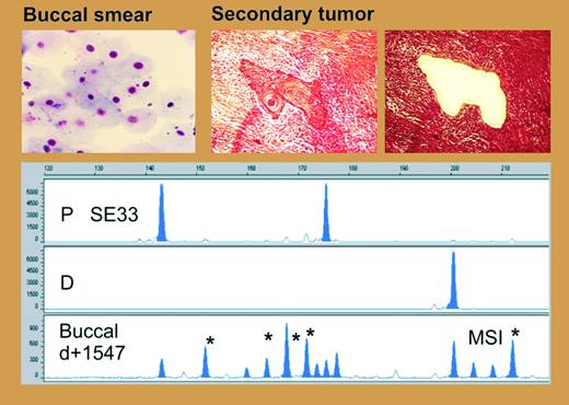
In this issue of Blood, Faber and colleagues found frequent microsatellite alterations in the colon and oral mucosa of patients who underwent allogeneic HCT (see figure). The authors examined 6 microsatellite loci by polymerase chain reaction (PCR). In colon biopsies, there were novel bands that were neither host specific nor donor specific in the laser-microdissected colonic crypts in 12 (75%) of 16 patients; these bands indicate the regular occurrence of MSI. In contrast to the allografted patients, Faber et al never found any indication of MSI in laser-microdissected crypts from patients after chemotherapy, or from control subjects. Additionally, the authors examined buccal cells for the presence of MSI. At the time of buccal sampling, no signs of oral GvHD were present. MSI was found in the buccal smears of 10 (42%) of 24 allografted patients. In contrast, no MSI was detected in the buccal smears obtained from patients after autologous HCT, from patients after chemotherapy, or from healthy controls. Finally, they identified 3 patients who developed secondary invasive squamous cell cancer. In all cases, both laser-microdissected tumor areas and surrounding normal epithelium showed evidence of MSI.
Replication errors during DNA synthesis may result in a change of the length of microsatellite loci, which are sequences particularly prone to DNA polymerase slippage because of their repetitive nature. If deficient DNA repair is coupled with a failure to elicit an apoptotic response, this association may result in a growth advantage sufficient to generate a clonal population. In the study of Faber and colleagues, all but one colon biopsy with detectable GvHD showed MSI, indicating a possible relationship between organ GvHD and MSI. Nitric oxide (NO) produced by TNF-alpha–stimulated epithelial cells has been described in GvHD. NO has been found to inhibit DNA repair and could thus be a link between chronic allogeneic stimulation, DNA repair mechanisms, and MSI.FIG1
Microsatellite instability in buccal mucosa and secondary tumors after allogeneic HCT. See the complete figure in the article beginning on page 3389.
Microsatellite instability in buccal mucosa and secondary tumors after allogeneic HCT. See the complete figure in the article beginning on page 3389.
Could MSI thus be used as a tool for early detection or prevention of secondary cancers after allogeneic HCT? As stated by the authors, the percentage of subjects with the MSI phenotype is much higher than the expected incidence of posttransplantation malignancies, and, although MSI was frequently found in colon tissue, colon carcinoma is currently an uncommon secondary malignancy. In my view, MSI should be viewed today as evidence of genomic instability, leaving cells that are, especially in a setting of chronic inflammation, prone to secondary genomic alterations (p53 mutation, transformation by viral oncogenesis, etc) and thus to fully malignant transformation. ▪


This feature is available to Subscribers Only
Sign In or Create an Account Close Modal