Nucleolin, originally described as a nuclear protein, was recently found to be expressed on the surface of endothelial cells during angiogenic. However, the functions of cell-surface nucleolin in angiogenic remain mysterious. Here we report that upon endothelial cells adhering to extracellular matrix components, vascular endothelial growth factor (VEGF) mobilizes nucleolin from nucleus to cell surface. Functional blockage or down-regulation of the expression of cell-surface nucleolin in endothelial cells significantly inhibits the migration of endothelial cells and prevents capillary-tubule formation. Moreover, nonmuscle myosin heavy chain 9 (MyH9), an actin-based motor protein, is identified as a nucleolin-binding protein. Subsequent studies reveal that MyH9 serves as a physical linker between nucleolin and cytoskeleton, thus modulating the translocation of nucleolin. Knocking down endogenous MyH9, specifically inhibiting myosin activity, or overexpressing functional deficient MyH9 disrupts the organization of cell-surface nucleolin and inhibits its angiogenic function. These studies indicate that VEGF, extracellular matrix, and intracellular motor protein MyH9 are all essential for the novel function of nucleolin in angiogenic.
Introduction
Angiogenesis, the generation of new capillaries from preexisting microvasculature, occurs in a variety of pathologic processes, such as tumor growth and metastasis and various inflammatory disorders.1,2 Angiogenic vessels differ from normal vessels in their morphologic and molecular characteristics. Some unique molecules have been identified specifically expressing on angiogenic vessels, including some types of endothelial growth factor receptors, integrins, proteolytic enzymes, and extracellular matrix, as well as membrane proteins of unknown functions.3,4 Recently, cell-surface nucleolin was reported as a specific marker of angiogenic endothelial cells.5
Nucleolin has been described as a nucleolar protein of eukaryotic cells, involved in regulation of cell proliferation, cytokinesis, replication, embryogenesis, and nucleogenesis.6,7 Recently, a number of studies have shown that it can also be expressed on the cell surface and serve as a receptor for several ligands such as matrix laminin-1, midkine, and lactoferrin.8-13 However, little information about the physiologic relevance of cell-surface nucleolin in angiogenic is known except that cell-surface nucleolin specifically appears in tumor-induced angiogenic vessels rather than mature vessels or capillaries.5 Here we report that cell-surface nucleolin is essential for the migration and tubule formation of endothelial cells. During the process of angiogenic, the up-regulation of cell-surface nucleolin is attributed to the shuttle of nucleolin from nucleus to cell membrane. Vascular endothelial growth factor (VEGF), a core regulator in tumor angiogenic and stroma generation,14,15 can significantly stimulate the translocation of nucleolin when endothelial cells adhere to extracellular matrix. Cell-surface nucleolin was reported to associate with actin cytoskeleton.9 In this study, we found that nonmuscle myosin heavy chain 9 (MyH9), an actin-based motor protein, provides a linker between cell-surface nucleolin and actin cytoskeleton and mediates its function in angiogenic. MyH9 is one isoform of the class II myosin family, which mediates a variety of cellular processes including protrusion, migration, and modulation of cell locomotion.16,17 Recent studies showed that MyH9 plays an important role in modulating T-cell motility and tissue organization during embryo development.18,19 The shuttle of nucleolin between nucleus and cell surface depends on anchoring to MyH9, which appears essential for the angiogenic function of cell-surface nucleolin.
Materials and methods
Antibodies, proteins, and chemicals
Polyclonal antibodies to nucleolin were from our lab storage. Plasma fibronectin was purified from human blood by gelatin affinity column. Other antibodies, proteins, and chemicals were from Sigma-Aldrich (Poole, United Kingdom; polyclonal antibodies of MyH9, antivinculin monoclonal antibody [mAb], ML7), Santa Cruz Biotechnology (Santa Cruz, CA; TRITC, FITC-2nd antibodies to Ra, Mo IgG, FITC–anti-CD31), Dako (Glostrup, Denmark; HRP-2nd antibodies to Ra, Mo IgG), and Protgen (VEGF-165; Beijing, China).
Identification of isolated proteins with MALDI-TOF mass spectrometry
The major band from coimmunoprecipitation was digested using sequencing grade porcine-modified trypsin (Promega, Madison, WI). The peptides produced were analyzed by matrix-assisted laser desorption/ionization–time-of-flight (MALDI-TOF) mass spectrometry using a Bruker Biflex linear time-of-flight spectrometer (Bruker Franzen, Bremen, Germany), equipped with a multiprobe SCOUT source (Bruker Daltonik, Bremen, Germany), ultra-nitrogen laser (337 nm), and a dual microchannel-plate detector. The MALDI-TOF data were searched against the Swiss-Prot protein data base for protein identification.
Cell-migration assay
Human microvascular endothelial cells (HMECs) or human umbilical vein endothelial cells (HUVECs), 2 × 10⁴ cells per well, were seeded into the upper chamber of a collagen precoated transwell filter (8-μm pores; Costar, Acton, MA) with Dulbecco modified Eagle medium (DMEM) containing 10% fetal calf serum (FCS). The same culture medium and reagents were added to the lower chamber. The endothelial cells were allowed to migrate for 4 hours at 37°C and 5% CO2. After being fixed and stained with ethanol and eosin, the cells migrated completely through the filter to the lower chamber and were counted in 3 different areas under the optical microscope, and the averaged number was obtained.
Tubule formation assay
The tubule formation assay was carried out as previously described.20,21 In brief, 24-well plates were coated with 100 μL/well of Matrigel (Becton Dickinson Labware, Bedford, MA). Endothelial cells on this matrix migrated and formed tubules within 6 hours of plating. ECV304 cells were seeded at 5 × 10⁴ cells per well and incubated for 6 hours in complete medium with 10 ng/mL VEGF. Wells were then fixed and visualized (at ×100 magnification), and tubule formation was assessed in 3 randomly selected fields of view per well using the image analysis package Scion Image (Scion, Frederick, MA).
Cell-invasion assay
ECV304 cells, transfected with MyH9nN–green fluorescent protein (GFP) or GFP, were seeded on 8-μm inserts (Costar) coated with Matrigel, using 5 ng/mL VEGF in 1% fetal bovine serum (FBS) as a chemoattractant. After a 24-hour incubation at 37°C, the invasive cells were fixed and observed and quantitated under fluorescence microscope at 488/530 nm.
RNA interference
The pBS-U6 vector was used to express a short, small interfering RNA (siRNA) sequence specific for the MYH9 gene or the nucleolin (NCL) gene, and the nonsilencing pBS-U6 vector was used as a control. The constructed plasmids were transfected into cells by lipofectin (Invitrogen, Carlsbad, CA). In GFP cotransfection, the GFP expression vector was combined with the MyH9 RNAi vector at a ratio of 10:1. At 48 hours after transfection, the inhibitory effect was detected by immunoblotting of MyH9.
Immunofluorescence and confocal microscopy
The cultured cells were fixed by 4% paraformaldehyde and then permeabilized with phosphate-buffered saline (PBS) containing 0.2% Triton X-100. After being washed twice with PBS, the cells were blocked with PBS containing 10% normal goat serum. After treatment, the cells were stained with different primary antibodies and FITC- or TRITC-conjugated secondary antibodies. Actin filaments were directly stained by FITC-phalloidin (Chemicon, Temecula, CA), and nuclei were stained by DAPI. Confocal fluorescence imaging was performed on an Olympus Fluoview laser scanning confocal imaging system (Olympus, Melville, NY). Images were captured using multiple photomultiplier tubes regulated by Fluoview 2.0 software (Olympus). All immunofluorescence images were obtained through an Olympus IX71-141 microscope equipped with either a 20 ×/0.4 or a 40 ×/0.65 objective lens (Olympus) and a Coolsnap HQ CCD camera (Photometrics, Tucson, AZ). Images were processed with Metamorph software (Universal Imaging, West Chester, PA).
Fluorescence-activated cell-sorting (FACS) analysis
Cells were briefly trypsinized and resuspended in PBS supplemented with 0.1% bovine serum albumin. Cells were incubated with antinucleolin polyclonal antibodies for 30 minutes on ice followed by 30 minutes incubation on ice with antirabbit secondary antibody conjugated to FITC. After the final wash, cells were directly analyzed using a FACScan (Becton Dickinson, San Jose, CA).
Results
Distribution of nucleolin in vivo
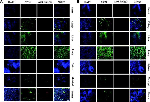
The presence of cell-surface nucleolin on angiogenic vessels, described by Christian et al,5 led us to further assess systematically the distribution of cell-surface nucleolin in different tissues. Since the majority of nucleolin is located in the nucleus in all eukaryotic cells, in order to detect cell-surface nucleolin, we injected antinucleolin polyclonal antibodies (Abs) into the tail vein of tumor-bearing or Matrigel-plus mice. At 3 hours after injection, mice were killed and their organs, heart, liver, kidney, spleen, lung, Matrigel plug, and tumor were removed for immunohistochemical analysis. As shown in Figure 1A, the normal organs, heart, liver, kidney, spleen, and lung bearing mature vessels showed negative staining for antinucleolin antibodies. However, in the tumor- or Matrigel-induced angiogenic vessels, antinucleolin Ab showed positive staining and colocalization with neo-vessels, which indicates that the cell-surface nucleolin selectively appears on tumor-induced anigogenic vessels or other inflammatory tissues but not mature vessels of normal organs. In order to exclude the accumulation of antibody for nonspecific effects, isotype Ab was used as a control (Figure 1B). Based on these observations, we propose that there must have some factors stimulating the translocation of nucleolin to the cell surface, and it is the cell-surface nucleolin that functionally contributes to angiogenic.
Angiogenic function of nucleolin
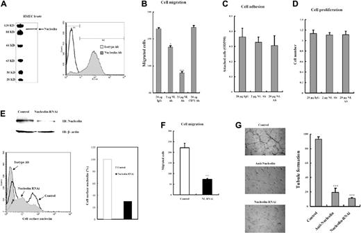
To elucidate the functions of cell-surface nucleolin in angiogenic, polyclonal antibodies (pAbs) against nucleolin were applied to neutralize cell-surface nucleolin of endothelial cells in vitro. The binding specificity of the pAb is shown in Figure 2A. Cell migration was measured by the transwell migration assay. It was observed that the migration of human microvascular endothelial cells (HMECs) was inhibited when cell-surface nucleolin was functionally blocked. As shown in (Figure 2B), the nucleolin Abs have a significant inhibitory effect on the migration of endothelial cells, which implies that cell-surface nucleolin regulates cell-matrix interaction and cell motility. Anti-CD71, a transferin receptor on the endothelial cell surface, was used as a control. However, antinucleolin Ab had only marginal effect on cell adhesion and cell proliferation (Figure 2C-D). Apparent inhibition of cell migration caused by the treatment of antinucleolin pAb may result in suppression of the network of endothelial cell formation. Human umbilic vein endothelial cell line ECV304 was seeded on Matrigel to assess its ability of forming tubule network. In consistent with our expectation, soluble antinucleolin pAb significantly inhibits tubule formation (Figure 2G). To further confirm this conclusion, RNA interference was subjected to suppress the expression level of nucleolin in endothelial cells, which also resulted in the reduction of the amount of cell-surface nucleolin (Figure 2E). It was observed that if nucleolin was down-regulated, cell migration and tubule formation were apparently inhibited (Figure 2F-G).
The distribution of cell-surface nucleolin in different tissues. (A) Purified antinucleolin Ab was injected intravenously into mice bearing S180 xenograft tumors or Matrigel plugs. Heart, kidney, liver, lung, spleen with tumors, and Matrigel plugs were removed 6 hours after injection for immunohistochemical assay. Cell-surface nucleolin was visualized by TRITC-secondary antibody against primary injected antibodies (red). The endothelial cells of blood vessels were stained with FITC–anti-CD31 (green), and nuclei were counterstained with DAPI (blue). Antinucleolin Ab specifically accumulated at angiogenic vessels of tumors or Matrigel plugs but did not appear in normal organs. (B) Nonspecific isotype Ab was injected as a control. Bar, 50 μm.
The distribution of cell-surface nucleolin in different tissues. (A) Purified antinucleolin Ab was injected intravenously into mice bearing S180 xenograft tumors or Matrigel plugs. Heart, kidney, liver, lung, spleen with tumors, and Matrigel plugs were removed 6 hours after injection for immunohistochemical assay. Cell-surface nucleolin was visualized by TRITC-secondary antibody against primary injected antibodies (red). The endothelial cells of blood vessels were stained with FITC–anti-CD31 (green), and nuclei were counterstained with DAPI (blue). Antinucleolin Ab specifically accumulated at angiogenic vessels of tumors or Matrigel plugs but did not appear in normal organs. (B) Nonspecific isotype Ab was injected as a control. Bar, 50 μm.
Translocation of nucleolin is mediated by VEGF and extracellular matrix
Nucleolin is normally located in nucleolus or nucleus; however, during the process of angiogenic, it mysteriously appears on the cell membrane of endothelial cells. However, how nucleolin expresses on the cell membrane remains unclear. We found that VEGF and extracellular matrix synergistically mediate the mobilization of nucleolin from nucleus to cell surface.
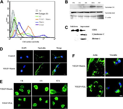
We analyzed the quantitative change of cell-surface nucleolin under stimulation of VEGF. HMECs, cultured on poly-l-lysine (PLL) or fibronectin, were stimulated by VEGF. After 12 hours, cells were harvested and their surface-expressed nucleolin was quantified by FACS. It was observed that under the stimulation of VEGF, the amount of nucleolin on the membrane of HMECs, cultured on fibronectin, significantly increased; however, when cultured on PLL, the amount of cell-surface nucleolin was only marginally elevated (Figure 3A). To further confirm the results, HMECs, grown on fibronectin-coated plates, were treated by VEGF (10 ng/mL) for different times: 0 hours, 1 hour, 2 hours, 6 hours, and 6 hours followed by being restarved for 2 hours. The 5 groups of HMECs were biotinlayted, and their membrane proteins were completely labeled by biotins. Then, HMECs were lysed and membrane proteins were pulled down by strepavidin-conjugated agarose. The fractions of cell lysate and membrane proteins were subjected to immnoblotting by antinucleolin Ab. During the treatment by VEGF, the quantity of nucleolin expressed on the cell membrane increased gradually, and without stimulation of VEGF, the cell-surface nucleolin quickly decreased (Figure 3B). The quantitative change of cell-surface nucleolin under stimulation of VEGF was also confirmed by immunofluorescence (Figure 3C). In this process, the quantity of total nucleolin did not change, which suggests that the cell-surface nucleolin come from the nucleolin originally located in the nucleus, and VEGF does not enhance the expression level of nucleolin but mobilizes it from nucleus to cell surface. In order to prove that the streptavidin sharply separates biotinylated from nonbiotinylated proteins, and to exclude the traces of nucleolar proteins, we tested typical cell membrane protein of CD31, cytoplasmic protein of cytochrome C, and nuclear protein of Histone-H1 in both streptavidin pull-down fraction and supernatant. It was observed that only cell membrane protein of CD31 could be pulled down by streptavidin. To capture the process of nucleolin mobilization, the cells were trypsinized from plates, and then replated on fibronectin or PLL-coated coverslips. At different time intervals (0 hours, 6 hours, or 12 hours), the cells were fixed and permeabilized to analyze the distribution of nucleolin. In HMECs grown on fibronectin, nucleolin translocated gradually from nucleus into cytoplasm and cell surface under stimulation of VEGF, while most of nucleolin remained in nucleolus in HMECs grown on PLL (Figure 3D).
Nucleolin mediates cell migration and tubule formation. The binding specificity of employed antinucleolin Ab was shown in panel A. Indicated concentrations of Ab were incubated with HMECs to neutralize cell-surface nucleolin, and the assays of cell adhesion (B), cell migration (C), and cell proliferation (D) were performed. Purified rabbit IgG served as a positive control. Neutralizing cell-surface nucleolin significantly inhibited cell migration but only marginally affected cell adhesion and did not affect cell proliferation. Anti-CD71 was employed in cell migration assay as a control. (E) The efficiency of RNAi was detected by immunoblotting, and the reduction of cell-surface nucleolin was quantified by FACS. (F) Knocking down the endogenous nucleolin in HMECs led to apparent reduction of cell migration. (G) Tubule formation of ECV304 on growth factors supplemented Matrigel in response to 20 μg/mL antinucleolin Abs or interfered with their endogenous expression of nucleolin. Bar, 100 μm. *P < .05; ***P < .01.
Nucleolin mediates cell migration and tubule formation. The binding specificity of employed antinucleolin Ab was shown in panel A. Indicated concentrations of Ab were incubated with HMECs to neutralize cell-surface nucleolin, and the assays of cell adhesion (B), cell migration (C), and cell proliferation (D) were performed. Purified rabbit IgG served as a positive control. Neutralizing cell-surface nucleolin significantly inhibited cell migration but only marginally affected cell adhesion and did not affect cell proliferation. Anti-CD71 was employed in cell migration assay as a control. (E) The efficiency of RNAi was detected by immunoblotting, and the reduction of cell-surface nucleolin was quantified by FACS. (F) Knocking down the endogenous nucleolin in HMECs led to apparent reduction of cell migration. (G) Tubule formation of ECV304 on growth factors supplemented Matrigel in response to 20 μg/mL antinucleolin Abs or interfered with their endogenous expression of nucleolin. Bar, 100 μm. *P < .05; ***P < .01.
During the mobilization of nucleolin, stress fibers and focal adhesion complex formed simultaneously (Figure 3E). Other matrix proteins such as collagen and laminin also exhibited a similar effect as fibronectin (data not shown), which indicates that the mobilization of nucleolin is not only regulated by VEGF signaling but is also correlated with reorganization of cytoskeleton modulated by ECM.
MyH9 provides an anchorage for nucleolin
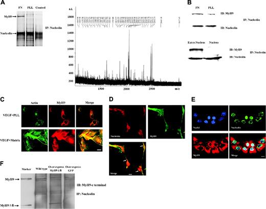
To further investigate how nucleolin modulates cell motility, coimmunoprecipitation (co-IP) was applied to search for the potential nucleolin-binding proteins. Finally, a 220-kDa protein was obtained from HMECs as a nucleolin-binding protein (Figure 4A). MALDI-TOF mass spectrometry identified it as nonmuscle myosin heavy chain 9 (MyH9), a motor protein expressed in endothelial cells. The identification was further confirmed by immunoblotting (Figure 4B). There may be some nucleolin from nucleus appearing in the whole-cell lysate. To exclude the possibility that MyH9 also binds to nucleolin from nucleus, we extracted nucleus from cells, then used both the nuclear fraction and extranuclear fraction to perform co-IP. We found that MyH9 can bind to nucleolin from cell surface and cytoplasm but not from nucleus (Figure 4B). Interestingly, the interaction between nucleolin and MyH9 is more pronounced in the cells grown on fibronectin than in those grown on PLL (Figure 4A-B), suggesting an essential role of matrix proteins in mediating the nucleolin-MyH9 interaction. To explain this phenomenon, the distribution of MyH9 in HMECs grown on fibronectin was investigated. MyH9 was observed as stress fibers, along the plasma membrane, and in membrane ruffles which colocalize with actin filaments. In contrast, MyH9 in the cells grown on PLL did not exhibit any stress fibers but formed centrally located punctuate filaments (Figure 4C). In order to reveal the relationship between MyH9-nucleolin interactions and angiogenic, the distributions of both nucleolin and MyH9 were studied under different conditions. ECV304 cells were cultured for 12 hours on Matrigel supplemented with 10 ng/mL VEGF or PLL without any supplement. Then nucleolin and MyH9 were stained by immnuofluorescence. ECV304 grown on Matrigel containing VEGF spread their cell shape and formed a network or tubule structure; moreover, nucleolin colocalized with MyH9 during tubule formation (Figure 4D). However, in ECV304 grown in PLL, nucleolin stays only in nucleus and MyH9 stays in cytoplasm (Figure 4E). Based on these observations, we propose that the matrix-induced conformational change of MyH9 facilitates the MyH9-nucleolin interaction, which contributes to the movement of nucleolin and subsequent angiogenic. Since the structure of myosin II consists of a globular head that can bind to actin filament and has ATPase activity, a neck domain that binds to myosin light chains or calmodulin, and a unique c-terminal tail domain of variable length that can either promote multimerization or bind to different cellular proteins,16,17 we propose that the c-terminal of MyH9 is the potential nucleolin-binding domain. To verify this hypothesis, we fused the c-terminal coil-coil domain of MyH9 with GFP (MyH9ΔN-GFP) and transfected it into HMECs. Then, co-IP was performed to detect whether there is any interaction between the c-terminal of MyH9 and nucleolin. As shown in Figure 4F, the MyH9ΔN-GFP was pulled down by antinucleolin Ab, which suggests that the c-terminal of MyH9 is indeed the binding site for nucleolin. Moreover, MyH9ΔN provides a useful framework to investigate the functional contribution of the interaction between MyH9 and nucleolin due to the lack of the N-terminal motor domain of MyH9.
Translocation of nucleolin is mediated by VEGF and extracellular matrix. (A) Cell-surface nucleolin of HMECs was determined by FACS as described in “Materials and methods.” For the histograms, NC indicates negative control; isotype Ab, isotype Ab control; ST, serum-starved cells; Matrix, HMECs grown on coated matrix protein-fibronectin; VEGF, HMECs cultured with serum-free medium containing 10 ng/mL VEGF. In HMECs grown on fibronectin, VEGF significantly enhanced the surface expression of nucleolin, but the 2 factors have only a marginal effect. (B) HMECs grown on fibronectin were stimulated by 10 ng/mL VEGF for 0 hours, 1 hour, 6 hours, or 12 hours, and then restarved by serum-free medium (SF). The membrane proteins were biotinylated and pulled down by strepavidin-conjugated agarose; meanwhile, total proteins were from whole-cell lysate. Both fractions were subjected to immunoblotting by antinucleolin Abs. The amount of nucleolin in cell membrane (nucleolin-CM) increased following VEGF stimulation and decreased without stimulation; however, during the process, the amount of nucleolin in whole cells (nucleolin-total) did not change. β-actin was used as a loading control. (C) Cell membrane protein CD31, cytoplasmic protein cytochrome C, and nuclear protein Histone-H1 were detected in both streptavidin pull-down fraction and supernatant, to prove that the streptavidin matrix sharply separates biotinylated from nonbiotinylated proteins, and to exclude the traces of nucleolar proteins. (D) After HMECs were plated on immobilized fibronectin (FN) or poly-L-lysine (PLL), the distribution of nucleolin (green) was visualized by immunofluorescence at different times. Bar, 10 μm. (E) Cell-surface nucleolin (green) and nuclei (blue) in HMECs grown on immobilized FN or PLL were stained. Cells were not permeabilized here to visualize cell surface proteins. Bar, 10 μm. (F) During the translocation of nucleolin, stress fibers and focal adhesion formed simultaneously. Actin filaments were stained by FITC-phalloidin (green) and focal adhesion complexes were visualized by indirect immunostaining of vinculin (green). Nuclei were counterstained with DAPI (blue). Bar, 10 μm.
Translocation of nucleolin is mediated by VEGF and extracellular matrix. (A) Cell-surface nucleolin of HMECs was determined by FACS as described in “Materials and methods.” For the histograms, NC indicates negative control; isotype Ab, isotype Ab control; ST, serum-starved cells; Matrix, HMECs grown on coated matrix protein-fibronectin; VEGF, HMECs cultured with serum-free medium containing 10 ng/mL VEGF. In HMECs grown on fibronectin, VEGF significantly enhanced the surface expression of nucleolin, but the 2 factors have only a marginal effect. (B) HMECs grown on fibronectin were stimulated by 10 ng/mL VEGF for 0 hours, 1 hour, 6 hours, or 12 hours, and then restarved by serum-free medium (SF). The membrane proteins were biotinylated and pulled down by strepavidin-conjugated agarose; meanwhile, total proteins were from whole-cell lysate. Both fractions were subjected to immunoblotting by antinucleolin Abs. The amount of nucleolin in cell membrane (nucleolin-CM) increased following VEGF stimulation and decreased without stimulation; however, during the process, the amount of nucleolin in whole cells (nucleolin-total) did not change. β-actin was used as a loading control. (C) Cell membrane protein CD31, cytoplasmic protein cytochrome C, and nuclear protein Histone-H1 were detected in both streptavidin pull-down fraction and supernatant, to prove that the streptavidin matrix sharply separates biotinylated from nonbiotinylated proteins, and to exclude the traces of nucleolar proteins. (D) After HMECs were plated on immobilized fibronectin (FN) or poly-L-lysine (PLL), the distribution of nucleolin (green) was visualized by immunofluorescence at different times. Bar, 10 μm. (E) Cell-surface nucleolin (green) and nuclei (blue) in HMECs grown on immobilized FN or PLL were stained. Cells were not permeabilized here to visualize cell surface proteins. Bar, 10 μm. (F) During the translocation of nucleolin, stress fibers and focal adhesion formed simultaneously. Actin filaments were stained by FITC-phalloidin (green) and focal adhesion complexes were visualized by indirect immunostaining of vinculin (green). Nuclei were counterstained with DAPI (blue). Bar, 10 μm.
MyH9 modulates the angiogenic function of nucleolin
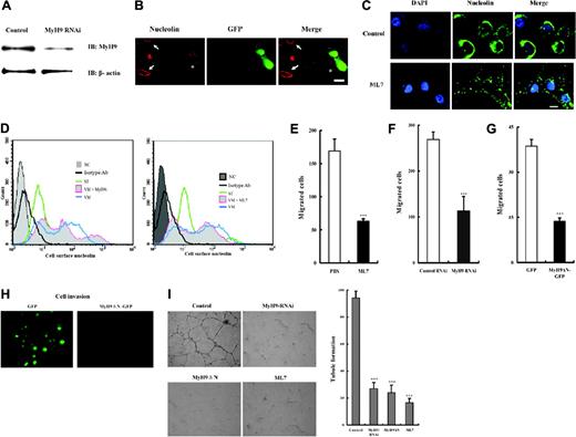
Different strategies were performed in the current study to elucidate the potential biologic relevance of the association between MyH9 and cell-surface nucleolin: (1) knocking down endogenous expression of MyH9 by RNAi (efficiency was shown in Figure 5A); (2) inhibiting the activity of MyH9 by ML-7, a specific myosin light chain kinase inhibitor; and (3) overexpressing the nucleolin-binding domain of MyH9 (MyH9ΔN), which is deficient in motor function in endothelial cells.
HMECs were cotransfected with MyH9-RNAi and GFP vectors, and then the cell-surface nucleolin was stained. We observed that the organization of nucleolin was disrupted and aggregated on the cell surface in those GFP-positive cells (Figure 5B), and a similar phenomenon was also observed in the ML7-treated cells (Figure 5C). Moreover, the amount of cell-surface nucleolin had a 10% to 20% reduction when MyH9 was suppressed (Figure 5D). We propose that MyH9 is critical for the organization of nucleolin on cell surface and may also play a potential role in regulating the recycling of cell-surface nucleolin. The following migration assay showed that suppressing MyH9 with RNAi and ML7 significantly inhibited the migration of HMECs (Figure 5E-F). Based on these observations, we propose that the shuttling trait of nucleolin, translocating between the cell surface and nucleus, is critical for angiogenic; furthermore, MyH9 not only provides an anchorage but also mediates the shuttle of nucleolin.
Recognizing that MyH9ΔN is the binding site for nucleolin (Figure 4F) but lacks of the N-terminal motor domain, we next overexpressed MyH9nN in endothelial cells to form a functional deficient complex with endogenous nucleolin. Then, its effect on angiogenic was evaluated. The overexpressed MyH9ΔN significantly suppressed the migration of HMECs (Figure 5G) and the invasive ability of those cells was totally impaired (Figure 5H). In another endothelial cell line, ECV304, the tubule formation assay showed that, similar to blockage of cell-surface nucleolin, overexpression of MyH9ΔN or suppressing MyH9 made ECV304 unable to form a network structure (Figure 5I).
Nucleolin anchors to intracellular MyH9 during angiogenic. (A) Cell-free lysate was prepared from HMECs grown on immobilized FN or PLL. Co-IP was performed with antinucleolin Ab and purified rabbit IgG as a control. The arrows indicate the precipitated protein band, which was identified as MyH9 by MALDI-TOF analysis. The SDS-PAGE gel was silver stained and the finger printing map was indicated. (B) Co-IP was performed as described for panel A, and the precipitated proteins were then blotted (IB) with antibodies against MyH9 or nucleolin. The nuclei of HMECs grown on immobilized FN were extracted; then both nuclear and extranuclear fraction were lysed to perform co-IP with the same condition. (C) Distribution of actin filament (green) and MyH9 (red) in HMECs grown on immobilized FN or PLL were analyzed. Bar, 10 μm. (D) Upon attachment to matrix and stimulation by growth factors, endothelial cells form a tubule structure; meanwhile, nucleolin (red) translocated to extra-nuclei and colocalized with MyH9 (green). Nucleolin and MyH9 were stained by indirect immunofluorescence. Bar, 10 μm. (E) Under normal conditions without matrix and growth factors supplements, nucleolin (green) stayed in nuclei and MyH9 (red) was located in cytosol. The nuclei (blue) were counterstained with DAPI. Bar, 10 μm. (F) The MyH9ΔN-GFP was pulled down by antinucleolin Ab.
Nucleolin anchors to intracellular MyH9 during angiogenic. (A) Cell-free lysate was prepared from HMECs grown on immobilized FN or PLL. Co-IP was performed with antinucleolin Ab and purified rabbit IgG as a control. The arrows indicate the precipitated protein band, which was identified as MyH9 by MALDI-TOF analysis. The SDS-PAGE gel was silver stained and the finger printing map was indicated. (B) Co-IP was performed as described for panel A, and the precipitated proteins were then blotted (IB) with antibodies against MyH9 or nucleolin. The nuclei of HMECs grown on immobilized FN were extracted; then both nuclear and extranuclear fraction were lysed to perform co-IP with the same condition. (C) Distribution of actin filament (green) and MyH9 (red) in HMECs grown on immobilized FN or PLL were analyzed. Bar, 10 μm. (D) Upon attachment to matrix and stimulation by growth factors, endothelial cells form a tubule structure; meanwhile, nucleolin (red) translocated to extra-nuclei and colocalized with MyH9 (green). Nucleolin and MyH9 were stained by indirect immunofluorescence. Bar, 10 μm. (E) Under normal conditions without matrix and growth factors supplements, nucleolin (green) stayed in nuclei and MyH9 (red) was located in cytosol. The nuclei (blue) were counterstained with DAPI. Bar, 10 μm. (F) The MyH9ΔN-GFP was pulled down by antinucleolin Ab.
Discussion
Nucleolin was originally described as a nucleolar protein involved in regulation of ribosome biogenesis, cell proliferation and growth, cytokinesis, replication, embryogenesis, and nucleogenesis.6,7 Recently, a number of studies showed that under some circumstances, nucleolin can also express on the cell surface.8-10 The cell-surface nucleolin has been described as a receptor for several ligands such as laminin-1, midkine, and lactoferrin.11-13 Christian and colleagues reported that the cell-surface nucleolin serves as a marker of endothelial cells in angiogenic blood vessels.5 However, unique roles of nucleolin in angiogenic and what mobilizes nucleolin, originally located in the nucleus, to the cell surface remain mysterious. Our studies reveal that without cell-surface nucleolin, endothelial cells partly lost their motility and cannot form a tubule structure. In the adult, angiogenic occurs in regenerating tissue and inflammatory conditions, which activates the expression of some unique molecules in endothelial cells.4 For instance, the levels of tyrosine kinase receptors for VEGF in endothelial cells elevate during tumorigenesis, and integrin αvβ3 and integrin α5β1 are selectively expressed in angiogenic vasculature.22-24 These marker proteins are functionally important in the angiogenic process. Previous studies showed that surface nucleolin could interact with laminin and internalize midkine. More recently, our group found that cell-surface nucleolin binds directly to collagen components (H.S. and Y.L., unpublished data, February 2005). The N-terminus of nucleolin (NL), composed of highly acidic stretches,7 is the proposed binding site for ligands rich in basic amino acids, such as midkine, bFGF, and heparan-binding domain of matrix proteins. Based on those studies and our results, we propose that the cell-surface nucleolin serves as an adhesion molecule modulating cell-matrix interaction, and internalizes potential stimulators of endothelial cells, as well as triggers their downstream signaling during angiogenic.
Normally, nucleolin stays totally at the nucleus in endothelial cells at quiescent state (Figure 4E).5,9 However, during tumorigenesis or other inflammatory conditions, the conditional expression of cell-surface nucleolin can be observed in angiogenic vessels. As far as is known, during the process of tumorigenesis, VEGF, a core regulator in tumor angiogenic and stroma generation,14,15 is released to increase endothelial cells motility and vascular permeability, thus promoting angiogenic.25,26 In sprouting vasculature, extravagated plasma proteins from permeable vasculature construct extravascular matrix surrounding endothelial cells.27 This circumstance is consistent with our observation: VEGF significantly stimulates the translocation of nucleolin in endothelial cells synergistically with extracellular matrix attachment, and therefore nucleolin specifically appears on the cell surface during angiogenic. Under stimulation of VEGF, a series of cellular events occur, including activation of several kinases,28 and extracellular matrix induces cells to remodel their actin cytoskeletons.29 Since the cellular localization of nucleolin correlates with the phosphorylation of its N-terminus and the organization of the cytoskeleton,9 we propose that the phosphorylation of nucleolin associated with the formation of myosin stress filaments results in subsequent mobilization of nucleolin.
MyH9 is essential for the angiogenic function of nucleolin. (A) MyH9-RNAi suppressed the expression of MyH9. After being transfected with control or MyH9 siRNA expression vectors, HMECs were lysed and subjected to immunoblotting analysis with anti-MyH9 Ab, and anti–β-actin as a sample loading control. (B) MyH9-RNAi diminished cell-surface nucleolin. After cotransfection with GFP and MyH9 siRNA expression vectors, HMECs were fixed without being permeabilized to visualize cell-surface nucleolin (red). Arrows indicate GFP-negative cells, and asterisks show GFP-positive cells in which expression of MyH9 was inhibited. Bar, 20 μm. (C) Without being permeabilized, the ML7-treated HMECs and cell nucleolin (green) were stained and the nuclei (blue) were counterstained with DAPI. PBS was added as a control. Bar, 10 μm. (D) The amount of cell-surface nucleolin of HMECs was determined by FACS as described in “Materials and methods.” For the histograms, NC indicates negative control; isotype Ab, isotype Ab control; ST, serum starved; VM, cultured on Matrigel supplemented with 10 ng/mL VEGF; MyH9i, RNAi against MyH9; ML7, treated cells with 5 μM ML7. Suppression of the endogenous MyH9 led to reduction of cell-surface nucleolin. HMECs were subjected to the following treatment: ML7-treated (E); down-regulated MyH9 by MyH9 RNAi (F); overexpressed MyH9ΔN-GFP (G), and then the migration of HMECs was analyzed as described in “Materials and methods.” ***P < .01. (H) MyH9ΔN-GFP was overexpressed in ECV304, and then a cell-invasive assay was performed; cell invasion was significantly inhibited by overexpressed, deficient MyH9. Overexpressed GFP was used as a control. Bar, 50 μm. (I) ECV304 cells were subjected to the following treatment: ML7-treated; down-regulated MyH9 by MyH9 RNAi; overexpressed MyH9ΔN-GFP; and then the tubule formation of each group was analyzed as described in “Materials and methods.” Bar, 100 μm. ***P < .01.
MyH9 is essential for the angiogenic function of nucleolin. (A) MyH9-RNAi suppressed the expression of MyH9. After being transfected with control or MyH9 siRNA expression vectors, HMECs were lysed and subjected to immunoblotting analysis with anti-MyH9 Ab, and anti–β-actin as a sample loading control. (B) MyH9-RNAi diminished cell-surface nucleolin. After cotransfection with GFP and MyH9 siRNA expression vectors, HMECs were fixed without being permeabilized to visualize cell-surface nucleolin (red). Arrows indicate GFP-negative cells, and asterisks show GFP-positive cells in which expression of MyH9 was inhibited. Bar, 20 μm. (C) Without being permeabilized, the ML7-treated HMECs and cell nucleolin (green) were stained and the nuclei (blue) were counterstained with DAPI. PBS was added as a control. Bar, 10 μm. (D) The amount of cell-surface nucleolin of HMECs was determined by FACS as described in “Materials and methods.” For the histograms, NC indicates negative control; isotype Ab, isotype Ab control; ST, serum starved; VM, cultured on Matrigel supplemented with 10 ng/mL VEGF; MyH9i, RNAi against MyH9; ML7, treated cells with 5 μM ML7. Suppression of the endogenous MyH9 led to reduction of cell-surface nucleolin. HMECs were subjected to the following treatment: ML7-treated (E); down-regulated MyH9 by MyH9 RNAi (F); overexpressed MyH9ΔN-GFP (G), and then the migration of HMECs was analyzed as described in “Materials and methods.” ***P < .01. (H) MyH9ΔN-GFP was overexpressed in ECV304, and then a cell-invasive assay was performed; cell invasion was significantly inhibited by overexpressed, deficient MyH9. Overexpressed GFP was used as a control. Bar, 50 μm. (I) ECV304 cells were subjected to the following treatment: ML7-treated; down-regulated MyH9 by MyH9 RNAi; overexpressed MyH9ΔN-GFP; and then the tubule formation of each group was analyzed as described in “Materials and methods.” Bar, 100 μm. ***P < .01.
In this work, a proteomic approach led us to identify MyH9, the carboxyl domain of which specifically binds to nucleolin and serves as an adaptor between nucleolin and actin cytoskeleton, consistent with a previous study on the association between cell-surface nucleolin and actin.9 Our studies demonstrate that MyH9 provides an anchorage to cell-surface nucleolin, which is critical for the angiogenic function of nucleolin. Subsequent investigation on the relationship between nucleolin and MyH9 provides a reasonable explanation on how cell-surface nucleolin carries out functions as an adhesion molecule. Nonmuscle myosin is responsible for the formation of intracellular contractility and tension by interacting with actin cytoskeleton.30 The fact that the interaction between a motor protein and nucleolin occurs in endothelial cells at angiogenic conditions suggests that this phenomenon has an important functional role in cell migration. Anchoring to actin filaments via MyH9, through this unique connection, cell-surface nucleolin can transmit information about the physical state of the ECM into cells and altering cytoskeleton dynamics. Actually, the accurate state and structure of nucleolin in the cell surface is still unclear. We found that the carboxyl terminal of surface-exposed nucleolin is located intracellularly and the N-terminus exposes extracellularly (data not shown), which might indicate that nucleolin is a transmembrane protein; meanwhile, intracellular nucleolin translocates to the cell surface, dependent on the interaction with cytoplasmic MyH9. Recently, a similar mechanism was found in integrins. Zhang and his colleagues reported that integrins facilitate cellular remodeling and filopodia elongation depending on myosin 10, which anchors to the b subunit of integrins.31 Our studies provide evidence for the connection between angiogenic function of nucleolin and the contractile forces generated by the cell myosin system. In addition, the interaction between them is apparently regulated by surrounding environments of endothelial cells. In conclusion, cell-surface nucleolin, associated with extracellular growth factors and the intracellular motor system, plays a critical role in the modulation of angiogenic.
Prepublished online as Blood First Edition Paper, January 10, 2006; DOI 10.1182/blood-2005-07-2961.
Supported by grants from the Major Program of National Science Foundation of China (no. 30 291 000), the National Science Fund for Distinguished Young Scholars in China (no. 30 225 014), and the National 863 Program in China (no. 2004AA2Z3802), Beijing, China.
Y.H. performed research and wrote the paper; H.S. and H.Z. performed research and analyzed data; X.S. and S.Y. contributed vital analytical tools; and Y.L. designed research and wrote the paper.
The publication costs of this article were defrayed in part by page charge payment. Therefore, and solely to indicate this fact, this article is hereby marked “advertisement” in accordance with 18 U.S.C. section 1734.
We thank Ying Li for critical reading of the manuscript and helpful advice, Yang Shi for the generous gift of pBS-U6 plasmid, and our group members for helpful discussion.

This feature is available to Subscribers Only
Sign In or Create an Account Close Modal