Activation of granzyme B, a key cytolytic effector molecule of natural killer (NK) cells, requires removal of an N-terminal pro-domain. In mice, cathepsin C is required for granzyme processing and normal NK cell cytolytic function, whereas in patients with Papillon-Lefèvre syndrome (PLS), loss-of-function mutations in cathepsin C do not affect lymphokine activated killer (LAK) cell function. Here we demonstrate that resting PLS NK cells do have a cytolytic defect and fail to induce the caspase cascade in target cells. NK cells from these patients contain inactive granzyme B, indicating that cathepsin C is required for granzyme B activation in unstimulated human NK cells. However, in vitro activation of PLS NK cells with interleukin-2 restores cytolytic function and granzyme B activity by a cathepsin C-independent mechanism. This is the first documented example of a human mutation affecting granzyme B activity and highlights the importance of cathepsin C in human NK cell function.
Introduction
Natural killer (NK) cells are cytolytic lymphocytes whose function is to control infection and malignancy. Granule exocytosis delivers granzymes, a family of pro-apoptotic serine proteases, to target cells leading to the induction of caspase-dependent and caspase-independent apoptosis.1 Mouse granzyme B is required for the rapid induction of apoptosis in target cells.2 However, other granzyme molecules contribute to the overall cytolytic phenotype,3 with human cells expressing granzymes A, B, H, K, and M.1,4
Activation of granzymes requires the removal of an N-terminal dipeptide.5 Cathepsin C, a cysteine protease also known as dipeptidyl peptidase I (DPPI), has been implicated in granzyme A and B processing.6,7 Cathepsin C-deficient mice fail to process granzymes A and B and hence lack both NK and T-cell-mediated cytolytic activity.7 Human cathepsin C deficiency is the cause of Papillon-Lefèvre syndrome (PLS).8-10 This is an extremely rare, autosomal recessive condition characterized by palmoplantar keratosis and severe periodontitis. In addition, skin infections and liver abscesses have been reported in PLS,10,11 strongly suggesting an immunodeficiency component to the disease. However, in contrast to the mouse model, interleukin-2 (IL-2)-generated lymphokine activated killer (LAK) cell activity in these patients appears to be normal, and granzyme B is active in PLS LAK cells.12 Here, we have tested the requirement for cathepsin C in the processing of granzyme B in unstimulated human NK cells from PLS patients. We describe an NK cell cytolytic defect and a failure of granzyme B activation.
Study design
Patients
PLS patients 1 and 2 are brother and sister from a consanguineous family, family 6 in Toomes et al.8 Both patients are homozygous for a mutation changing glycine 277 of cathepsin C to a serine (G277S). This nomenclature is that used in the structural study13 and refers to cathepsin C with the 24 amino-acid signal peptide removed. A third family member is unaffected by PLS and has one G277S allele and one wild-type allele and is referred to as the heterozygote. Blood samples from patients, the heterozygote, and healthy control donors were collected with informed consent and approval from the Leeds Teaching Hospital NHS Trust UK ethics committee.
Functional studies
NK cells were phenotyped in whole blood using flow cytometry and isolated from peripheral blood mononuclear cells (PBMCs) using the NK isolation kit II (Miltenyi Biotec, Bergisch Gladbach, Germany). NK cells were used directly or cultured for 10 to 14 days with 50 units/mL human recombinant IL-2 (Sigma, St Louis, MO). Whole cell lysates were assayed for cathepsin C or granzyme B activity using the colorimetric substrates Gly-Phe-pNA (Sigma) and Ac-IEPD-pNA (Calbiochem, San Diego, CA), respectively,14,15 or used in immunoblotting experiments. Aprotinin agarose binding was performed as described.16,17 Killing and caspase activation assays were performed using flow cytometry, using CellTracker dye (Molecular Probes, Eugene, OR)-labeled K562 target cells and either propidium iodide18 (PI) or PhiPhiLux (Calbiochem) staining.19
Results and discussion
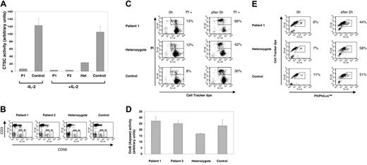
The PLS patients exhibited very low levels of cathepsin C substrate hydrolysis in unstimulated and IL-2-stimulated NK cells compared with controls (Figure 1A), consistent with the proximity of the mutated residue to the active site of the enzyme.13 The CD56bright and CD56dim NK subsets20 from the patients and controls were indistinguishable (Figure 1B), and expression of CD158a, CD158b, CD94, and CD16 was also normal in the patients (Figure S1, available on the Blood website; see the Supplemental Figures link at the top of the online article). In a previous report, LAK cells from PLS patients were found to have normal cytolytic activity.12 Our results confirmed this since control, heterozygote, and PLS NK cells cultured in IL-2 were able to kill K562 target cells (Figure 1C). The heterozygote had an intermediate level of cathepsin C activity (Figure 1A). However, this dosage effect was not evident in the cytoxicity assay (Figure 1C) since, in multiple experiments, the cytolytic activity of the PLS LAK cells was never less than the controls. Furthermore, cell lysates from IL-2-activated PLS NK cells and control NK cells contained similar levels of granzyme B activity (Figure 1D).
These results confirm that human LAK cells, unlike those from the mouse,7 do not require cathepsin C for cytolytic activity or granzyme B activation. An early event in granzyme B-mediated apoptosis is the induction of the caspase cascade.1 To verify that PLS LAK cells were capable of activating the caspase cascade, we modified the killing assay to include the fluorescent caspase 3 substrate, PhiPhiLux, in place of PI staining.19 The results indicate that IL-2-stimulated NK cells from PLS patients and controls are able to rapidly induce the caspase cascade in target cells (Figure 1E).
Cathepsin C is not required for granzyme B activation and cytolytic function in IL-2-activated NK cells. (A) Cathepsin C (CTSC) activity in unstimulated and IL-2-stimulated NK cells from the PLS patients (P1 and P2), heterozygote (Het), and healthy control. Hydrolysis of the colorimetric substrate Gly-Phe-pNA14 is expressed as arbitrary units of activity (based on absorbance at 405 nm). The activity observed in the patient samples is equivalent to the background absorbance of lysate alone. Each reaction contained lysate from 6 × 10⁵ cells and were performed in triplicate; standard deviation is shown. (B) Flow cytometric analysis of CD3neg, CD56dim (dim), and CD56bright (br) NK cell subsets20 in PLS patients 1 and 2, the heterozygote, and a healthy control. (C) Cytolytic activity of IL-2-activated NK cells from PLS patient 1, heterozygote, and healthy control. CellTracker Green-labeled K562 target cells were cocultured with IL-2-activated NK cells at an E/T ratio of 2:1 (2 × 10⁵ cells in 200 μL) for 5 hours at 37°C. Propidium iodide (PI) staining is shown at 0 hours and after 5 hours of coculture. Zero-hour time points were incubated at the same cell density but were not mixed until the 5 hours' time point. Dead or dying cells are detected as a PI+, CellTracker Green+ population by flow cytometry. The percentage of target cells in this gate is shown. This experiment is representative of 3 independent experiments and has been confirmed in patient 2 (data not shown). (D) Granzyme B activity in IL-2-activated NK cells from PLS patients, heterozygote, and healthy control. Activity was measured by hydrolysis of the colorimetric substrate Ac-IEPD-pNA as described.15 Each reaction contained lysate derived from 6 × 10⁵ cells and was performed in triplicate (standard deviation shown); activity is indicated in arbitrary units. Hydrolysis of the substrate was unaffected by the caspase inhibitor z-VAD-fmk (data not shown). (E) Activation of the caspase cascade in K562 cells by IL-2-activated NK cells. The assay was performed as in Figure 1C except that targets were labeled with CellTracker Orange and cocultured with effector cells for 2 hours. Cells were then stained with PhiPhiLux according to the manufacturer and as described.19 Caspase activation results in cleavage of a peptide spacer in PhiPhiLux and increased fluorescence.19 The percentage of cells with increased caspase activity is shown. This experiment is representative of 2 separate experiments and was confirmed using patient 2 (data not shown).
Cathepsin C is not required for granzyme B activation and cytolytic function in IL-2-activated NK cells. (A) Cathepsin C (CTSC) activity in unstimulated and IL-2-stimulated NK cells from the PLS patients (P1 and P2), heterozygote (Het), and healthy control. Hydrolysis of the colorimetric substrate Gly-Phe-pNA14 is expressed as arbitrary units of activity (based on absorbance at 405 nm). The activity observed in the patient samples is equivalent to the background absorbance of lysate alone. Each reaction contained lysate from 6 × 10⁵ cells and were performed in triplicate; standard deviation is shown. (B) Flow cytometric analysis of CD3neg, CD56dim (dim), and CD56bright (br) NK cell subsets20 in PLS patients 1 and 2, the heterozygote, and a healthy control. (C) Cytolytic activity of IL-2-activated NK cells from PLS patient 1, heterozygote, and healthy control. CellTracker Green-labeled K562 target cells were cocultured with IL-2-activated NK cells at an E/T ratio of 2:1 (2 × 10⁵ cells in 200 μL) for 5 hours at 37°C. Propidium iodide (PI) staining is shown at 0 hours and after 5 hours of coculture. Zero-hour time points were incubated at the same cell density but were not mixed until the 5 hours' time point. Dead or dying cells are detected as a PI+, CellTracker Green+ population by flow cytometry. The percentage of target cells in this gate is shown. This experiment is representative of 3 independent experiments and has been confirmed in patient 2 (data not shown). (D) Granzyme B activity in IL-2-activated NK cells from PLS patients, heterozygote, and healthy control. Activity was measured by hydrolysis of the colorimetric substrate Ac-IEPD-pNA as described.15 Each reaction contained lysate derived from 6 × 10⁵ cells and was performed in triplicate (standard deviation shown); activity is indicated in arbitrary units. Hydrolysis of the substrate was unaffected by the caspase inhibitor z-VAD-fmk (data not shown). (E) Activation of the caspase cascade in K562 cells by IL-2-activated NK cells. The assay was performed as in Figure 1C except that targets were labeled with CellTracker Orange and cocultured with effector cells for 2 hours. Cells were then stained with PhiPhiLux according to the manufacturer and as described.19 Caspase activation results in cleavage of a peptide spacer in PhiPhiLux and increased fluorescence.19 The percentage of cells with increased caspase activity is shown. This experiment is representative of 2 separate experiments and was confirmed using patient 2 (data not shown).
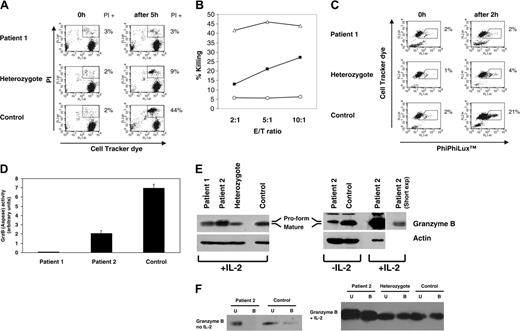
The proliferation and activation induced by IL-2 provides a convenient method to generate large numbers of highly cytolytic NK cells for functional studies. NK cells are activated by T-cell-derived IL-2.21 However, this is likely to be a late event in a primary immune response. We therefore analyzed PLS NK cells directly, without provision of exogenous IL-2. Unstimulated PLS NK cells were not cytolytic (Figure 2A). Lower levels of target cell killing by heterozygote NK cells could be increased using higher effector-target cell ratios. However, this was not the case with the PLS NK cells, which are clearly defective (Figure 2B). Furthermore, unstimulated PLS NK cells were unable to initiate the caspase cascade in target cells (Figure 2C), and lysates from the unstimulated PLS NK cells possess little granzyme B activity (Figure 2D). Lack of cytolytic activity was not due to loss of perforin expression, since this was normal in PLS NK cells (Figure S2).
A lack of cathepsin C activity in unstimulated PLS NK cells coupled with deficient granzyme B activity suggests a failure of granzyme B processing. On immunoblots, granzyme B from unstimulated PLS NK cells is slightly larger than granzyme B from unstimulated control cells, consistent with the retention of the pro-domain in unstimulated PLS NK cells (Figure 2E). In IL-2-stimulated NK cells, control samples possess the smaller species, whereas the corresponding samples from PLS patients contain both forms. The presence of both species in lysates from PLS IL-2-stimulated NK cells suggest that IL-2 activation allows the granzyme pro-domain to be removed, albeit with lower efficiency.
A cytolytic defect in unstimulated NK cells from PLS patients is associated with defective granzyme B processing. (A) Cytolytic activity of unstimulated NK cells derived from PLS patient 1, heterozygote, and healthy control. The killing assay was performed as in Figure 1C except that exogenous IL-2 was not used. The percentage of PI+ target cells is indicated. This experiment is representative of 3 separate killing assays performed and has been confirmed in patient 2. (B) Cytolytic activity of control, heterozygote, and PLS patient NK cells at different effector-target (E/T) ratios. Percentage killing is indicated. ○ indicates patient 1; ▪, heterozygote; ▵, control. (C) Caspase activation by unstimulated NK cells. This experiment was performed as in Figure 1E except that exogenous IL-2 was not used for NK stimulation. Percentages of cells with increased caspase activity are indicated. (D) Granzyme B activity in unstimulated NK cell samples (performed as in Figure 1D); activity is displayed in arbitrary units (standard deviation is shown). A lysate derived from IL-2-activated NK cells contained approximately 4-fold more granzyme B activity than unstimulated NK cells, as in Figure 1D (data not shown). The level of substrate hydrolysis detected in a B-cell line (BJAB), which does not express granzyme B, is similar to the level of activity found in patient 2 (data not shown). (E) Granzyme B immunoblot (using anti-granzyme B antibody clone 2C5/F5) contains lysate from 1 × 10⁶ IL-2-stimulated NK cells per lane or 2 × 10⁶ unstimulated NK cells boiled in SDS-containing loading buffer. A second, shorter exposure of the IL-2-stimulated sample is shown to clearly reveal the different species of granzyme B present in the samples. The unlabeled band at the top of the granzyme B blot highlights the small size difference between granzyme B in the patient and control samples. The mature and pro-form of granzyme B are indicated. Actin was used as a loading control. (F) Aprotinin agarose binding16,17 of granzyme B from unstimulated NK cells and IL-2-activated NK cells from patient 2, heterozygote, and control. Lysates were mixed with aprotinin-agarose beads, the beads were pelleted, and the supernatant removed. The beads were washed and bound (B) and unbound (U) fractions analyzed by granzyme B immunoblotting (as in Figure 2E).
A cytolytic defect in unstimulated NK cells from PLS patients is associated with defective granzyme B processing. (A) Cytolytic activity of unstimulated NK cells derived from PLS patient 1, heterozygote, and healthy control. The killing assay was performed as in Figure 1C except that exogenous IL-2 was not used. The percentage of PI+ target cells is indicated. This experiment is representative of 3 separate killing assays performed and has been confirmed in patient 2. (B) Cytolytic activity of control, heterozygote, and PLS patient NK cells at different effector-target (E/T) ratios. Percentage killing is indicated. ○ indicates patient 1; ▪, heterozygote; ▵, control. (C) Caspase activation by unstimulated NK cells. This experiment was performed as in Figure 1E except that exogenous IL-2 was not used for NK stimulation. Percentages of cells with increased caspase activity are indicated. (D) Granzyme B activity in unstimulated NK cell samples (performed as in Figure 1D); activity is displayed in arbitrary units (standard deviation is shown). A lysate derived from IL-2-activated NK cells contained approximately 4-fold more granzyme B activity than unstimulated NK cells, as in Figure 1D (data not shown). The level of substrate hydrolysis detected in a B-cell line (BJAB), which does not express granzyme B, is similar to the level of activity found in patient 2 (data not shown). (E) Granzyme B immunoblot (using anti-granzyme B antibody clone 2C5/F5) contains lysate from 1 × 10⁶ IL-2-stimulated NK cells per lane or 2 × 10⁶ unstimulated NK cells boiled in SDS-containing loading buffer. A second, shorter exposure of the IL-2-stimulated sample is shown to clearly reveal the different species of granzyme B present in the samples. The unlabeled band at the top of the granzyme B blot highlights the small size difference between granzyme B in the patient and control samples. The mature and pro-form of granzyme B are indicated. Actin was used as a loading control. (F) Aprotinin agarose binding16,17 of granzyme B from unstimulated NK cells and IL-2-activated NK cells from patient 2, heterozygote, and control. Lysates were mixed with aprotinin-agarose beads, the beads were pelleted, and the supernatant removed. The beads were washed and bound (B) and unbound (U) fractions analyzed by granzyme B immunoblotting (as in Figure 2E).
Removal of the dipeptide pro-domain from several serine proteases exposes the active site to inhibitors such as aprotinin, allowing processed forms to be identified by binding to aprotinin-agarose.16,17 Granzyme B (which is inhibited by aprotinin22 ) was detected in the aprotinin-bound fraction in all the IL-2-stimulated samples (Figure 2F), consistent with the presence of enzymatically active granzyme B (Figure 1D). However, no granzyme B was detected in the aprotinin-bound fraction in unstimulated PLS NK cells. Instead, granzyme B from unstimulated PLS NK cells is found only in the unbound fraction (Figure 2F). Furthermore, in cell lysates, active granzyme B forms a sodium dodecyl sulfate (SDS)-resistant complex with the protease inhibitor PI-9.23 This complex was observed in IL-2-stimulated NK cell lysates from patient and control samples as well as lysates from unstimulated NK cells from control samples. However, the complex was not detectable in the unstimulated PLS NK cell lysates, indicating that granzyme B was not activated in these cells (Figures S3). Collectively, these results show that although granzyme B is present in the unstimulated PLS NK cells, it is inactive because of a processing defect. However, IL-2 does not induce cathepsin C activity in human NK cells (Figure 1A). Interestingly, some granzyme C is processed in cathepsin C-deficient mice,7 indicating that cathepsin C-independent pathways of granzyme processing are present in mouse and human.
The cytolytic defect in PLS NK cells is likely to be due, in part, to a failure in granzyme B processing. However, cathepsin C is likely to be important for the activation of other granzymes. Thus, at present, we cannot unequivocally link the NK cytolytic defect to a failure of granzyme B activation, since other cytolytic components also might require cathepsin C-mediated activation. In addition, we have studied a single mutation, and the role of cathepsin C in human NK cell function should be confirmed in unrelated PLS patients. In PLS, neutrophil defects12 and impaired NK activity may increase susceptibility to particular infections. NK cell deficiencies are frequently associated with herpesvirus susceptibility,24 and this family of viruses may play a role in periodontitis.25 While IL-2 therapy might improve NK cell function, it is unlikely to overcome defects in other cell types.26 In vivo, the cytolytic defect is presumably overcome once IL-2 (or possibly other cytokines) is produced.
Prepublished online as Blood First Edition Paper, January 12, 2006; DOI 10.1182/blood-2005-03-1140.
Supported by grants from The West Riding Medical Research Trust, the Yorkshire Cancer Research, the Wellcome Trust, the Candlelighters, and Cancer Research UK.
The online version of this article contains a data supplement.
The publication costs of this article were defrayed in part by page charge payment. Therefore, and solely to indicate this fact, this article is hereby marked “advertisement” in accordance with 18 U.S.C. section 1734.
We are indebted to the PLS family for participating in this study. We thank Carmel Toomes for genetic information on the PLS family, Mushtaq Ahmed for help with patient liaison, and colleagues for numerous blood samples. Finally, we thank Gina Doody and Yasser El-Sherbiny for help and advice, and Peter Selby for support and encouragement.


This feature is available to Subscribers Only
Sign In or Create an Account Close Modal