Abstract
Type 1 von Willebrand disease (VWD) is characterized by a partial quantitative deficiency of von Willebrand factor (VWF). Few VWF gene mutations have been identified that cause dominant type 1 VWD. The decreased survival of VWF in plasma has recently been identified as a novel mechanism for type 1 VWD. We report 4 families with moderately severe type 1 VWD characterized by low plasma VWF:Ag and FVIII:C levels, proportionately low VWF:RCo, and dominant inheritance. A decreased survival of VWF in affected individuals was identified with VWF half-lives of 1 to 3 hours, whereas the half-life of VWF propeptide (VWFpp) was normal. DNA sequencing revealed a single (heterozygous) VWF mutation in affected individuals, S2179F in 2 families, and W1144G in 2 families, neither of which has been previously reported. We show that the ratio of steady-state plasma VWFpp to VWF:Ag can be used to identify patients with a shortened VWF half-life. An increased ratio distinguished affected from unaffected individuals in all families. A significantly increased VWFpp/VWF:Ag ratio together with reduced VWF:Ag may indicate the presence of a true genetic defect and decreased VWF survival phenotype. This phenotype may require an altered clinical therapeutic approach, and we propose to refer to this phenotype as type-1C VWD.
Introduction
Von Willebrand factor (VWF) is a large, adhesive, multimeric plasma glycoprotein that serves as the carrier of factor VIII (FVIII) and mediates platelet adhesion and aggregation at the site of vessel wall injury.1,2 The pre–pro-VWF protein is synthesized exclusively in megakaryocytes and endothelial cells with a 22-amino acid signal peptide, 741-amino acid propeptide, and 2050-amino acid mature VWF protein.3 The pre–pro-VWF protein undergoes an extensive series of intracellular modifications, including signal peptide cleavage, C-terminal dimerization, carbohydrate processing, sulfation, and amino-terminal multimerization.4-8 In the acidic compartment of the trans-Golgi, proteolytic processing yields the propeptide (VWFpp) and mature VWF multimers. The VWFpp noncovalently associates with mature VWF multimers, and both proteins are stored in α-granules (megakaryocytes) or Weibel-Palade bodies (endothelial cells) for release through the regulated pathway.9-12 After secretion into plasma (pH ∼ 7.4), VWFpp dissociates from VWF and circulates as a homodimer at a concentration of about 1 μg/mL with a half-life of 2 to 3 hours, whereas mature VWF circulates at about 10 μg/mL with a half-life of 8 to 12 hours.13-15
Defects in the function or synthesis of VWF are identified in patients with von Willebrand disease, a common inherited bleeding disorder with an estimated prevalence as high as 1%.16,17 Current classification of von Willebrand disease (VWD) recognizes 3 types. Type 3 VWD is characterized by a complete deficiency of VWF. Type 2 VWD refers to qualitative deficiency of VWF and is subdivided into types 2A, 2B, 2M, and 2N. Type 1 VWD is characterized by partial quantitative deficiency of VWF with autosomal dominant inheritance, variable penetrance, and a highly variable phenotype. In contrast to other VWD types, very few mutations in the VWF gene have been linked to type 1 VWD.18-21 Although some type 1 VWD phenotypes have been linked to defects in the VWF gene, others do not cosegregate with genetic markers at the VWF gene locus.22,23 A novel mechanism for type 1 VWD has been linked to increased clearance of VWF from plasma in humans.24,25
Here, we report the assay of propeptide and mature VWF in the plasma of patients with type 1 VWD to predict those with decreased VWF survival. We examined 4 unrelated families with the diagnosis of type 1 VWD. In affected individuals we identified an accelerated clearance of their endogenously produced VWF with a half-life of 1 to 3 hours. The half-life of FVIII in these individuals was also significantly reduced and closely paralleled VWF half-life. We identified either a W1144G mutation in the VWF D3 domain or a S2179F mutation in the VWF D4 domain in the affected family members. An increased ratio of plasma VWFpp to mature VWF differentiated affected individuals from unaffected individuals in all families. In summary, these observations could be of clinical importance because DDAVP administration may not be the treatment of choice in patients with a decreased VWF survival phenotype as predicted by an increased VWFpp/VWF ratio.
Patients, materials, and methods
Patients
Patients and family members were studied after we received their written informed consent. The research protocol for this study was approved by the IRBs of Children's Hospital of Wisconsin, the General Clinical Research Center of the Medical College of Wisconsin, the Medical College of Wisconsin, and the BloodCenter of Wisconsin's Blood Research Institute. Propositi were identified by the Comprehensive Center for Bleeding Disorders of the BloodCenter of Wisconsin; available family members from multiple generations were seen through the Pediatric General Clinical Research Center at Children's Hospital of Wisconsin. Plasma and platelet VWF:Ag, VWF:RCo, VWF multimers, and FVIII levels were determined by the Hemostasis Reference Laboratory at BloodCenter of Wisconsin, Milwaukee, WI, as previously described.26
Antibodies
The following antibodies were produced by our laboratory: (1) anti-VWF monoclonal antibodies AVW-1, AVW-5, AVW-17, and 105.4; (2) an anti-VWF polyclonal antibody Edwina; (3) anti-VWFpp monoclonal antibodies 239.2 and 239.3; and (4) an anti-VWFpp polyclonal antibody (Mango). A polyclonal anti–human VWF was purchased from DAKO (Carpinteria, CA).
Reverse ABO blood group typing
Patient plasma was added to saline-washed red blood cells obtained from individuals with blood type A and blood type B. The samples were gently mixed and observed for hemagglutination.
ELISA assays
The concentrations of VWF and VWFpp in patient plasma were determined by using standard enzyme-linked immunoabsorbent assay (ELISA) assays. VWF was captured using the monoclonal antibody, AVW-1, and detected using a rabbit anti–human VWF polyclonal antibody. VWFpp was captured using monoclonal antibodies, 239.2 and 239.3, and detected using a rabbit anti-VWFpp polyclonal antibody.
Statistical analysis
One-way analysis of variance, Sidak test, Mann-Whitney test, and Student t test were performed as appropriate.
Determination of half-life after desmopressin (DDAVP) or Humate-P infusion
DDAVP was infused intravenously at a dose of 0.3 μg/kg. Some patients were infused with Humate-P (30 U/kg). Samples were taken prior to the start of the treatment and at multiple time points after infusion. Whole venous blood was collected, and platelet-poor plasma was prepared and stored at –80°C until assayed as previously described.26 To prevent inadvertent cryoprecipitation of VWF, frozen samples were warmed at 37°C and thoroughly mixed prior to assay. The half-lives of VWF, FVIII, or VWFpp were determined by calculation of the first-order rate constant for the elimination phase from the slope of the VWF:Ag, VWF:RCo activity, FVIII concentration, or VWFpp concentration against time.24,27 The following formula was used: C(t) = C0 + Ae–kEt, where C(t) = VWF:Ag as a function of time, C0 = baseline VWF:Ag, A = y-axis intercept, kE = first-order rate constant for the elimination phase, and t = time. Half-life, t1/2, was determined by the equation: t1/2 = ln2/kE.
Isolation of genomic DNA and sequencing
Total cellular RNA and genomic DNA were prepared from platelets and leukocytes, respectively, as previously described.28,29 Reverse transcription of purified platelet RNA was performed as previously described.29 Polymerase chain reaction (PCR) amplification and direct sequencing was performed on each VWF exon. The VWF DNA sequence is numbered starting from the ATG of the initiating Met codon, and VWF protein sequence numbering starts from the initiating Met.
Complex multimer analysis
VWF in patient plasma was analyzed by electrophoresis through a 0.8% (wt/vol) HGT(P) agarose (DMC Bioproducts, Rockland, ME) stacking gel and 3% (wt/vol) HGT(P) agarose running gel containing 0.1% SDS for 16 hours at 40 V using the Laemmli buffer system as previously described.30 Western blotting was performed as previously described.30
Results
Four families diagnosed with type 1 VWD
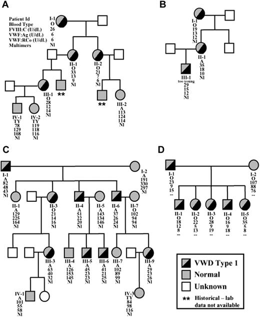
Recently, the decreased survival of VWF in plasma has been identified as a mechanism causing type 1 VWD in a subset of patients.24,25,31 We had the opportunity to study members from several generations of 4 families with the diagnosis of type 1 VWD. The pedigrees are shown in Figure 1 along with phenotypic data. Patients AI-1, AII-1, and BI-1 had lifelong histories of mild mucocutaneous bleeding, characterized by easy bruising, moderately severe epitaxis, prolonged bleeding after tooth extraction, and bleeding following tonsillectomy. Patients BII-1, CII-3, CIII-9, and CIII-3 all reported easy bruising, menorrhagia, and multiple instances of prolonged bleeding after injury or tooth extraction. All affected family members had decreased plasma VWF:Ag, VWF: RCo, and FVIII levels, consistent with a mode of dominant inheritance. Affected patients had VWF:Ag levels ranging from 5 to 48 U/dL and exhibited a normal VWF:RCo/VWF:Ag ratio. The platelet VWF:Ag for patient AI-1 was 40.0 U/1011 platelets, within the normal range (22-47 U/1011 platelets for the population served by BloodCenter of Wisconsin). Analysis of plasma multimers by low-resolution gel (1.2% agarose) showed the presence of normal VWF multimers in the affected patients, but with reduced intensity (Figure 2A, inset). Thus, affected individuals in these families have low VWF:Ag levels, proportionally low VWF:RCo levels, and a normal VWF multimer, consistent with type 1 VWD.
DDAVP infusion
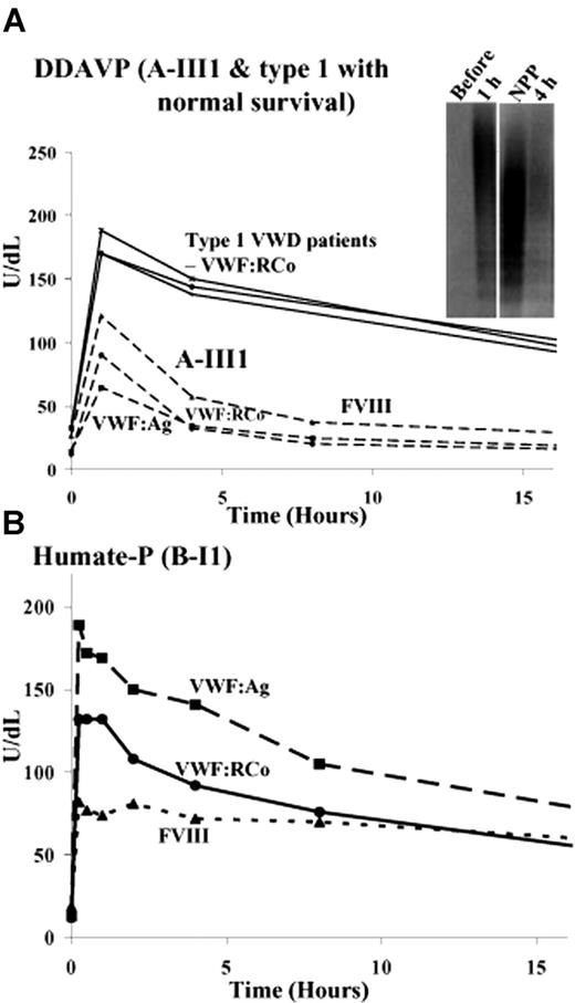
Affected members in the 4 families required 1-desamino-8-D-arginine vasopressin (DDAVP) or VWF replacement therapy on multiple occasions. In some individuals a detailed time course of VWF:Ag, VWF:RCo, and FVIII plasma levels was assessed. DDAVP administration resulted in a significant increase in FVIII and VWF plasma levels. However, after reaching normal levels, both proteins disappeared rapidly from the circulation (Figure 2A). The half-life (t1/2) of VWF for patient AIII-1 was 3 hours, significantly less than normal. In other affected family members VWF half-lives ranged from 1.0 to 3.6 hours (Table 1). The t1/2 of FVIII closely parallels that of VWF and was significantly reduced compared with the normal t1/2 of 11 to 16 hours (Table 1).32 In contrast, when DDAVP was administered to 3 other, unrelated patients with type 1 VWD, a normal VWF survival was observed (Figure 2A; Table 1). To determine whether the full range of multimers was affected, the multimer structure of VWF in plasma from patient AIII-1 was examined by low-resolution (1.2%) SDS-agarose gel. One hour after DDAVP, an increase in high molecular weight multimers was observed (Figure 2A). At 4 hours, normal multimers were present with reduced intensity, indicating increased clearance of all VWF oligomers. Plasma VWFpp levels were determined after DDAVP for patients AI-1 and AIII-1 (data not shown). The t1/2 of VWFpp was within the normal range (Table 1).14 These data are consistent with decreased survival of VWF in plasma with normal survival of VWFpp.
Half-lives of VWF, FVIII, and VWFpp in affected family members after administration of DDAVP or Humate-P
Family member . | t1/2 (VWF) after DDAVP, h . | t1/2 (FVIII) after DDAVP, h . | t1/2 (VWFpp) after DDAVP, h . | t1/2 (VWF) after Humate-P, h . |
|---|---|---|---|---|
| A-I-1 | 1.0 | 1.3 | 2.5 | 12.7 |
| A-II-2 | 1.2 | 1.9 | ND | ND |
| A-III-1 | 3.1 | 2.3 | 2.4 | ND |
| B-I1 | 1.4 | 1.6 | ND | 11.8 |
| B-II1 | 1.7 | ND | ND | ND |
| C-II-3 | 1.4 | 1.3 | ND | 9.9 |
| C-III-3 | 3.6 | ND | ND | ND |
| Type 1-1 | 9.2 | ND | ND | ND |
| Type 1-2 | 11.0 | ND | ND | ND |
| Type 1-3 | 12.5 | ND | ND | ND |
| Healthy individuals,13,14,32 range | 8-12 | 11-18 | 2-3 | 8-12 |
Multimers were normal in all family members and healthy individuals.
ND indicates not done.
Pedigrees and laboratory data of 4 families with type 1 VWD. Pedigrees are shown for 4 families, A, B, C, and D. Affected individuals are represented by half-filled symbols, unaffected individuals by gray symbols, and those with an unknown phenotype are denoted by white, unfilled symbols. In some cases, diagnosis was based on historical data and the laboratory data are unavailable, and these patients are denoted by **. The laboratory test results are listed as follows: Patient ID; blood type (TY = too young); FVIII:C, VWF:Ag, VWF:RCo, and multimer status (Nl = normal). Laboratory parameters are characteristic of type 1 VWD in all 4 families.
Pedigrees and laboratory data of 4 families with type 1 VWD. Pedigrees are shown for 4 families, A, B, C, and D. Affected individuals are represented by half-filled symbols, unaffected individuals by gray symbols, and those with an unknown phenotype are denoted by white, unfilled symbols. In some cases, diagnosis was based on historical data and the laboratory data are unavailable, and these patients are denoted by **. The laboratory test results are listed as follows: Patient ID; blood type (TY = too young); FVIII:C, VWF:Ag, VWF:RCo, and multimer status (Nl = normal). Laboratory parameters are characteristic of type 1 VWD in all 4 families.
In contrast, when affected patients received VWF replacement therapy, the infused VWF had a normal survival. Patient BI-1 was infused with Humate-P and a detailed time-course was assessed (Figure 2B). The t1/2 of the infused VWF was 11.8 hours. A normal VWF half-life was observed for other patients who received Humate-P (Table 1). Plasma multimers were found to be nearly normal in samples obtained over 24 hours (data not shown). These results show that the elimination of VWF multimers from the plasma of the affected family members with type 1 VWD is significantly increased and may be linked to the patients' endogenously synthesized VWF. Together these results suggest reduced survival of a VWF protein that is synthesized and secreted at a normal rate.
Abnormal multimer satellite band structure
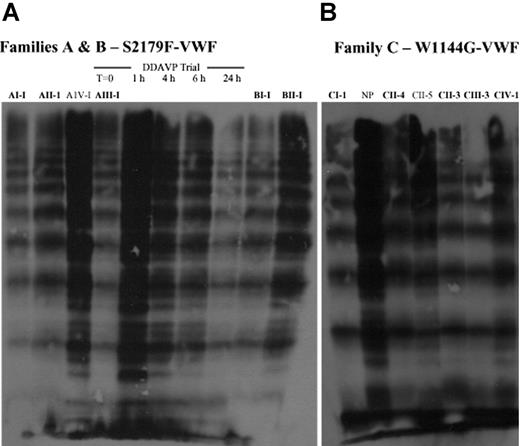
We further examined the plasma VWF multimer structure in family members by using a high-resolution (3%) SDS-agarose gel (Figure 3). In affected members of families A, B, and C (Figure 3A-B), we observed a significant decrease in intensity of the VWF satellite bands (eg, AI-1, AIII-1) when compared with an unaffected family member (AIV-1) or pooled normal human plasma (NP). These VWF satellite bands are the products of ADAMTS13 limited proteolysis of VWF multimers.33,34 Strikingly, analysis of a plasma sample obtained 1 hour after DDAVP administration in patient AIII-1 showed the presence of satellite bands (Figure 3A). After 6 hours, the intensity of these bands had decreased, and after 24 hours they were significantly diminished. These data are not consistent with increased ADAMTS13 degradation that results in a VWD type 2A phenotype characterized by the presence of only the smallest VWF multimers in plasma. However, these data do suggest that the ADAMTS13 degradation products are perhaps cleared from circulation more quickly than the intact multimer.
Identification of a mutation in affected family members
We examined the VWF gene sequence of members of the 4 families to determine whether a mutation in the VWF gene was present. All VWF exons were sequenced. In all affected individuals in families A and B we detected a mutation in exon 37, a C to T transition at position 6536 (C6536T) of the VWF cDNA, predicting the substitution of Ser by Phe at amino acid 2179 (S2179F). This mutation was identified in both families, although no historical link between families could be established. The affected patients are heterozygous for the substitution, because both normal and mutant alleles were identified. No other mutations were found in the VWF coding region. This mutation was not found in the unaffected family members.
In all affected individuals from families C and D we identified a (heterozygous) mutation in exon 26 (T3430G), predicting the substitution of Trp by Gly at amino acid 1144 (W1144G). No other mutations were identified, and this mutation was not found in unaffected family members. The W1144G mutation was identified in both C and D families, although no historical link between families could be established. Neither of these 2 mutations has been previously reported and is not listed in the VWD database.35
Time course of VWF:Ag, FVIII:C, and VWF:RCo levels after administration of DDAVP or Humate-P. (A) DDAVP administration to patient A-III-1 and 3 unrelated patients with type 1 VWD. In all patients, VWF:Ag, VWF:RCo, and FVIII levels rose significantly after DDAVP administration. Although VWF levels in the 3 patients with “type 1 VWD” slowly diminished over time, these levels rapidly decreased in patient AIII-1. Similar results were observed in other affected patients. Inset: Multimer analysis before, 1 hour, and 4 hours after DDAVP administration compared with pooled normal human plasma (NP). (B) VWF:Ag, VWF:RCo, and FVIII levels after Humate-P treatment in patient BI-1 slowly decrease over time. The elimination of plasma VWF multimers in affected family members is significantly increased and may be linked to the patients' endogenously synthesized VWF.
Time course of VWF:Ag, FVIII:C, and VWF:RCo levels after administration of DDAVP or Humate-P. (A) DDAVP administration to patient A-III-1 and 3 unrelated patients with type 1 VWD. In all patients, VWF:Ag, VWF:RCo, and FVIII levels rose significantly after DDAVP administration. Although VWF levels in the 3 patients with “type 1 VWD” slowly diminished over time, these levels rapidly decreased in patient AIII-1. Similar results were observed in other affected patients. Inset: Multimer analysis before, 1 hour, and 4 hours after DDAVP administration compared with pooled normal human plasma (NP). (B) VWF:Ag, VWF:RCo, and FVIII levels after Humate-P treatment in patient BI-1 slowly decrease over time. The elimination of plasma VWF multimers in affected family members is significantly increased and may be linked to the patients' endogenously synthesized VWF.
VWFpp levels in healthy individuals
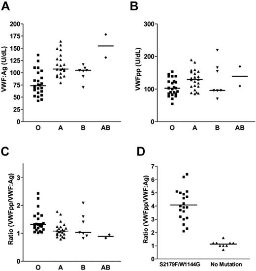
We hypothesized that the ratio between VWFpp and VWF plasma concentrations could be used to predict those with abbreviated VWF survival. The free VWFpp circulates in plasma with a t1/2 of 2 to 3 hours, whereas mature VWF has a t1/2 of 8 of 12 hours. Their concentrations in plasma are expressed in units, and by definition 1 mL normal plasma contains 1 U each of VWFpp and VWF with a VWFpp/VWF ratio of 1. We sought to verify this ratio in our pool of 55 healthy individuals. VWF:Ag and VWFpp were measured by ELISA assays, and plasma samples were ABO typed as described in “Patients, materials, and methods.” Both ELISA assays were referenced against an ISTH VWF standard curve that has been standardized for VWF:Ag, but it has not been cross-standardized for VWFpp. We assumed that the ISTH standard accurately represents the average steady-state concentration of VWFpp, as well as VWF:Ag. Table 2 summarizes the range, arithmetic mean, and standard deviation for VWF:Ag, VWFpp level, and VWFpp/VWF:Ag ratio determined for each blood group. Our pooled normal human plasma (NP) consisted of 43.6% blood group O, 40% group A, 12.7% group B, and 3.6% group AB, similar to the distribution observed in the population served by BloodCenter of Wisconsin (Table 2).36 The range of VWF:Ag for each blood type in our normal pool was found to be similar to the range previously reported (Figure 4A; Table 2).36 Within our data set, VWF:Ag levels for individuals in the group with type O blood (78.2 U/dL) were significantly different (P < .001) than those in the groups wit type A (115.6 U/dL) and type AB (154.8 U/dL) blood. Group B (100.4 U/dL) VWF:Ag levels significantly differed from group AB (P < 0.04).
Influence of ABO blood group on VWF:Ag, VWFpp, and VWFpp/VWF:Ag in healthy individuals
. | Group O . | Group A . | Group B . | Group AB . | Overall . |
|---|---|---|---|---|---|
| Percentage in BCW population | 45 | 45 | 7 | 4 | NA |
| Percentage in NP (N) | 43.6 (24) | 40.0 (22) | 12.7 (7) | 3.6 (2) | NA |
| “Normal range” (IU/dL)36 | 41-179 | 55-267 | 64-275 | 73-271 | 50-240 |
| VWF:Ag, range in NP | 43-136 | 79-164 | 70-117 | 131-178 | 43-162 |
| VWF:Ag (IU/dL), mean ± SD | 78 ± 25 | 116 ± 23 | 100 ± 15 | 155 ± 28 | 99 ± 30 |
| VWFpp, range in NP | 55-154 | 85-189 | 70-219 | 109-169 | 55-219 |
| VWFpp (U/dL), mean ± SD | 105 ± 26 | 128 ± 30 | 128 ± 53 | 139 ± 43 | 119 ± 35 |
| Ratio, range in NP | 1.04-2.43 | 0.84-1.78 | 0.82-2.09 | 0.83-0.95 | 0.82-2.43 |
| Ratio, mean ± SD | 1.41 ± 0.37 | 1.12 ± 0.25 | 1.26 ± 0.46 | 0.89 ± 0.08 | 1.26 ± 0.36 |
BCW indicates BloodCenter of Wisconsin; NP, pooled normal human plasma; NA, not applicable.
Plasma VWF multimer pattern obtained by high-resolution SDS-agarose gel electrophoresis for affected and unaffected family members. (A) Plasma VWF multimer distribution of members of families A and B. Affected family members harbor a S2179F VWF mutation and are denoted in bold type above each lane. Multimers in plasma from Patient AIII-1 were obtained before (T = 0) and at various time points after DDAVP administration. (B) Plasma VWF multimer distribution of members of family C. Affected family members harbor a W1144G VWF mutation and are denoted in bold type above each lane. At steady-state, VWF multimer satellite bands are nearly absent from the plasma of affected family members.
Plasma VWF multimer pattern obtained by high-resolution SDS-agarose gel electrophoresis for affected and unaffected family members. (A) Plasma VWF multimer distribution of members of families A and B. Affected family members harbor a S2179F VWF mutation and are denoted in bold type above each lane. Multimers in plasma from Patient AIII-1 were obtained before (T = 0) and at various time points after DDAVP administration. (B) Plasma VWF multimer distribution of members of family C. Affected family members harbor a W1144G VWF mutation and are denoted in bold type above each lane. At steady-state, VWF multimer satellite bands are nearly absent from the plasma of affected family members.
We also measured VWFpp levels in the healthy individuals (Figure 4B). A normal range of VWFpp for each blood group has not been previously established. The range for all individuals was found to be 55 to 219 U/dL with an average value of 118.5 U/dL (Table 2). The differences in VWFpp levels between blood groups was not found to be statistically significant (P = .07). However, when blood group O (105.2 U/dL) was compared with all others combined, group O was significantly lower (P < .01).
The ratio of VWFpp to VWF:Ag was calculated for each individual (Figure 4C), and the values for each blood group are reported in Table 2. Within our data set, the ratio of VWFpp to VWF:Ag in individuals in group O (1.41 ± 0.37) was significantly different (P < .04) from group A (1.12 ± 0.25). The average ratio for all individuals was found to be 1.26 ± 0.36, corresponding to a 2 standard deviation range of 0.54 to 1.98. This ratio appears to be in agreement with the expected value of 1.0 for healthy individuals.
Prediction of decreased VWF survival
We predicted that individuals with a decreased VWF survival would have an altered steady-state ratio of plasma VWFpp and VWF. All affected individuals in the 4 families had an increased VWFpp/VWF ratio, ranging from 2.1 to 6.4 with an average ratio of 4.1 (Figure 4D). These ratios fall outside of the 2 standard deviation normal range of 0.54 to 1.98. Unaffected family members were easily discriminated from the affected family members because unaffected individuals had ratios in the normal range, ranging from 0.7 to 1.6 (Figure 4D). We also examined the correlation of the VWFpp/VWF:Ag ratio with the presence or absence of a VWF gene mutation as summarized in Table 3. All affected individuals harbored a VWF gene mutation and showed an increased ratio, whereas no mutation was detected in unaffected individuals. Individuals within our type 1 VWD families had blood types of either group O or group A. The mean VWF:Ag in affected individuals of blood group O (16.25 ± 5.6 U/dL) was found to be statistically different (P = .013) from individuals in blood group A (27.13 ± 14.3 U/dL). However, no statistical difference in VWFpp/VWF:Ag ratio between affected individuals of blood group O (4.4 ± 0.93) and group A (3.6 ± 1.4) could be found. Furthermore, no subjects were found to be misclassified if the ABO-specific ranges for VWFpp/VWF:Ag ratio (Table 2) were used to distinguish affected individuals from healthy individuals. Together, these results indicate that the steady-state ratio of plasma VWFpp and VWF can be used to easily identify patients with type 1 VWD with an increased plasma VWF clearance phenotype.
Characterization of affected and unaffected family members by VWF:Ag, VWFpp, VWF mutation, and VWFpp/VWF ratio
ID (blood group) . | VWF:Ag, U/dL . | VWFpp, U/dL . | Mutation . | VWFpp/VWF ratio . |
|---|---|---|---|---|
| AI-1 (O) | 24 | 77 | S2179F | 3.2 |
| AII-1 (O) | 12 | 63 | S2179F | 5.1 |
| AII-2 (O) | 9 | 56 | S2179F | 6.4 |
| AIII-1 (O) | 16 | 67 | S2179F | 4.2 |
| AIV-2 (*) | 53 | 65 | None | 1.2 |
| BI-1 (O) | 12 | 60 | S2179F | 4.9 |
| BII-1 (A) | 21 | 130 | S2179F | 6.2 |
| CI-1 (A) | 59 | 122 | W1144G | 2.1 |
| CII-1 (A) | 158 | 112 | None | 0.7 |
| CII-3 (O) | 12 | 55 | W1144G | 4.6 |
| CII-4 (A) | 31 | 80 | W1144G | 2.6 |
| CII-5 (A) | 112 | 107 | None | 1.0 |
| CII-6 (A) | 23 | 69 | W1144G | 3.0 |
| CII-7 (O) | 60 | 70 | None | 1.2 |
| CIII-3 (A) | 32 | 72 | W1144G | 2.3 |
| CIII-4 (A) | 106 | 106 | None | 1.0 |
| CIII-5 (A) | 16 | 80 | W1144G | 5.0 |
| CIII-6 (A) | 17 | 66 | W1144G | 3.9 |
| CIII-7 (A) | 55 | 68 | None | 1.2 |
| CIII-9 (A) | 18 | 66 | W1144G | 3.7 |
| CIV-1 (A) | 59 | 92 | None | 1.6 |
| CIV-3 (*) | 75 | 93 | None | 1.2 |
| DI-1 (O) | 28 | 142 | W1144G | 5.1 |
| DI-2 (O) | 80 | 79 | None | 1.0 |
| DII-1 (O) | 16 | 57 | W1144G | 3.6 |
| DII-2 (O) | 13 | 61 | W1144G | 4.7 |
| DII-3 (O) | 22 | 69 | W1144G | 3.1 |
| DII-4 (O) | 15 | 61 | W1144G | 4.0 |
| DII-5 (O) | 16 | 63 | W1144G | 3.9 |
Too young to obtain blood type.
Influence of ABO blood group on VWF:Ag, VWFpp, and ratio of VWFpp to VWF:Ag in healthy individuals. (A) VWF:Ag levels in healthy individuals of blood group O, A, B, or AB. The horizontal line represents the median VWF:Ag value. (B) VWFpp levels in healthy individuals of blood group O, A, B, or AB. The horizontal line represents the median VWFpp value. (C) Ratio of VWFpp to VWF:Ag in healthy individuals of blood group O, A, B, or AB. The horizontal line represents the median value of the ratio. (D) Ratio of VWFpp to VWF:Ag in family members with a VWF mutation or without a mutation. The horizontal line represents the median value of the ratio.
Influence of ABO blood group on VWF:Ag, VWFpp, and ratio of VWFpp to VWF:Ag in healthy individuals. (A) VWF:Ag levels in healthy individuals of blood group O, A, B, or AB. The horizontal line represents the median VWF:Ag value. (B) VWFpp levels in healthy individuals of blood group O, A, B, or AB. The horizontal line represents the median VWFpp value. (C) Ratio of VWFpp to VWF:Ag in healthy individuals of blood group O, A, B, or AB. The horizontal line represents the median value of the ratio. (D) Ratio of VWFpp to VWF:Ag in family members with a VWF mutation or without a mutation. The horizontal line represents the median value of the ratio.
Discussion
The affected individuals in the 4 families studied showed a characteristic type 1 VWD phenotype (Figure 1) with a significantly reduced VWF t1/2 following the administration of DDAVP (Figure 2; Table 1). In contrast, in individuals treated with Humate-P, the VWF from concentrate had a normal t1/2 (Table 1). These data indicate reduced survival of the patient's endogenously synthesized VWF. We observed a robust response to DDAVP with a 5- to 15-fold increase in plasma VWF:Ag (data not shown). Together with the normal VWFpp t1/2, these data are consistent with normal synthesis and regulated VWF (and VWFpp) storage and release in endothelial cells.37,38 A single, heterozygous VWF gene mutation, W1144G or S2179F, was identified in each of the affected individuals in the 4 families (Table 3). A historical link was not identified between the 2 families with the W1144G or families with the S2179F mutations. However, analysis of markers within intron 40 and the promoter region of the VWF gene showed that each of the 2 family groups with each mutation shared a common haplotype (data not shown). Although the common background suggests that each of the mutations may have arisen from a common founder, the analysis of additional families with each mutation will be necessary to define a common founder chromosome. Together, the data are consistent with type 1 VWD resulting from reduced VWF plasma survival in families with either a S2179F or W1144G VWF gene mutation.
We predicted that the VWFpp/VWF:Ag ratio could identify patients with type 1 VWD with a reduced VWF survival phenotype. We first determined the levels of VWF:Ag and VWFpp in a group of healthy individuals. Our analysis of VWF:Ag levels in healthy individuals confirms previous studies from our center reporting the effect of the ABO blood group on VWF:Ag levels: those within blood group O had the lowest levels (Table 2; Figure 4).36 In many previous studies, blood group O has been associated with the lowest VWF:Ag levels.36,39,40 Although the normal range for VWF:Ag and the effect of ABO blood group has been firmly established, the normal range and influence of blood group on VWFpp levels have not been well defined. In our present study of 55 healthy individuals, VWFpp levels ranged from 55 to 219 U/dL. We did not find a statistical difference in VWFpp levels between blood groups, only when group O was compared with all others was a statistical difference apparent. The limitation of our study is that our data set comprised a relatively small number of individuals. However, the distribution of blood groups in our pool was similar to that found in the population served by the BloodCenter of Wisconsin, indicating that our data set, as a whole, represents a normal distribution of individuals. To firmly establish VWFpp normal ranges for each blood group, a much larger study will be necessary.
We observed variability in VWF:Ag and VWFpp levels in affected family members. VWF:Ag levels in affected individuals of blood group O were significantly lower than blood group A (P = .013). This is consistent with a previous study of heterozygous carriers of a VWF null allele, a type 2N mutation, or a missense mutation which determined that VWF:Ag levels were significantly lower in individuals with blood group O, regardless of their mutation.41 In addition to ABO blood group, many other variables, including age, sex, or environmental factors, could have an influence on VWF:Ag and VWFpp levels.
The steady-state levels of plasma VWF:Ag and VWFpp represent the equilibrium between VWF secretion and clearance. Our purpose in establishing a normal range for VWFpp level was to use plasma concentration of VWFpp as a gauge of VWF synthesis/secretion. The plasma level of VWFpp has been used to distinguish acute and chronic endothelial cell perturbation and as a marker of acquired von Willebrand syndrome.13,42,43 We hypothesized that a patient with VWD with decreased VWF survival would have normal VWF (and VWFpp) secretion and an unaffected steady-state concentration of VWFpp; only the clearance of VWF from plasma would be increased and would be reflected by an altered ratio of plasma VWFpp/VWF:Ag. Our normal pool had a range of 2 standard deviations for VWFpp/VWF:Ag ratio of 0.54 to 1.98, in agreement with the expected ratio of 1. Only 3 of the healthy individuals had a ratio that fell outside of this range. Affected individuals in the 4 families were easily identified by their increased VWFpp/VWF:Ag ratio (2.1-6.4) that fell outside the normal range. Similarly, the unaffected family members were also easily differentiated because their ratios fell within the normal range. Affected individuals with a known reduced VWF half-life, a robust response to DDAVP, and a VWF gene mutation had a significantly increased VWFpp/VWF:Ag ratio. In all of the affected family members, a markedly increased ratio was found to correlate with the presence of a VWF mutation. Collectively, these data confirm the utility of the steady-state plasma VWFpp/VWF:Ag ratio in identifying patients with a reduced VWF survival phenotype.
The ratio of VWFpp to VWF:Ag may also have utility in discriminating other types of VWD. Using a different method of VWFpp determination, the correlation of VWFpp and VWF:Ag has been used to discriminate between type 1 and type 2 VWD.44 That previous study did not discriminate between type 2A-1 and type 2A-2, nor were mutations identified in any of the patients with VWD. In a pilot study, we have now examined VWFpp/VWF:Ag ratio in patients with type 2B and 2M with known mutations (data not shown). Four patients with type 2B had an average VWFpp/VWF:Ag ratio of 2.6 that fell outside of the normal range. In contrast, 4 patients with confirmed type 2M had an average ratio of 1.8, within the normal range. These data are consistent with increased platelet and VWF clearance in patients with type 2B VWD, whereas patients with type 2M often have normal VWF:Ag levels. A comprehensive study of VWFpp/VWF:Ag ratio in patients with type 2A-I and 2A-II (as well as additional type 2M and 2B) in which mutations have been identified and phenotype confirmed by expression studies would enhance the diagnostic utility and applicability of this assay.
Although we used the plasma level of VWFpp as an indicator of VWF synthesis and secretion in this study, others have used platelet VWF:Ag level. The subclassification of type 1 VWD based on platelet level (platelet “normal,” “low,” and “discordant”) has been suggested.45 This assay could also potentially be used to identify patients with type 1 VWD with a reduced plasma survival phenotype. These patients, including the “Vicenza” patients, would be included in the platelet normal subgroup that is characterized by normal platelet VWF:Ag and VWF:RCo, low plasma VWF:Ag, normal platelet VWF multimers, and DDAVP responsiveness.24,46
The advantage of the assay of plasma VWFpp/VWF:Ag is the clinical utility of an ELISA assay using only a patient plasma sample. Although beyond the scope of this report, a thorough comparison of platelet VWF:Ag and VWFpp/VWF:Ag assays in the identification of patients with type 1 VWD with either a reduced survival or a reduced secretion phenotype would be worthwhile. The clearance mechanism for VWF is not well defined. In 2 murine models of low VWF levels, aberrantly glycosylated VWF was shown to have reduced survival, resulting in a significantly decreased VWF half-life.47,48 Similar mechanisms involving aberrant glycosylation/carbohydrate processing could potentially affect VWF clearance in humans as well as in mice. VWF has several N-linked and O-linked glycosylation sites. Analysis of VWFpp sequence predicted a few N-linked glycosylation sites, but no O-linked sites. We observed increased VWFpp/VWF:Ag ratios in healthy individuals with type O blood (Figure 4), suggesting that reduced VWF survival could potentially contribute to the reduced levels of VWF:Ag in individuals with type O blood. However, we did not find the VWFpp/VWF:Ag ratios in affected family members of blood group O to be significantly higher than in individuals with type A blood, although clearance due to the patient's mutation is most likely the dominant mechanism. In this study we found 2 distinct novel VWF mutations, S2179F, located in the D4 domain, and W1144G, located in the D3 domain of VWF. However, the glycosylation of these variants theoretically should not be altered because there are no glycosylation sites near these 2 amino acids.
Very few VWF mutations have been associated with reduced VWF survival. The first report was in patients with the Vicenza variant, an R1205H mutation in VWF that was found to cause increased clearance from plasma.24,25,46,49 Recently, 3 VWF cysteine-mutation variants, C1130F, C1149R, and C2671Y, have been associated with increased clearance of VWF from plasma.31 In general, the mechanisms governing the normal clearance of VWF or VWFpp from plasma are not well defined. The clearance mechanism has been examined by Lenting et al25 using an experimental murine model to study the in vivo survival of VWF. The researchers determined that the D4-CK region may play a regulatory role and the D′-D3 region may contain a receptor-recognition site. We examined the alignment of the D3 and D4 domain protein sequence and found the domains to be moderately homologous with 26% identical amino acids and 37% similar amino acids (data not shown). The VWF variants in the present study are located in the D4 domain (S2179F) and D3 domain (W1144G), whereas previously reported variants, R1205H, C1130F, C1149R, are located in the D3 domain. Collectively, these data suggest that these domains may contain determinants of VWF survival or clearance.
VWF serves as the carrier protein for FVIII in plasma. Recent reports investigating the clearance mechanism for FVIII indicate a role for the low-density lipoprotein-related protein (LRP) in the clearance of FVIII.50,51 An LRP-independent pathway of FVIII clearance has also been identified that involves binding of FVIII to heparin sulfate proteoglycans.52 Because FVIII circulates in plasma with VWF, it is difficult to assess clearance of these proteins independently. A study of VWF:Ag, FVIII:Ag levels, and ABO blood group in monozygotic and dizygotic twin pairs suggested that the effect of the ABO blood group on plasma levels of FVIII is primarily mediated through an effect on VWF:Ag level.53 In our current study, the half-life of FVIII after DDAVP administration was found to be greatly reduced and closely paralleled the shortened VWF half-life in affected family members, indicating that FVIII clearance is secondary to VWF clearance. Together, these studies suggest that the clearance of FVIII depends primarily on the clearance of VWF.
The role of ADAMTS13 in the degradation of VWF has been well established.54,55 The clearance of the variants in our study does not appear to be due to increased susceptibility to ADAMTS13 degradation because all VWF oligomers are removed from circulation. However, the plasma VWF multimer structure in affected individuals was not entirely normal although high-molecular weight multimers were present. We did not observe the presence of supranormal multimers in affected individuals as has been identified in patients with type Vicenza VWD.46 The shift toward an ultralarge multimer distribution in patients with type Vicenza has been described by Sadler56 and others57 as the result of clearance dominating over proteolysis: the newly released, ultralarge VWF multimers that are secreted into plasma have limited exposure to ADAMTS13. A possible explanation for the absence of supranormal multimers in patients with the S2179F and W1144G mutations is that these mutations could affect the assembly of multimers, as well as the clearance of the variant VWF. The affected individuals in our current study showed an absence of VWF satellite bands (Figure 3). Interestingly, robust satellite bands were observed 1 hour after DDAVP administration to an affected individual, indicating degradation of the newly secreted VWF by ADAMTS13. The intensity of the satellite bands had decreased significantly after 6 hours and were nearly absent after 24 hours, suggestive of increased clearance of the ADAMTS13 degradation products in the affected individuals. The relations between ADAMTS13 cleavage, the diminished intensity of VWF satellite bands, and the increased VWF clearance remain unclear.
In this report we have shown the utility of the propeptide to mature VWF assay to predict a decreased plasma VWF survival phenotype in patients with type 1 VWD. An increased VWFpp/VWF:Ag ratio distinguished affected individuals from unaffected individuals in 4 VWD families. In other studies, an increased clearance of VWF:Ag after DDAVP has been observed in a subset of patients with type 1 VWD.24,27,31 This observation is of clinical importance because DDAVP may not be the treatment of choice in this subset of patients because of the short residence time in plasma. In these patients, VWF replacement therapy may be a more appropriate treatment choice. This phenotype results from a mechanism that is distinctly different from a reduced secretion/intracellular retention mechanism. This novel phenotype clearly affects the clinical therapy of patients with reduced VWF survival; thus, as more patients with this phenotype are identified, we propose a future new designation for these patients, VWD type 1C (1-Clearance). In summary, the assay that we present here is of clinical significance because an increased VWFpp/VWF:Ag ratio in combination with a borderline VWF level may indicate the presence of a true genetic defect and decreased VWF survival phenotype.
Prepublished online as Blood First Edition Paper, July 11, 2006; DOI 10.1182/blood-2006-04-015065.
Supported by National Institutes of Health grants HL-44612 and HL-33721 (R.R.M.) and grant HL-081588 (R.R.M. and S.L.H.); American Heart Association grant SDG 0435466N (S.L.H.); and Clinical Research Center of the Medical College of Wisconsin grant M01 RR00058 (R.R.M.).
The authors declare no competing financial interests.
The publication costs of this article were defrayed in part by page charge payment. Therefore, and solely to indicate this fact, this article is hereby marked “advertisement” in accordance with 18 USC section 1734.


This feature is available to Subscribers Only
Sign In or Create an Account Close Modal