Abstract
We investigated the role of the breast cancer resistance protein (BCRP/ABCG2) in drug resistance in multiple myeloma (MM). Human MM cell lines, and MM patient plasma cells isolated from bone marrow, were evaluated for ABCG2 mRNA expression by quantitative polymerase chain reaction (PCR) and ABCG2 protein, by Western blot analysis, immunofluorescence microscopy, and flow cytometry. ABCG2 function was determined by measuring topotecan and doxorubicin efflux using flow cytometry, in the presence and absence of the specific ABCG2 inhibitor, tryprostatin A. The methylation of the ABCG2 promoter was determined using bisulfite sequencing. We found that ABCG2 expression in myeloma cell lines increased after exposure to topotecan and doxorubicin, and was greater in logphase cells when compared with quiescent cells. Myeloma patients treated with topotecan had an increase in ABCG2 mRNA and protein expression after treatment with topotecan, and at relapse. Expression of ABCG2 is regulated, at least in part, by promoter methylation both in cell lines and in patient plasma cells. Demethylation of the promoter increased ABCG2 mRNA and protein expression. These findings suggest that ABCG2 is expressed and functional in human myeloma cells, regulated by promoter methylation, affected by cell density, up-regulated in response to chemotherapy, and may contribute to intrinsic drug resistance.
Introduction
The development of drug resistance to chemotherapeutic agents remains one of the primary obstacles in cancer treatment. Membrane drug-efflux pumps such as P-glycoprotein (MDR1), multidrug resistance protein (MRP), and ABCG2 have been shown to produce resistance to several commonly used chemotherapeutic agents. Breast cancer resistance protein (BCRP), or ATP-binding cassette protein G2 (ABCG2), is a 655–amino-acid polypeptide transporter that forms a homodimer1 and has been reported as a tetramer in plasma membranes.2 ABCG2 is a half-transporter, containing a single N-terminal ATP-binding cassette and 6 transmembrane segments. ABCG2 was first described in drug-resistant MCF-7/AdrVp cells1 and has been the subject of recent reviews.3-5 Like other members of the ATP-binding cassette family of membrane transporters, such as MDR1 and MRP1, ABCG2 is expressed in a variety of malignancies, where it may produce resistance to chemotherapeutic agents. Among cultured human cell lines that express high levels of ABCG2 are fibrosarcoma, ovarian cancer, breast cancer, and myeloma cell lines.6 Human neoplasms frequently found to express ABCG2 protein include adenocarcinomas arising from the digestive tract, the endometrium, and the lung; melanoma; soft tissue sarcomas7,8 ; and hematologic malignancies such as acute myeloid leukemia (AML)9 and acute lymphoblastic leukemia (ALL).10 Several studies have been performed to investigate potential correlations between ABCG2 expression and clinical outcomes. Studies from patients with AML demonstrated significantly increased expression of ABCG2 mRNA in the relapsed/refractory samples compared with pretreatment samples.11 AML patients with high levels of ABCG2 expression had significantly shorter overall survival rates,12 while decreased ABCG2 was found to be a prognostic factor in adult patients who achieved complete remission of AML.13 The substrate specificity of ABCG2 includes the antineoplastic drugs primarily targeting topoisomerases, including anthracyclines and camptothecins. Topoisomerase I and II inhibitors that are substrates of ABCG2 include topotecan, SN-38, CPT-11, mitoxantrone, daunomycin, doxorubicin, and epirubicin.1,14 Topotecan in particular is an excellent substrate for ABCG2. In addition, flavopiridol resistance is mediated by ABCG2.15 Recently, several potent and specific inhibitors of ABCG2 have been developed, potentially opening the door to clinical applications of ABCG2 inhibition. These inhibitors include the targeted agents gefitinib (Iressa) and imatinib mesylate (Gleevec),16 as well as the more specific inhibitors fumitremorgin C,17 tryprostatin A,18,19 and GF120918 (Glaxo, Philadelphia, PA).20
The normal tissue localization of ABCG2 is in hematologic stem cells, placenta, bile canaliculi, colon, small bowel, and brain microvessel endothelium.21 Given the specific tissue localizations, the role of ABCG2 in healthy tissues may be to protect an organism or tissue from potentially harmful toxins. ABCG2 expression has been associated with Akt signaling,22 and its promoter contains an estrogen-response element.23 However, regulation by the microenvironment or in direct response to chemotherapeutics has not been reported in multiple myeloma (MM). It has been shown that the ABCG2 promoter contains a potential CpG island, which may be regulated by methylation.24 Another ABC family transporter, MDR1, has a promoter with a similar CpG island that has been shown to regulate gene expression via methylation of this site.25-28
In the current study, we determined that ABCG2 is present and functional in human MM cells. Using quantitative polymerase chain reaction (QPCR), cytologic staining, Western blot analyses, and functional efflux of chemotherapeutic drugs we found that ABCG2 may be involved in drug resistance. In vitro, we found that ABCG2 expression increased in response to exposure to the ABCG2 substrates doxorubicin and topotecan. Cell density also affected ABCG2 expression, as myeloma cells grown at log-phase densities had greater levels of ABCG2 than cells cultured at higher (plateau) densities. Myeloma patients treated with a high-dose chemotherapy (HDC) regimen that included topotecan had an increase in ABCG2 mRNA and protein expression after treatment and at relapse when compared with pretreatment samples. Expression of ABCG2 is regulated at least in part by promoter methylation both in cell lines and in plasma cells from patients. Demethylation of the promoter using 5-aza-2′-deoxycytidine increased ABCG2 expression. Thus, ABCG2 may contribute to intrinsic drug resistance in human MM, and this may be augmented by exposure to chemotherapeutic agents that are substrates for ABCG2.
Materials and methods
Cell lines
Human MM cell lines, RPMI-8226 (8226) and NCI-H929 (H929), were obtained from the American Type Culture Collection (Manassas, VA). Mitoxantrone-resistant 8226 (8226MR) cells were isolated by Dr William Dalton at the H. Lee Moffitt Cancer Center (Tampa, FL).29 All cell lines were grown in RPMI-1640 media containing penicillin/streptomycin (Gibco, Gaithersburg, MD) and 10% fetal bovine serum (Hyclone, Logan, UT) at 37°C and 5% CO2.
Clinical trial with high-dose melphalan and topotecan
Human myeloma cells were obtained from patients enrolled in a phase 1/2 HDC protocol using melphalan, VP-16 phosphate, and dose-escalated topotecan (MTV trial) followed by peripheral blood stem cell transplantation. This protocol was approved by the University of South Florida institutional review board, and signed informed consent was obtained from all patients prior to their participation in the study, in accordance with the Declaration of Helsinki. Patients were infused for 3 consecutive days with melphalan (50 mg/m2 per day intravenously over 30 minutes), followed immediately by topotecan (from 0 to 9 mg/m2 per day intravenously over 30 minutes), followed by VP-16 phosphate (1200 mg/m2 per day VP-16 equivalents intravenously over 4 hours) for 2 days. The dose-escalation scheme for topotecan was as follows: dose level 1 (DL 1), 0-mg/m2 total dose over 3 days; DL 2, 10-mg/m2 total dose; DL 3, 15-mg/m2 total dose; DL 4, 20-mg/m2 total dose; and DL 5, 27-mg/m2 total dose over 3 days. Bone marrow aspirates were taken before the start of HDC, on the day after completion of 3 days of melphalan/topotecan infusion (before the first of 2 days of VP-16), and in patients who had relapsed from this HDC protocol. Plasma cells were isolated from bone marrow aspirates by Ficoll gradient separation followed by CD138 antibody/magnetic bead (Miltenyi Biotec, Auburn, CA) purification according to the manufacturer's instructions. Percent purity of CD138-selected cells for all patient samples was routinely between 80% and 99%. The analysis of patient plasma cells for ABCG2 mRNA expression was not an original end point of the MTV trial. An IRB-approved amendment allowed us to analyze aliquots of residual bone marrow aspirates for ABCG2 expression.
Real-time QPCR
A quantitative primer/probe set was designed to evaluate and to quantify ABCG2 mRNA. Total RNA was extracted from human myeloma cell lines and patient CD138-selected cells by using the guanidine isothiocyanate and phenol/chloroform method30 (Trizol; Gibco) with the addition of 20 μg glycogen as a carrier for the RNA. Reverse transcription of RNA was performed using Omniscript reverse transcriptase (Qiagen, Germantown, MD), according to the manufacturer's protocol.
Primers and probes for real-time PCR were designed using Primer Express software (Applied Biosystems, Foster City, CA). Each primer set consisted of standard PCR primers (Tm 58°C-60°C) designed to span gene introns in order to exclude any possible genomic DNA contamination. Detection and quantitation of each gene were accomplished using an amplicon-specific fluorescent oligonucleotide probe (Tm 68°C-70°C), with a 5′ reporter dye (carboxyfluorescein) and a downstream 3′ quencher dye (carboxytetramethylrhodamine). The sequences of the primers used for ABCG2 detection were 5′-TTT CCA AGC GTT CAT TCA AAA A-3′ (forward primer), 5′-TAC GAC TGT GAC AAT GAT CTG AGC-3′ (reverse primer), and 5′-TTG CTG GGT AAT CCC CAG GCC TCT-3′ (fluorescent probe) (Integrated DNA Technologies, Coralville, IA). cDNA (2 μL/well) was assayed and the QPCR performed as previously described.31 The ABCG2 expression data were found to be log normally distributed. Consequently, the geometric means were used to average both within and between patient data. Changes due to treatment were assessed by taking the logarithm of the ratio of the data compared with baseline level. Statistical significance was assessed using the Wilcoxon signed rank test. P values less than .05 were considered to be statistically significant.
Western blot for cell lines and patient myeloma samples
Human 8226 and H929 myeloma cells were harvested by centrifugation, washed with cold phosphate-buffered saline (PBS), and lysed by sonication in 2% SDS buffer. Protein from 2 × 10⁵ cells per lane was separated on 8% sodium dodecyl sulfate–polyacrylamide gel electrophoresis (SDS-PAGE) gels and transferred to nitrocellulose membranes (Amersham, Arlington Heights, IL) using a Biorad Mini-Transblot Apparatus (Biorad, Hercules, CA). Membranes were blocked for 1 hour at ambient temperature in a blocking buffer containing 0.1 M Tris-HCl, 0.9% NaCl, 0.5% Tween-20 (TBST), and 5% nonfat dry milk. ABCG2 was identified by incubation in a 1:1000 dilution of BXP-21 antibody (Kamiya, Seattle, WA) in blocking buffer overnight at 4°C. Membranes were washed 3 times for 10 minutes in TBST, and incubated for 1 hour with a goat anti–mouse IgG antibody linked to horseradish peroxidase (Sigma-Aldrich, St Louis, MO) in blocking buffer at a 1:2000 dilution. Antibody binding was visualized by enhanced chemiluminescence (Amersham) on autoradiography film (Kodak, Rochester, NY).
Protein loading on gels was assessed by Coomassie blue staining of the Western blots. Blots were incubated at room temperature in a shaker apparatus with 250 mg/L Coomassie blue in 50% methanol and 10% glacial acetic acid. Blots were then destained for 2 hours in a solution containing 50% methanol and 10% glacial acetic acid. Protein staining was compared visually to ensure equal loading in each lane, and unless otherwise noted was equivalent in each immunoblot.
Flow cytometry/ABCG2 functional assay
ABCG2 expression was assayed by flow cytometry using an antibody that specifically recognizes only membrane-bound ABCG2 epitopes (Bcrp1-PE; Chemicon, Billerica, MA). Human myeloma patient cells and myeloma cell lines were fixed with 4% paraformaldehyde for 10 minutes and washed in phosphate-buffered saline (PBS). Approximately 105 cells were labeled with 10 μL Bcrp1-PE antibody in 190 μL 1% bovine serum albumin (BSA) in PBS at 37°C for 30 minutes. Labeled cells were washed in PBS and assayed by flow cytometry on a FACScan (Becton Dickinson, Franklin Lakes, NJ). ABCG2 function was assayed as the efflux of the ABCG2 substrates doxorubicin and topotecan or as the efflux of Hoechst 33342 (Sigma-Aldrich).32 ABCG2 function was assayed in myeloma patient bone marrow aspirates obtained before and after exposure to 1 μM topotecan (aspirates obtained prior to HDC on the MTV protocol), and in H929, 8226, and 8226MR cell lines. The plasma cells were isolated from bone marrow aspirates (frozen in liquid nitrogen) using CD138 magnetic bead–antibody conjugates (Miltenyi Biotec) after separation by a Ficoll gradient. Topotecan is very fluorescent and accumulates in cells that do not express ABCG2. Myeloma cell lines and CD138-purified patient samples were incubated with 40 μM topotecan or 1 μM doxorubicin for 20 minutes at 37°C, washed twice in ice-cold PBS, and analyzed by flow cytometry for topotecan and doxorubicin fluorescence.
14C-mitoxantrone uptake was also used to assess ABCG2 function. Cells were incubated for 2 hours with 14C-mitoxantrone with and without a large molar excess of unlabeled mitoxantrone. Radioactivity was measured by liquid scintillation counting (Perkin-Elmer, Wellesley, MA). Controls used were identical cell samples without drug or coincubated with the specific ABCG2 inhibitor tryprostatin A.18,19 Tryprostatin A was synthesized by Dr Chunchun Zhang and Dr James M. Cook, at the University of Wisconsin–Milwaukee. To determine if decreased drug uptake was due to ABCG2 activity, both patient samples and cell lines were coincubated with the specific ABCG2 inhibitor tryprostatin A. This drug blocks the drug efflux function of ABCG2, resulting in increased fluorescence due to intracellular topotecan or doxorubicin accumulation. ABCG2 function was expressed as the change in relative fluorescence in topotecan-treated versus untreated control cells.
Immunofluorescence microscopy and quantitative measurement of ABCG2
Patient plasma cell samples and myeloma cell lines (1 × 10⁵ cells) were plated on double cytoslides (Shandon, Pittsburgh, PA) by cytocentrifugation at 500 rpm, and fixed and stained with anti-ABCG2 (BXP-21; Kamiya) according to the protocol in Engel et al.33 Slides were washed with PBS and air-dried, and the nuclei stained with 4′,6-diamidino-2-phenylindole (DAPI; Vector laboratories, Burlingame, CA). Cellular membrane ABCG2 staining was performed directly on paraformaldehyde-fixed cells using the membrane-specific antibody Bcrp1-FITC (Chemicon). Immunofluorescence microscopy for Figures 1B, 4B, 5A-B, and 6A-C was observed with a Leitz Orthoplan 2 fluorescent microscope (Leica, Wetzlar, Germany) with a 40×/0.70 objective lens, and the images were captured by a charge-coupled device (CCD) camera (Photometrics, Tucson, AZ) with Smart Capture 3.0 software (Vysis, Downers Grove, IL). Quantitation of FITC fluorescence in Figure 5C and general image arrangements were performed using Adobe Photoshop 7.0 (Adobe Systems, San Jose, CA).
Cell density and low-dose drug treatment
The model used to assess possible microenvironmental effects involved incubating cells at high- and low-density culture conditions, assuming that high-density conditions mimic the in vivo bone marrow environment. We have shown previously that myeloma cells grown at different densities exhibit specific characteristics, including drug resistance to topoisomerase I and II inhibitors that depends on the nuclear to cytoplasmic trafficking of topoisomerases.31,33,34 Myeloma cell lines (8226, H929, 8226MR) grown at 2 × 10⁵ cells/mL media were defined as low density (log phase), and cells grown at 2 × 10⁶ cells/mL were defined as high density (plateau phase). Cell lines were placed at log and plateau density conditions and grown for 24 hours at 37°C in 5% CO2. Cells were harvested and assayed for ABCG2 expression by flow cytometry, immunostaining, mRNA analysis (QPCR), and Western blot as described in the 4 previous sections. In addition, log- and plateau-phase cells were further incubated in the presence of 1 μM topotecan or 0.1 μM doxorubicin for 20 hours at 37°C in a 5% CO2 incubator and harvested for the determination of ABCG2 expression.
Bisulfite sequencing and demethylation by 5-aza-2′-deoxycytidine of the ABCG2 promoter in patient samples and myeloma cell lines
Genomic DNA from patient biopsies or cell lines was extracted using the DNeasy Tissue Kit (Qiagen). DNA (2 μg) was subjected to bisulfite conversion according to methods published in Warnecke et al.35 Primers were designed to clone the bisulfite-converted CpG island–rich portions of the human ABCG2 promoter using standard PCR conditions (ABCG2 forward, GGA TAA TAT TAG GTA AGG TTG AGT AA; ABCG2 reverse, TCA AAA TAA CTC CCT CCA AAC AAA AC).
Low ABCG2–expressing H929 cells, which had highly methylated promoter CpG islands, were treated with the demethylating agent 5-aza-2′-deoxycytidine to determine if ABCG2 promoter demethylation allowed increased ABCG2 expression. Cells were incubated for 72 hours in media containing 5-aza-2′-deoxycytidine (Sigma-Aldrich) at a concentration of 100 nM and harvested to detect ABCG2 expression by flow cytometry, immunostaining, QPCR, and Western analysis.
Methylation-specific quantitative PCR
Genomic DNA samples that were extracted from patient myeloma cells for bisulfite sequencing were further analyzed to determine the percentage of methylated alleles. Primers were designed to anneal specifically to methylated and nonmethylated CpG dinucleotides in a region of the ABCG2 promoter. This area of the ABCG2 promoter was previously found to be methylated by bisulfite sequencing (data not shown). The primers used had the following sequences: 5′-TGA TTG GGT AAT TTG TGC GTT AGC G-3′, methylated forward primer; 5′-TGA TTG GGT AAT TTG TGT GTT AGT GTT-3′, unmethylated forward primer; and 5′-AAA TAA ACC AAA ATA ATT AAC TAC-3′, reverse primer that was used for both PCR reactions. The PCR reaction was performed in a 96-well optical reaction plate. The reaction mixture consisted of 2 μL bisulfite DNA, 0.2 μM each primer, and 23 μL SYBR green PCR mix (Biorad) according to the manufacturer's protocol. QPCR was performed in an ABI 5700 sequence detection system (Applied Biosystems, Foster City, CA). For each sample, the exact number of alleles that was methylated and nonmethylated was assayed, and the data were expressed as the percentage of methylated alleles.
Results
Quantitative PCR of ABCG2
Table 1 and Figure 1 show the relative expression of ABCG2 mRNA in several human cancer cell lines. High levels of expression were found in mitoxantrone-resistant 8226MR cells29 and MCF-7/mitox cells,36 while parental 8226 cells (827.4 RU) and H929 cells (56.1 RU) had intermediate and low levels of expression, respectively. ABCG2 mRNA copy numbers were normalized to housekeeping gene GAPDH copy numbers and expressed as relative units (RU). Plasma cells obtained from patients prior to high-dose chemotherapy also had intermediate levels of expression, with a geometric mean of 118.4 RU (Table 2).
ABCG2 mRNA expression determined by QPCR in human cancer cell lines
Cell/tissue type . | Mean ABCG2 mRNA (SD) . |
|---|---|
| Normal PBMCs | 1.1 (1.0) |
| 8226MR myeloma* | 4402.7 (195.5) |
| 8226 myeloma | 827.4 (30.6) |
| H929 myeloma | 56.1 (3.1) |
| CCRF leukemia | 0.0 (0.0) |
| HL-60 leukemia | 0.0 (0.0) |
| KG1A leukemia | 0.6 (0.1) |
| MCF-7 breast cancer | 37.9 (1.1) |
| MCF-7/mitox† | 5040.4 (589.3) |
| MDA 231 breast cancer | 16.9 (0.2) |
| MDA 361 breast cancer | 0.4 (0.1) |
| A375 melanoma | 13.2 (0.4) |
| SK5 melanoma | 19.7 (2.8) |
| SK28 melanoma | 9.2 (0.3) |
| CRL 1974 melanoma | 29.0 (1.8) |
ABCG2 mRNA expression determined by QPCR in CD138 selected human plasma cells from bone marrow aspirates obtained from patients with multiple myeloma prior to and during high-dose chemotherapy, and at the time of relapse
. | . | ABCG2 mRNA expression/cell† . | . | . | ||
|---|---|---|---|---|---|---|
| Patient . | Dose level MTV* . | Before HDC . | During HDC . | Relapse from HDC . | ||
| 1 | 1 | 143.5 | 421.3 | 282.5 | ||
| 2 | 1 | 7.4 | 12.3 | 29.6 | ||
| 3 | 1 | 87.9 | 29.8 | ND | ||
| 4 | 1 | 140.8 | 67.5 | 38.9 | ||
| 5 | 1 | 20.6 | 49.0 | ND | ||
| 6 | 1 | 2.4 | 24.5 | 0.1 | ||
| 7 | 1 | 37.6 | ND | 40.4 | ||
| 8 | 2 | 8.6 | 18.7 | 12.4 | ||
| 9 | 2 | 8.7 | 111.8 | 62.3 | ||
| 10 | 3 | 33.2 | 86.4 | 422.6 | ||
| 11 | 3 | 30.0 | 35.6 | 38.9 | ||
| 12 | 3 | 253.2 | 154.7 | ND | ||
| 13 | 3 | 9.5 | ND | 44.1 | ||
| 14 | 3 | 10.2 | ND | 67.7 | ||
| 15 | 4 | 193.8 | 122.3 | 1234.8 | ||
| 16 | 4 | 882.6 | 1133.4 | ND | ||
| 17 | 4 | 443.9 | 1908.0 | ND | ||
| 18 | 4 | 202.8 | 1791.1 | ND | ||
| 19 | 4 | 813.6 | 1595.6 | ND | ||
| 20 | 4 | 446.0 | 852.5 | ND | ||
| 21 | 4 | 45.9 | 14.3 | ND | ||
| 22 | 4 | 342.2 | 164.0 | ND | ||
| 23 | 4 | 1604.1 | 1877.3 | ND | ||
| 24 | 4 | 558.1 | 316.2 | ND | ||
| 25 | 4 | 42.9 | 97.4 | ND | ||
| 26 | 4 | 176.3 | ND | 354.1 | ||
| 27 | 4 | 100.2 | ND | 140.6 | ||
| 28 | 5 | 44.2 | 183.1 | ND | ||
| 29 | 5 | 78.6 | 146.6 | ND | ||
| 30 | 5 | 53.9 | 31.6 | 103.0 | ||
| 31 | 5 | 24.7 | 2893.8 | 238.2 | ||
The QPCR was repeated twice for each patient, and the value is the geometric mean of those 2 observations (see “Real-time QPCR”).
MTV indicates melphalan + topotecan + VP-16 phosphate; HDC, high-dose chemotherapy; ND, not done either because of insufficient CD138 cells isolated or because the patient has not relapsed from HDC.
Patients on dose level 1 received 3 days of melphalan followed by 2 days of VP-16 phosphate. Those on dose levels 2 to 5 received 3 days of melphalan followed immediately by dose-escalated topotecan each day for 3 days, followed by 2 days of VP-16 phosphate.
The expression of ABCG2 is normalized to that of GAPDH.
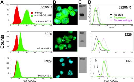
ABCG2 expression and function in myeloma cell lines. (A) Flow cytometric analysis of MM cell lines for ABCG2 membrane expression was performed. 8226MR cells express more ABCG2 than wild-type 8226 cells, and H929 cells express very little, as shown by the shift in fluorescence. (B) Immunostaining of myeloma cell lines using an anti-ABCG2 FITC (Chemicon)–labeled antibody (ABCG2 is green and DAPI is blue). (C) Western blot of protein (25 μg/lane) extracted from myeloma cell lines for ABCG2. (D) Functional analysis of ABCG2 using topotecan as a substrate and the ABCG2-specific inhibitor tryprostatin A. Topotecan, a very good ABCG2 substrate and a naturally fluorescent molecule, is effluxed in high (8226MR) and moderate (8226) ABCG2 expressers, but is accumulated by H929 cells (which do not express ABCG2). Tryprostatin A (trypA) blocks the efflux of topotecan, demonstrating that topotecan efflux is ABCG2 dependent.
ABCG2 expression and function in myeloma cell lines. (A) Flow cytometric analysis of MM cell lines for ABCG2 membrane expression was performed. 8226MR cells express more ABCG2 than wild-type 8226 cells, and H929 cells express very little, as shown by the shift in fluorescence. (B) Immunostaining of myeloma cell lines using an anti-ABCG2 FITC (Chemicon)–labeled antibody (ABCG2 is green and DAPI is blue). (C) Western blot of protein (25 μg/lane) extracted from myeloma cell lines for ABCG2. (D) Functional analysis of ABCG2 using topotecan as a substrate and the ABCG2-specific inhibitor tryprostatin A. Topotecan, a very good ABCG2 substrate and a naturally fluorescent molecule, is effluxed in high (8226MR) and moderate (8226) ABCG2 expressers, but is accumulated by H929 cells (which do not express ABCG2). Tryprostatin A (trypA) blocks the efflux of topotecan, demonstrating that topotecan efflux is ABCG2 dependent.
Patient bone marrow aspirates obtained prior to HDC, after 3 days of melphalan alone (DL 1) or after 3 days of melphalan and topotecan (DL 2-5), or at relapse from HDC were analyzed for ABCG2 mRNA expression (Tables 2 and 3). These were unused aliquots of bone marrow aspirates from MTV trial patients,37,38 for which we obtained IRB approval for ABCG2 analysis. All possible residual samples from this trial were analyzed: 42 paired samples from 31 patients (paired either before HDC and during HDC, or before HDC and relapse). Ten patients had samples from all 3 time points. The frozen samples were thawed, selected using CD138 immunomagnetic beads, and analyzed by QPCR.
ABCG2 mRNA expression in plasma cells from subjects enrolled in the MTV trial
. | . | ABCG2 mRNA expression/cell* . | . | . | . | ||
|---|---|---|---|---|---|---|---|
| Paired sample . | n . | Before HDC . | During HDC . | Relapse from HDC . | P† . | ||
| Before HDC versus during HDC | |||||||
| Dose level 1 | 6 | 29.3 | 48.2 | NA | .56 | ||
| Dose levels 2 to 5 | 20 | 118.4 | 232.5 | NA | .033 | ||
| Before HDC versus relapsed from HDC | |||||||
| Dose level 1 | 5 | 26.6 | NA | 17.6 | .999 | ||
| Dose levels 2 to 5 | 11 | 31.7 | NA | 117.1 | .001 | ||
The analyses presented are for paired samples only, that is, before + during and before + relapse.
NA indicates not applicable.
The expression of ABCG2 is normalized to that of GAPDH.
Wilcoxon signed rank test.
Patients who had received 3 days of melphalan followed by 2 days of VP-16 (Table 2; 6 patients, DL 1) had no significant change in ABCG2 expression when compared with pre-HDC plasma cells (P = .56), nor when relapse values were compared with pre-HDC plasma cells (5 patients; P = .999; Table 3). In contrast, those patients who received melphalan and topotecan (Table 2; 20 patients, DL 2-5) had a significant increase in ABCG2 expression compared with pre-HDC samples (P = .033; Table 3). In addition, patient samples from relapse in DLs 2 to 5 (Table 2) also had a statistically significant increase in ABCG2 when relapse samples were compared with pre-HDC values (11 patients; P = .001). A statistical analysis comparing ABCG2 mRNA levels and patient clinical outcome (best response to high-dose chemotherapy) failed to find any statistically significant prognostic value of mRNA levels in this limited number of samples.
ABCG2 protein expression determined by Western analysis, flow cytometry, and immunofluorescence
The protein expression of ABCG2 in human myeloma cell lines was assayed by Western blot, immunofluorescence staining, and flow cytometry, and found to correlate well with the QPCR data (Figure 1). 8226MR cells expressed very high levels of ABCG2, while parental 8226 cells and H929 cells expressed intermediate and low levels, respectively. Flow cytometric analysis of human myeloma cell lines for ABCG2 membrane expression also showed high levels of expression. 8226MR expressed more ABCG2 than wild-type 8226, and H929 expressed very little as shown by the shift in fluorescence (Figure 1A). Immunostaining of myeloma cell lines using an anti-ABCG2 Bcrp1-FITC–labeled antibody (Chemicon) demonstrated high levels of ABCG2 expression in 8226MR and 8226 cells, but not in H929 cells (Figure 1B). A Western analysis of protein extracted from myeloma cell lines for ABCG2 also followed the same pattern of protein expression (Figure 1C).
ABCG2 functional assay: topotecan efflux
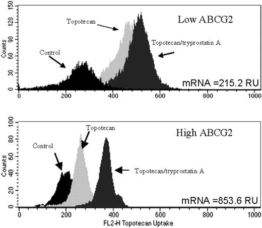
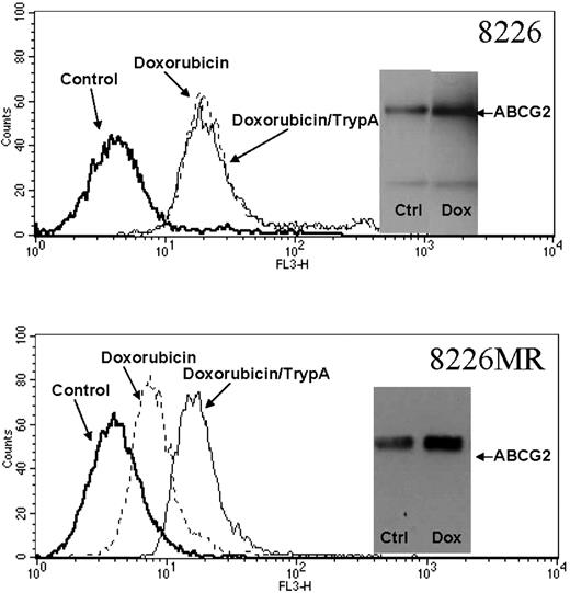
The purpose of the functional assay was to evaluate ABCG2-mediated drug efflux in myeloma cell lines. Topotecan, an exceptional ABCG2 substrate and a naturally fluorescent molecule, was effluxed in high (8226MR) and moderate (8226) ABCG2 expressers, but was not effluxed by H929 cells (low-expressing cells). To show that efflux was specific to ABCG2 and not other transporters, the inhibitor tryprostatin A was used as a control (Figure 1D). Tryprostatin A efficiently blocked the efflux of topotecan in 8226 and 8226MR cells, demonstrating that in these cell lines topotecan efflux was ABCG2 dependent (Figure 1D).18,19 ABCG2 function was expressed as the change in relative fluorescence in topotecan-treated versus untreated control cells. This analysis showed that ABCG2 protein and mRNA levels correlated well with function (Figure 1A-D). Similar results were found in patient bone marrow samples, where a high ABCG2-expressing sample was found to efflux topotecan more efficiently than a low expresser (Figure 2). In addition, doxorubicin was effluxed more efficiently by high ABCG2-expressing 8226MR than 8226 parental cells (Figure 3). ABCG2 efflux of doxorubicin was inhibited by the addition of the ABCG2 inhibitor tryprostatin A (Figure 3).
Functional assay in patient myeloma cells. Patient samples with high ABCG2 and low ABCG2 mRNA (as measured by QPCR) were assayed for ABCG2 function. The high ABCG2 expresser effluxed topotecan more efficiently than the lower expresser. Efflux was shown to be ABCG2 specific by the addition of tryprostatin A.
Functional assay in patient myeloma cells. Patient samples with high ABCG2 and low ABCG2 mRNA (as measured by QPCR) were assayed for ABCG2 function. The high ABCG2 expresser effluxed topotecan more efficiently than the lower expresser. Efflux was shown to be ABCG2 specific by the addition of tryprostatin A.
14C-mitoxantrone uptake was also used to assay ABCG2 function, as we have previously described.39 Human myeloma cells were incubated for 2 hours with 14C-mitoxantrone with and without a large molar excess of unlabeled mitoxantrone. 14C-mitoxantrone uptake corroborated the findings of topotecan uptake; high ABCG2-expressing 8226MR cells effluxed labeled drug more efficiently than low-expressing 8226 parental cells. 8226MR cells had an equilibrium cellular radioactivity of 5374 ± 65.6 cpm/mg cellular protein, while parental 8226 cells had 13 187 ± 102.9 cpm/mg cellular protein. Thus, the ABCG2-expressing 8226MR cells are able to efflux mitoxantrone more efficiently.
Effect of the microenvironment and topoisomerase inhibitors on ABCG2 expression
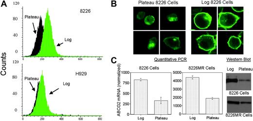
We also examined the expression of ABCG2 as a function of cell density (Figure 4) and found that log-phase (low density) 8226 and 8226MR cells express significantly more ABCG2 than plateauphase (high density) cells, as shown by flow cytometry (Figure 4A), immunofluorescence microscopy (Figure 4B), QPCR (Figure 4C), and Western blot analysis (Figure 4C). Changes in cell density failed to induce ABCG2 protein expression in H929 cells (Figure 4A).
ABCG2 expression increases in response to doxorubicin exposure. ABCG2 functional expression was assayed by flow cytometry in 8226 and 8226MR MM cells after exposure to 1 μM doxorubicin for 20 minutes. Higher ABCG2-expressing 8226MR cells were able to efflux doxorubicin more efficiently than parental 8226 cells. The ABCG2-specific inhibitor tryprostatin A decreased efflux in the 8226MR cell line but not the 8226 parental cell line, indicating that doxorubicin efflux was mediated by ABCG2. Myeloma cells treated with low-dose doxorubicin, 0.1 μM in 8226MR cells and 1.0 μM in 8226 cells, exhibit an increase in protein expression as determined by Western analysis (inset of each graph). Equal amounts of protein (25 μg) were assayed. Both 8226 and 8226MR cells demonstrated a 1.7-fold increase in ABCG2 protein after low-dose doxorubicin treatment.
ABCG2 expression increases in response to doxorubicin exposure. ABCG2 functional expression was assayed by flow cytometry in 8226 and 8226MR MM cells after exposure to 1 μM doxorubicin for 20 minutes. Higher ABCG2-expressing 8226MR cells were able to efflux doxorubicin more efficiently than parental 8226 cells. The ABCG2-specific inhibitor tryprostatin A decreased efflux in the 8226MR cell line but not the 8226 parental cell line, indicating that doxorubicin efflux was mediated by ABCG2. Myeloma cells treated with low-dose doxorubicin, 0.1 μM in 8226MR cells and 1.0 μM in 8226 cells, exhibit an increase in protein expression as determined by Western analysis (inset of each graph). Equal amounts of protein (25 μg) were assayed. Both 8226 and 8226MR cells demonstrated a 1.7-fold increase in ABCG2 protein after low-dose doxorubicin treatment.
ABCG2 expression is elevated in log-phase myeloma cells. (A) Flow cytometric data using an ABCG2 antibody (Chemicon) demonstrate that ABCG2-expressing 8226 cells have increased ABCG2 at log-phase density compared with log-phase H929 cells. (B-C) The FACScan data are confirmed by immunostaining for ABCG2 (B), and by QPCR and Western analyses (C). Densitometry analysis of the immunoblot shows a 4:1 ratio of log-plateau ABCG2 in 8226 cells, and a 2:1 ratio of log-plateau 8226MR ABCG2. Error bars represent the standard deviation for 3 separate experiments. Note, 8226 and 8226MR Western blots were exposed for different time intervals and do not reflect relative protein levels.
ABCG2 expression is elevated in log-phase myeloma cells. (A) Flow cytometric data using an ABCG2 antibody (Chemicon) demonstrate that ABCG2-expressing 8226 cells have increased ABCG2 at log-phase density compared with log-phase H929 cells. (B-C) The FACScan data are confirmed by immunostaining for ABCG2 (B), and by QPCR and Western analyses (C). Densitometry analysis of the immunoblot shows a 4:1 ratio of log-plateau ABCG2 in 8226 cells, and a 2:1 ratio of log-plateau 8226MR ABCG2. Error bars represent the standard deviation for 3 separate experiments. Note, 8226 and 8226MR Western blots were exposed for different time intervals and do not reflect relative protein levels.
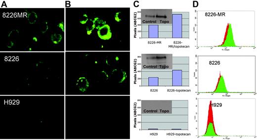
8226MR and 8226 cells were also found to increase the expression of ABCG2 in response to low-dose topotecan exposure (Figure 5). This was shown by immunofluorescence microscopy (Figure 5A-B), Western blot analysis (Figure 5C inset), and protein expression measured as pixel intensity from immunofluorescence (Figure 5C). In addition, ABCG2 protein expression was measured by flow cytometry using the membrane epitope-specific antibody Bcrp1-PE (Chemicon), and showed an increase in ABCG2 protein in log-phase 8226MR and 8226 cells after exposure to low-dose topotecan (1 μM) for 20 hours (Figure 5D). The low ABCG2-expressing myeloma cell line H929 had no ABCG2 antibody binding (Figure 5D). 8226 parental and 8226MR cells treated with low-dose topotecan exhibit an increase in membrane ABCG2 compared with the no-drug control, as shown by immunostaining with ABCG2 antibody (MXB-21) (Figure 5A-B).
8226MR and 8226 parental cell cultures treated with low-dose doxorubicin for 20 hours also demonstrated an increase in ABCG2 expression by Western blot analysis (Figure 3 insets).
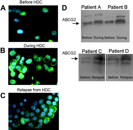
As was seen at the mRNA level (Table 2), patient bone marrow aspirates taken before, during, and after HDC with melphalan and topotecan showed changes inABCG2 expression (Figure 6). Alimited number of patient samples from the MTV trial were available for these Western and immunofluorescence analyses. The same patient sample from dose level 5 showed increased ABCG2 expression after 3 days of exposure in vivo to topotecan (Figure 6B), as well as at relapse (Figure 6C). Four different patient samples analyzed by immunoblotting (from dose levels 4 and 5 of the MTV trial) also demonstrated an increase in ABCG2 protein expression after 3 days of topotecan or at relapse from high-dose chemotherapy (Figure 6D).
ABCG2 expression increases in response to topotecan chemotherapy. (A-B) Multiple myeloma 8226MR, 8226, and H929 cells treated with low-dose topotecan (B) exhibit an increase in membrane ABCG2 over the no-drug control (A), as shown by immunostaining with ABCG2 antibody (Bcrp1 FITC). (C) Protein expression of ABCG2 was quantified as pixel intensity (from immunofluorescence microscopy), and also assessed by Western analysis (inset of each graph). (D) ABCG2 expression was measured by flow cytometry and showed an increase in log-phase 8226MR and 8226 cells after exposure to low-dose topotecan (1 μM) for 24 hours. The ABCG2-nonexpressing cell line (H929) shows no increase in ABCG2 antibody binding. Red indicates control cells in the absence of topotecan, and green represents myeloma cells exposed to topotecan.
ABCG2 expression increases in response to topotecan chemotherapy. (A-B) Multiple myeloma 8226MR, 8226, and H929 cells treated with low-dose topotecan (B) exhibit an increase in membrane ABCG2 over the no-drug control (A), as shown by immunostaining with ABCG2 antibody (Bcrp1 FITC). (C) Protein expression of ABCG2 was quantified as pixel intensity (from immunofluorescence microscopy), and also assessed by Western analysis (inset of each graph). (D) ABCG2 expression was measured by flow cytometry and showed an increase in log-phase 8226MR and 8226 cells after exposure to low-dose topotecan (1 μM) for 24 hours. The ABCG2-nonexpressing cell line (H929) shows no increase in ABCG2 antibody binding. Red indicates control cells in the absence of topotecan, and green represents myeloma cells exposed to topotecan.
ABCG2 promoter methylation
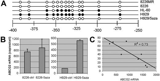
The methylation status of a previously described CpG island in the ABCG2 promoter was examined via bisulfite sequencing. Figure 7 shows the CpG dinucleotides that were methylated in 4 cell lines tested. The promoter region of ABCG2-overexpressing cells, 8226MR, was completely unmethylated, whereas the H929 cells, which express very little ABCG2, had 13 methylated CpG dinucleotide groups (Figure 7A). We found that protein, mRNA, and topotecan efflux function in live cells correlated with the methylation of CpG dinucleotides in the ABCG2 promoter (Figure 1). Therefore it is likely that ABCG2 expression is controlled in part by methylation of its promoter.
We also tried to increase ABCG2 expression by treating low-expressing H929 cells with 100 nM 5-aza-2′-deoxycytidine. This agent has been shown to increase gene expression by inhibiting DNA–methyltransferase I, thereby decreasing epigenetic methylation of DNA. After treatment of cells for 72 hours, we found that ABCG2 mRNA and protein increased approximately 6-fold in low-expressing H929 cells, whereas moderately expressing 8226 cells were unaffected (Figure 7B). Bisulfite sequencing of 5-aza-2′-deoxycytidine–treated H929 DNA showed that the ABCG2 promoter CpG island was fully demethylated (Figure 7A).
ABCG2 increases in patient plasma cells after high-dose chemotherapy and at relapse. (A-B) A patient bone marrow aspirate taken before (A) and during (B) HDC with melphalan and topotecan exhibited an increase in immunofluorescence of ABCG2 (green). (C) This same patient demonstrated an increase in ABCG2 expression by immunofluorescence at relapse as well. (D) ABCG2 protein expression before HDC and at relapse assessed by Western blot in 4 patients on the MTV study. Laser densitometry analysis of the immunoblots shows a 1.54- and 1.94-fold increase in ABCG2 during HDC for patients A and B, respectively, and a 3.68- and 1.34-fold increase in ABCG2 at relapse for patients C and D, respectively. In all cases, this is relative to the pre-HDC ABCG2 protein expression.
ABCG2 increases in patient plasma cells after high-dose chemotherapy and at relapse. (A-B) A patient bone marrow aspirate taken before (A) and during (B) HDC with melphalan and topotecan exhibited an increase in immunofluorescence of ABCG2 (green). (C) This same patient demonstrated an increase in ABCG2 expression by immunofluorescence at relapse as well. (D) ABCG2 protein expression before HDC and at relapse assessed by Western blot in 4 patients on the MTV study. Laser densitometry analysis of the immunoblots shows a 1.54- and 1.94-fold increase in ABCG2 during HDC for patients A and B, respectively, and a 3.68- and 1.34-fold increase in ABCG2 at relapse for patients C and D, respectively. In all cases, this is relative to the pre-HDC ABCG2 protein expression.
Methylation-specific quantitative PCR of patient myeloma cells
The percentage of ABCG2 promoter alleles that were methylated was assayed using SYBR green–based real-time quantitative PCR. These 8 patient samples were all obtained before HDC from the MTV trial. The data are expressed in Figure 7C and Table 4 as the percentage of alleles methylated for each patient sample compared with ABCG2 mRNA levels assayed by fluorescent probe–based real-time quantitative PCR (as described previously in “Real-time QPCR”). Figure 7C and Table 4 show that ABCG2 mRNA expression correlates inversely with the percentage of alleles methylated. High levels of methylated alleles resulted in a decrease in ABCG2 mRNA transcripts, whereas patient samples with low levels of methylated alleles had much higher amounts of ABCG2 mRNA. These data, along with cell culture methylation data, indicate that promoter methylation may contribute to the control of ABCG2 expression in human myeloma cells.
ABCG2 promoter methylation in plasma cells from subjects enrolled in the MTV trial
. | Patient† . | . | . | . | . | . | . | . | |||||||
|---|---|---|---|---|---|---|---|---|---|---|---|---|---|---|---|
. | A . | B . | C . | D . | E . | F . | G . | H . | |||||||
| ABCG2 mRNA expression* | 882.6 | 257.6 | 384.5 | 1439.7 | 692.1 | 1321.8 | 1597.3 | 1650.7 | |||||||
| Methylated alleles, % | 71.6 | 65.7 | 69.5 | 40.7 | 39.8 | 28.8 | 13.3 | 9.6 | |||||||
The expression of ABCG2 is normalized to that of GAPDH.
A to H represent 8 different patient samples.
Discussion
We have found that ABCG2 is present and functional in human myeloma cell lines and patient plasma cells. Using QPCR, protein assays, and functional efflux of chemotherapeutic drugs, we have found that ABCG2 is potentially involved in drug resistance to specific agents in human myeloma cells.
The principal findings of our in vitro experiments are that (1) human multiple myeloma cell lines have a wide range of ABCG2 expression and function, (2) low-density 8226 cells express more ABCG2 than high-density cells, (3) human myeloma cell lines with moderate or high baseline levels of ABCG2 expression further increase this expression upon exposure to the ABCG2 substrates topotecan and doxorubicin, and (4) methylation of the CpG island of the ABCG2 promoter correlates inversely with ABCG2 expression. In vivo, we found that myeloma patients treated with topotecan had an increase in ABCG2 mRNA expression, both after 3 days of topotecan exposure and at the time of relapse. In addition, we found that a CpG island in the ABCG2 promoter is heavily methylated in cells that do not express ABCG2. This promoter region was completely demethylated in cells that expressed high to intermediate levels of ABCG2. Therefore, expression of ABCG2 was regulated, at least in part, by promoter methylation, both in cell lines and in patient plasma cells.
The ability to up-regulate ABCG2 in response to chemotherapy could confer a selective survival advantage to malignant plasma cells. Plasma cells are derived from hematologic stem cells that have demonstrated an intrinsic ability to produce ABCG2, and therefore, myeloma cells may come by this ability naturally.40-42 However, clonal selection may play a part in the further development of ABCG2 expression in myeloma. Rapidly growing log-phase myeloma cells also increased ABCG2 expression in vitro. Increased drug resistance conferred to rapidly growing cells could possibly produce an additive result with the unlimited replicative potential of cancer cells, one of the “hallmarks of cancer.”43 ABCG2 may contribute to intrinsic drug resistance in myeloma, and its effect is likely increased by exposure to chemotherapeutic drugs that are substrates for ABCG2.
In general, we observed the same results in the limited number of patient plasma cells available from the HDC MTV trial. Patient cells exposed to topotecan in vivo had increased ABCG2 expression, as did plasma cells from relapse bone marrow aspirates. In addition, patient plasma cells with increased CpG island methylation of the ABCG2 promoter had decreased ABCG2 mRNA expression. In studies performed in another hematologic malignancy, AML, patients demonstrated significantly increased expression of ABCG2 mRNA in the relapsed/refractory samples compared with before treatment.11 AML patients with high levels of ABCG2 expression had significantly shorter overall survival rates,12 and decreased ABCG2 expression predicted a complete remission of AML in adult patients.13
In a recent study, Raaijmakers et al44 isolated plasma cells from healthy bone marrow donors and from 10 patients with myeloma prior to treatment with VAD chemotherapy using flow cytometry and an anti-CD38 antibody. The authors observed that ABCG2 expression was relatively high in both normal and malignant plasma cells, but that ABCG2-mediated efflux of mitoxantrone was significantly impaired in the malignant plasma cells. These results differ from ours, however; our functional assay was limited to only a high and a low expresser of ABCG2 (Figure 2) and was not compared with drug efflux in normal plasma cells, and the plasma cells were isolated using an anti-CD138 antibody and immunomagnetic beads from patients previously treated with chemotherapy. Thus, the 2 studies may be comparing different populations of plasma cells.
ABCG2 promoter methylation. (A) Cells were harvested at plateau phase, and the DNA was extracted and assayed by bisulfite DNA sequencing analysis. The figure shows the methylation status of the putative CpG island of the ABCG2 promoter in 4 cell lines. Filled circles represent methylated groups and the open circles, demethylated CpG. (B) H929/5aza are cells treated with 100 nM 5-aza-2′-deoxycytidine for 72 hours and the ABCG2 promoter was assayed by bisulfite sequencing. 5-Aza-2′-deoxycytidine was able to augment ABCG2 transcription in low ABCG2-expressing H929 cells but had no effect in moderate-expressing 8226 cells. (C) CD138-selected MM patient samples were assayed for ABCG2 promoter methylation after bisulfite conversion using real-time quantitative PCR. The percentage of alleles that was methylated inversely correlated with ABCG2 mRNA expression. Error bars represent the standard deviation for 3 separate experiments.
ABCG2 promoter methylation. (A) Cells were harvested at plateau phase, and the DNA was extracted and assayed by bisulfite DNA sequencing analysis. The figure shows the methylation status of the putative CpG island of the ABCG2 promoter in 4 cell lines. Filled circles represent methylated groups and the open circles, demethylated CpG. (B) H929/5aza are cells treated with 100 nM 5-aza-2′-deoxycytidine for 72 hours and the ABCG2 promoter was assayed by bisulfite sequencing. 5-Aza-2′-deoxycytidine was able to augment ABCG2 transcription in low ABCG2-expressing H929 cells but had no effect in moderate-expressing 8226 cells. (C) CD138-selected MM patient samples were assayed for ABCG2 promoter methylation after bisulfite conversion using real-time quantitative PCR. The percentage of alleles that was methylated inversely correlated with ABCG2 mRNA expression. Error bars represent the standard deviation for 3 separate experiments.
In previous studies, ABCG2 overexpression has been observed in drug-resistant cell lines.6,7,14,15,36,45-47 This overexpression of ABCG2, in the majority of cases, has been attributed to heavy amplification of the gene locus.36,45,47 Also, significant increases in function have been found to occur due to specific mutations, but without a concurrent increase in gene transcription or translation.46-49 Analysis of a putative ABCG2 promoter region presents a TATA-less promoter with several putative transcription factor–binding sites. In addition, the promoter has an estrogen response element, all of which may contribute to increased gene expression levels.23,24 It has been reported that the ABCG2 promoter contains a potential CpG island, which may regulate expression by methylation.24 The MDR1 promoter has a similar CpG island. In a recent publication, it was found that hypermethylation of CpG dinucleotides in the MDR1 promoter region strongly contributed to differences in gene expression in related cell lines.26 In our study, we examined the methylation status of the ABCG2 promoter region in cell lines that differed in their respective ABCG2 expression. We found that the promoter of very low level–expressing cells was almost completely methylated, whereas high and medium ABCG2 expressers were either completely or almost completely unmethylated. Analysis of the ABCG2 promoter via bisulfite sequencing showed that methylation occurred precisely at the putative CpG island as described by Bailey-Dell et al.24 In addition, when low ABCG2-producing H929 cells were exposed to the demethylating agent, 5-aza-2′-deoxycytidine, the cells were induced to express ABCG2 mRNA and protein (Figure 7B). Methylation was also shown to be important in human myeloma patient samples. The percentage of methylated alleles inversely correlated with ABCG2 mRNA expression (Figure 7C and Table 4). Therefore, our data suggest that promoter methylation contributes to gene expression of ABCG2.
In summary, our data suggest that ABCG2 may be involved in the resistance of human myeloma cells, both in vitro and in vivo, to chemotherapeutic agents that are substrates of ABCG2. Doxorubicin, VP-16, and topotecan are all substrates of ABCG2. Doxorubicin is commonly used in the treatment of myeloma (vincristine + adriamycin + decadron, or VAD regimen). In this study, we found that doxorubicin is actively effluxed by ABCG2 in vitro. Single-agent topotecan has been shown to have activity in relapsed and refractory multiple myeloma patients in a SWOG trial.50 The overall response rate in these highly pretreated patients was 16%. We have recently combined melphalan and VP-16 phosphate with dose-escalated topotecan in a phase 1/2 high-dose chemotherapy trial in high-risk myeloma37,38 and found this to be an active and tolerable regimen. Future trials that incorporate ABCG2 transport inhibitors, such as GF120918, may increase the efficacy of topoisomerase I inhibitors in this disease.20,51,52
Authorship
J.G.T. designed research, performed research, analyzed data, and wrote the paper; J.L.G. and D.M. performed research; C.Z., J.M.C., L.H., and W.S.D. contributed vital new reagents or analytical tools; M.J.S. and P.M. analyzed data; and D.M.S. designed research, analyzed data, and wrote the paper.
The authors declare no competing financial interests.
Prepublished online as Blood First Edition Paper, August 17, 2006; DOI 10.1182/blood-2005-10-009084.
The publication costs of this article were defrayed in part by page charge payment. Therefore, and solely to indicate this fact, this article is hereby marked “advertisement” in accordance with 18 USC section 1734.
This work was supported in part by National Institutes of Health (NIH) grant CA82533 and by the Biostatistics Core, Flow Cytometry Core, Microscopy Core, Imaging Core, and Molecular Biology Core Facilities at the H. Lee Moffitt Cancer Center and Research Institute.

This feature is available to Subscribers Only
Sign In or Create an Account Close Modal