Abstract
Lymphomagenesis in Eμ-Myc mice is opposed by the Arf tumor suppressor, whose inactivation compromises p53 function and accelerates disease. Finding nascent Eμ-Myc–induced tumors in which p19Arf causes cell-cycle arrest or apoptosis is problematic, since such cells will be eliminated until Arf or p53 function is lost. Knock-in mice expressing a green fluorescent protein (GFP) in lieu of Arf coding sequences allow analysis of Arfpromoter regulation uncoupled from p19Arf action. Prior to frank lymphoma development, unexpectedly low levels of Eμ-Myc–induced p19Arf or GFP were expressed. However, as lymphomas arose in Arf+/GFP heterozygotes, additional oncogenic events synergized with Eμ-Myc to further induce the functionally null Arf-Gfp allele. Concomitant up-regulation of p19Arf was not observed; instead, the wild-type allele was inactivated. We infer that very low levels of Arf are tumor suppressive, and that further induction provides the selective pressure for the emergence of tumors that have inactivated the gene.
Introduction
The CDKN2A (INK4A/ARF) locus, which encodes 2 distinct tumor suppressors (p16INK4a and p14ARF) from partially overlapping reading frames, is commonly inactivated in human cancers.1,2 Mice homozygous or heterozygous for an Arf-null allele are predisposed to cancer, and tumors arising in Arf+/− mice inactivate the wild-type Arf allele, indicating that Arf behaves as a classical “2-hit” tumor suppressor gene. Arf is induced by abnormally sustained, increased thresholds of mitogenic signals emanating from mutationally activated or dysregulated oncogenes. In turn, the Arf protein (p19Arf in mice) antagonizes the activity of the p53-negative regulator Mdm2 to induce a p53 transcriptional response that leads to cell-cycle arrest or apoptosis.2,3
Consequences of Arf inactivation have been widely studied in Eμ-Myc transgenic mice4-6 in which Myc, regulated by the immunoglobulin heavy chain enhancer-promoter, is expressed in B lymphocytes.7-9 Eμ-Myc mice exhibit an initial preneoplastic phase characterized by polyclonal expansion of pre-B cells already evident before birth.8 Within their first year of life, these mice develop malignant monoclonal lymphomas (mean latency of 25 weeks). Biallelic loss of Arf occurs in approximately 25% of these animals, whereas another 33% display p53 mutations.4 Arf+/− Eμ-Myc mice more rapidly develop tumors (mean latency, 7-10 weeks depending on strain background) in which the wild-type Arf allele is commonly inactivated, thus canceling selective pressure for p53 mutations. Arf−/− Eμ-Myc mice are even more prone to lymphoma development and die of aggressive lympholeukemias by only 4 to 7 weeks of age. Although the evidence that Arf acts as a tumor suppressor gene in this model is unequivocal, no longitudinal study of Arf regulation has yet been undertaken. Aided by use of a high-affinity monoclonal antibody to p19Arf and a knock-in Arf-Gfp reporter mouse,10,11 we have now evaluated the response of Arf throughout the course of lymphoma development in Eμ-Myc mice.
Materials and methods
Work with mice has been approved by the St Jude Children's Research Hospital Animal Care and Use Committee under Animal Protocol no. 225 (function of INK4a/ARF in pediatric neoplasia). A purified monoclonal antibody (5-C3-1) directed to mouse p19Arf, which is 25-fold more sensitive than previously derived reagents,10 was conjugated to AlexaFluor-647 and used to score Arf expression in fixed B lymphoid cells by fluorescence-activated flow cytometry (FC). All other procedures for analysis of B cells from Eμ-Myc mice of various Arf genotypes have been described in detail.4,11,12
Results and discussion
Spleen and bone marrow cells were harvested from groups of overtly healthy Eμ-Myc and nontransgenic control mice of various ages, and B lineage cells, isolated using magnetic beads coated with the B220 antibody to CD45R, were lysed and immunoblotted using the monoclonal antibody (mAb) 5-C3-1 to p19Arf. Low levels of p19Arf were detected in B220+ cells from Eμ-Myc (M) but not control (Con) mice of 2 weeks of age and did not increase thereafter as long as disease was latent (Figure 1A). The p53 protein and p21Cip1, a p53-responsive gene product, were also weakly induced, compared with more robust p53 induction in Arf-null B cells exposed to ionizing radiation (lane 2 vs lane 1). The levels of p19Arf expressed were about 20-fold less than those observed in p53-null lymphomas, in which Arf is induced to high levels but is without effect,4 and at least 200-fold less than those in cultured, immortalized p53-null B cells (Figure 1A, lane 3; Figure 1B). Dual-color FC analysis performed on gated B220+ cells using the mAb 5-C3-1 conjugated to AlexaFluor-647 revealed that the entire population of prelymphomatous B cells exhibited modestly increased fluorescence (Figure 1C). Given that Arf inactivation greatly accelerates lymphomagenesis,4-6,11 these very low levels of p19Arf and p53 expressed throughout the B220+ population must contribute to tumor suppression. However, protection is by no means absolute because the proliferative fraction of B cells in the spleen and bone marrow is significantly increased in the presence of Eμ-Myc,8,12 and all such animals ultimately develop lymphomas.
Arf induction in prelymphomatous Eμ-Myc mice. (A) B220+ cells were purified from the bone marrow (BM) and spleens (Sp) of nontransgenic control C57BL/6 (Con) mice or from syngeneic animals expressing the Eμ-Myc (M) transgene. Cells were harvested from healthy mice at biweekly (w) intervals after birth. Cell lysates (25 μg protein per lane from B220+ cells purified from BM or Sp) were separated on denaturing gels, and proteins were immunoblotted with antibodies directed to p19Arf (5-C3-1),10 c-Myc (N-262; Santa Cruz Biotechnology, Santa Cruz, CA), p53 (NCL-p53-505; Novocastra, Newcastle upon Tyne, United Kingdom), p21 (F5; Santa Cruz), or beta-actin (A-5441; Sigma, St Louis, MO), as indicated at the left of the panels. Cultured Arf-null B cells were untreated (lane 1) or irradiated with 5 Gy 2 hours prior to lysis (lane 2) to induce higher levels of p53. Cultured p53-null B cells express very high levels of p19Arf (lane 3). (B) Lysates (in micrograms of total protein as indicated at the bottom of the panel) of B220+ BM cells and splenocytes (Sp) purified from control (Con) or Eμ-Myc (M) transgenic mice were immunoblotted with antibodies to p19Arf. The signals were compared with those generated with various quantities of lysate protein obtained from a representative p53-null lymphoma and from cultured p53-null B cells. (C) Cultured B cells from p53-null mice were stained with an isotype-matched control mAb (panel Ci, negative control) or with mAb 5-C3-1 to p19Arf (panel Cii; p53-null cells as positive control) conjugated to AlexaFluor-647. Arf-null cells served as an additional negative control for mAb 5-C3-1 staining (Ciii). BM and Sp cells from control [Myc (−)] or transgenic [Myc (+)] mice of 2 weeks of age were assayed by dual-color FC using antibodies to B220 and to p19Arf (panels Civ-Cix). Gated B220+ cells in BM (Civ-Cvi) and Sp cells (Cvii-Cix) were analyzed for reactivity with AlexaFluor-647–conjugated mAb 5-C3-1 directed to p19Arf. Vertical dashed lines indicate fluorescence intensities in negative control panels (Ci, Civ, Cviii).
Arf induction in prelymphomatous Eμ-Myc mice. (A) B220+ cells were purified from the bone marrow (BM) and spleens (Sp) of nontransgenic control C57BL/6 (Con) mice or from syngeneic animals expressing the Eμ-Myc (M) transgene. Cells were harvested from healthy mice at biweekly (w) intervals after birth. Cell lysates (25 μg protein per lane from B220+ cells purified from BM or Sp) were separated on denaturing gels, and proteins were immunoblotted with antibodies directed to p19Arf (5-C3-1),10 c-Myc (N-262; Santa Cruz Biotechnology, Santa Cruz, CA), p53 (NCL-p53-505; Novocastra, Newcastle upon Tyne, United Kingdom), p21 (F5; Santa Cruz), or beta-actin (A-5441; Sigma, St Louis, MO), as indicated at the left of the panels. Cultured Arf-null B cells were untreated (lane 1) or irradiated with 5 Gy 2 hours prior to lysis (lane 2) to induce higher levels of p53. Cultured p53-null B cells express very high levels of p19Arf (lane 3). (B) Lysates (in micrograms of total protein as indicated at the bottom of the panel) of B220+ BM cells and splenocytes (Sp) purified from control (Con) or Eμ-Myc (M) transgenic mice were immunoblotted with antibodies to p19Arf. The signals were compared with those generated with various quantities of lysate protein obtained from a representative p53-null lymphoma and from cultured p53-null B cells. (C) Cultured B cells from p53-null mice were stained with an isotype-matched control mAb (panel Ci, negative control) or with mAb 5-C3-1 to p19Arf (panel Cii; p53-null cells as positive control) conjugated to AlexaFluor-647. Arf-null cells served as an additional negative control for mAb 5-C3-1 staining (Ciii). BM and Sp cells from control [Myc (−)] or transgenic [Myc (+)] mice of 2 weeks of age were assayed by dual-color FC using antibodies to B220 and to p19Arf (panels Civ-Cix). Gated B220+ cells in BM (Civ-Cvi) and Sp cells (Cvii-Cix) were analyzed for reactivity with AlexaFluor-647–conjugated mAb 5-C3-1 directed to p19Arf. Vertical dashed lines indicate fluorescence intensities in negative control panels (Ci, Civ, Cviii).
We next performed longitudinal studies with Arf+/GFP heterozygotes in which one Arf allele has been replaced by a cDNA cassette encoding green fluorescent protein (GFP) under the control of the Arf promoter.11 The Arf-Gfp allele behaves as a null and accelerates Eμ-Myc–induced lymphomagenesis. Tumors arising in Arf+/GFP mice inactivate the wild-type Arf allele, retain functional p53, and are brightly green fluorescent.
In Eμ-Myc mice, lymphoblastic lymphoma is accompanied by lympholeukemia, so an increase in the peripheral blood white cell count precedes the appearance of clinically palpable tumors. Animals were considered to be in an intermediate phase of disease when their white blood cell counts, monitored weekly, rose to 30 × 109/L (30 000/μL); moribund mice with frank tumor masses were classified as terminal. Expression of GFP and p19Arf in lymphoid organs was monitored throughout.
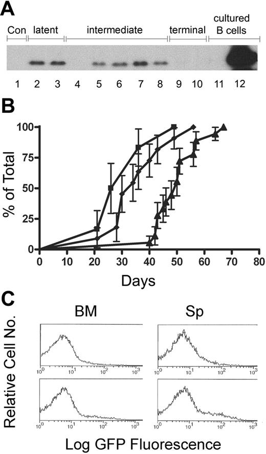
During the prelymphomatous latent phase, we detected low levels of p19Arf in splenocytes from Eμ-Myc Arf+/GFP mice, but not in nontransgenic control animals (Figure 2A, lanes 1-3). At this time, GFP expression could not be detected by FC or immunoblotting in these same tissues, most likely due to the relatively reduced sensitivities of these assays. However, GFP-positive cells appeared in peripheral blood and accumulated as total white cell counts rose (Figure 2B), implying that at least one additional oncogenic event synergizes with Eμ-Myc to further activate the Arf promoter. In direct contrast to the Arf-Gfp allele, expression of p19Arf protein encoded by the wild-type Arf allele was reduced (Figure 2A, lanes 5, 6, and 8) or completely absent (Figure 2A, lane 4) in splenocytes from Arf+/GFP mice in the intermediate phase of disease and was almost invariably undetectable in 22 mice during their terminal phase (examples in Figure 2A, lanes 9 and 10). Thus, while further Arf induction might transiently enhance tumor suppression during the intermediate phase (as can only be inferred from the behavior of the Arf-Gfp allele), this process must also provide a concomitant selective pressure favoring the outgrowth of lymphoma cells that no longer express the wild-type Arf allele.
Analysis of p19Arf and Arf-Gfp reporter expression in Eμ-Myc transgenic mice. (A) Levels of p19Arf were determined in splenocytes from control (lane 1) and Eμ-Myc transgenic animals at the designated stages of disease (lanes 2-10). Cultured Arf-null (lane 11) and p53-null B cells (lane 12) served as controls. Whereas splenocytes from mice in the latent phase do not express GFP, significant proportions of splenocytes (more than 20%) expressed the Arf-Gfp allele at later stages of disease. The overall levels of p19Arf were diminished or absent in 4 of 5 intermediate-phase mice (lanes 4-8) and almost invariably absent in terminal-phase mice (2 examples of 22 mice are shown in lanes 9 and 10). (B) High-level expression of the Arf-Gfp allele was initially detected by FC analysis in a subset of peripheral white blood cells (▪) just prior to the development of lympholeukemia, as manifested by white blood cell counts of more than 30 × 109/L (30 000/μL) (♦). Animals that became moribund were killed approximately 2 weeks later (▴; terminal phase). Error bars indicate SD from the mean. (C) B220+ BM and Sp cells taken during the intermediate phase of disease from Eμ-Myc ArfGFP/GFP mice initially generated a subpopulation of cells that express high levels of the Arf-Gfp allele.
Analysis of p19Arf and Arf-Gfp reporter expression in Eμ-Myc transgenic mice. (A) Levels of p19Arf were determined in splenocytes from control (lane 1) and Eμ-Myc transgenic animals at the designated stages of disease (lanes 2-10). Cultured Arf-null (lane 11) and p53-null B cells (lane 12) served as controls. Whereas splenocytes from mice in the latent phase do not express GFP, significant proportions of splenocytes (more than 20%) expressed the Arf-Gfp allele at later stages of disease. The overall levels of p19Arf were diminished or absent in 4 of 5 intermediate-phase mice (lanes 4-8) and almost invariably absent in terminal-phase mice (2 examples of 22 mice are shown in lanes 9 and 10). (B) High-level expression of the Arf-Gfp allele was initially detected by FC analysis in a subset of peripheral white blood cells (▪) just prior to the development of lympholeukemia, as manifested by white blood cell counts of more than 30 × 109/L (30 000/μL) (♦). Animals that became moribund were killed approximately 2 weeks later (▴; terminal phase). Error bars indicate SD from the mean. (C) B220+ BM and Sp cells taken during the intermediate phase of disease from Eμ-Myc ArfGFP/GFP mice initially generated a subpopulation of cells that express high levels of the Arf-Gfp allele.
In further support of the idea that Arf inactivation is itself insufficient to trigger lymphomagenesis in Eμ-Myc transgenic mice, GFP was not detectably expressed in the lymphoid tissues of 2-week-old Eμ-Myc ArfGFP/GFP homozygous mice that are even more highly tumor-prone. When GFP-positive cells later appeared in their peripheral blood, FC analysis of gated B220+ spleen and bone marrow cells revealed the emergence of subpopulations exhibiting high GFP fluorescence, as would result from an expanding clonal malignancy (Figure 2C). Tumor masses and invasive malignant cells at terminal phase were uniformly and brightly fluorescent.11 In short, cryptic mutations other than Arf loss accompany the conversion of premalignant Eμ-Myc B cells to tumor cells; this precedes robust Arf induction, but also precipitates Arf inactivation.
The nature of these additional mutations remains unknown. Various genetic alterations that attenuate apoptosis have been documented in cultured tumor cells and in bone marrow recovered from Eμ-Myc mice,13-17 but this process is difficult to detect in vivo, probably because dying B cells are very rapidly eliminated.18 Activating mutations in Myc itself16 or those affecting other collaborating oncogenes15,17 could well exert similar inductive effects on the Arf promoter.
Together, these findings argue that seemingly negligible levels of p19Arf or p53 observed in incipient tumor cells sustaining initiating oncogenic mutations exert significant tumor-suppressive effects. This may be particularly important in the analysis of human tumor material, where antibodies capable of detecting equally low levels of p14ARF are not yet available and where ARF may therefore not be inferred to play any tumor suppressive role.
The publication costs of this article were defrayed in part by page charge payment. Therefore, and solely to indicate this fact, this article is hereby marked “advertisement” in accordance with 18 USC section 1734.
Conflict-of-interest statement: The authors declare no competing financial interests.
Contributions: D.B. performed the experiments. D.B. and C.J.S. designed the research, analyzed the data, and wrote the paper.
Acknowledgments
We thank the members of the Sherr-Roussel laboratory for advice and criticism and Richard A. Ashmun for assistance with FC analysis.
This work was supported by Howard Hughes Medical Institute, Cancer Center Core Grant CA-21765, and by the American Lebanese Syrian Associated Charities of St Jude Children's Research Hospital. C.J.S. is an Investigator of the Howard Hughes Medical Institute.

![Figure 1. Arf induction in prelymphomatous Eμ-Myc mice. (A) B220+ cells were purified from the bone marrow (BM) and spleens (Sp) of nontransgenic control C57BL/6 (Con) mice or from syngeneic animals expressing the Eμ-Myc (M) transgene. Cells were harvested from healthy mice at biweekly (w) intervals after birth. Cell lysates (25 μg protein per lane from B220+ cells purified from BM or Sp) were separated on denaturing gels, and proteins were immunoblotted with antibodies directed to p19Arf (5-C3-1),10 c-Myc (N-262; Santa Cruz Biotechnology, Santa Cruz, CA), p53 (NCL-p53-505; Novocastra, Newcastle upon Tyne, United Kingdom), p21 (F5; Santa Cruz), or beta-actin (A-5441; Sigma, St Louis, MO), as indicated at the left of the panels. Cultured Arf-null B cells were untreated (lane 1) or irradiated with 5 Gy 2 hours prior to lysis (lane 2) to induce higher levels of p53. Cultured p53-null B cells express very high levels of p19Arf (lane 3). (B) Lysates (in micrograms of total protein as indicated at the bottom of the panel) of B220+ BM cells and splenocytes (Sp) purified from control (Con) or Eμ-Myc (M) transgenic mice were immunoblotted with antibodies to p19Arf. The signals were compared with those generated with various quantities of lysate protein obtained from a representative p53-null lymphoma and from cultured p53-null B cells. (C) Cultured B cells from p53-null mice were stained with an isotype-matched control mAb (panel Ci, negative control) or with mAb 5-C3-1 to p19Arf (panel Cii; p53-null cells as positive control) conjugated to AlexaFluor-647. Arf-null cells served as an additional negative control for mAb 5-C3-1 staining (Ciii). BM and Sp cells from control [Myc (−)] or transgenic [Myc (+)] mice of 2 weeks of age were assayed by dual-color FC using antibodies to B220 and to p19Arf (panels Civ-Cix). Gated B220+ cells in BM (Civ-Cvi) and Sp cells (Cvii-Cix) were analyzed for reactivity with AlexaFluor-647–conjugated mAb 5-C3-1 directed to p19Arf. Vertical dashed lines indicate fluorescence intensities in negative control panels (Ci, Civ, Cviii).](https://ash.silverchair-cdn.com/ash/content_public/journal/blood/109/2/10.1182_blood-2006-07-033985/4/m_zh80020706700001.jpeg?Expires=1767763129&Signature=qNOM5Aqr33kEDuSBYoNQZ64J5GbBKw0VjYgKiKnKunq8jzkVFbiM9ji7CN~UEWDlTBhNBTGmOjMLuTMmeHweEGe0NIy2Sswr5DRuqwlAX20IVptO3sTpqC8fa1e7DxbqvDFeJp13PeehD4u5n5GJF7tNvkCqvRBGr1ZgGflb3DYIK-XCgch80DG~V9dxkZalC4zpAxMwx8jWUF6HgdQOBAh6coYkjvQ0UhLurqRxe7DVbcd9jTjJpe2WfnGq1n1R6oE2rKl8HtGzD48B7SZojcSHXSTfCsf2Kgbfjz4mbs25PMrTj9Nlae4d8dGdsKyRGOCNE2OPNOnSymQOs5~9dg__&Key-Pair-Id=APKAIE5G5CRDK6RD3PGA)

This feature is available to Subscribers Only
Sign In or Create an Account Close Modal