Abstract
Hepatocyte growth factor (HGF) promotes cell growth and motility and also increases neovascularization. Multiple myeloma (MM) cells produce HGF, and the plasma concentration of HGF is significantly elevated in patients with clinically active MM, suggesting that HGF might play a role in the pathogenesis of MM. NK4, an antagonist of HGF, is structurally homologous to angiostatin, and our previous report showed that NK4 inhibited the proliferation of vascular endothelial cells induced by HGF stimulation. The purposes of this study were to elucidate the contribution of HGF to the growth of MM cells as well as to investigate the possibility of the therapeutic use of NK4. In vitro study showed that NK4 protein stabilized the growth of MM cell lines and regulated the activation of c-MET, ERK1/2, STAT3, and AKT-1. Recombinant adenovirus containing NK4 cDNA (AdCMV.NK4) was injected intramuscularly into lcr/scid mice bearing tumors derived from HGF-producing MM cells. AdCMV.NK4 significantly inhibited the growth of these tumors in vivo. Histologic examination revealed that AdCMV.NK4 induced apoptosis of MM cells, accompanied by a reduction in neovascularization in the tumors. Thus, NK4 inhibited the growth of MM cells via antiangiogenic as well as direct antitumor mechanisms. The molecular targeting of HGF by NK4 could be applied as a novel therapeutic approach to MM.
Introduction
Multiple myeloma (MM) is a hematologic malignancy that occurs in terminally differentiated B cells or plasma cells. Although several chemotherapeutic regimens and high-dose therapy combined with autologous hematopoietic stem cell transplantation have improved the survival of patients with MM, this disease still remains incurable.1 At present, most patients with MM become resistant to therapy, and the disease is ultimately fatal. Thus, new potential therapies should be extensively explored. Recent studies have shown that bone marrow microvessel density and the plasma concentrations of angiogenic growth factors, including hepatocyte growth factor (HGF), fibroblast growth factor 2 (FGF-2), and vascular endothelial growth factor (VEGF), are significantly elevated in patients with clinically active MM.2-5 These angiogenic growth factors also modulate the interaction of MM cells with bone marrow stromal cells via both paracrine and autocrine mechanisms. The results obtained thus far have suggested that angiogenic growth factors provide MM cells with a growth advantage and could serve as novel therapeutic targets, with the expectation of achieving direct antitumor as well as antienvironmental effects
HGF is a multifunctional protein that promotes tumor cell growth, activates osteoblasts function, and contributes to tumor angiogenesis via its receptor, c-MET. c-MET is widely expressed in epithelial cells, in solid tumors, and in MM cells.6 Recent observations have demonstrated that MM cells occasionally produce HGF, and plasma concentrations of HGF have been shown to be significantly elevated in approximately 30% of patients with clinically active MM.7,8 In some patients, the plasma HGF level has been shown to dramatically decrease in response to therapy.9 Such results have suggested that HGF plays a critical role in the progression of MM. However, previous reports have only provided circumstantial evidence of this hypothesis, and to date, only a limited amount of information has been reported demonstrating that HGF induces MM cell growth.10
Recently, the HGF antagonist, NK4, was developed; it encodes the NH2-terminal hairpin and 4 kringle domains of the HGFα subunit.11,12 NK4 competitively antagonizes HGF to bind to its receptor, c-MET, and abrogates HGF-induced tyrosine phosphorylation of c-MET. NK4 does not inhibit the binding of other growth factors such as the FGFs, VEGF, and epidermal growth factor (EGF) to their receptors, and NK4 is an HGF-specific antagonist. It has been reported that NK4 inhibits invasion and metastasis by various cancer cells including colon cancer, pancreatic cancer, gallbladder cancer, lung cancer, prostate cancer, and glioma cells, to name a few.13 Furthermore, NK4 is structurally homologous to other kringle-containing proteins such as angiostatin, which is a fragment of plasminogen, a potent angiogenic inhibitor.14
It was also found that NK4 abrogates endothelial cell growth induced by stimulation with angiogenic growth factors including HGF.15,16 Thus, NK4 is expected to serve as a novel antineoplastic modality via an antiangiogenic mechanism. These multifunctional properties of NK4 with respect to antitumor cells and antitumor angiogenesis are suggestive of the therapeutic potential of NK4 for the treatment of MM.
In this report, we describe the growth inhibition of HGF-producing MM cells by NK4 in vitro and in vivo. Namely, recombinant NK4 stabilized the proliferation of MM cells in vitro and inhibited intracellular signaling. When an adenoviral vector containing cDNA for NK4 was injected in vivo into lcr/scid mice bearing HGF-producing MM cells, tumor growth was significantly delayed. Thus, HGF is likely a target molecule for the control of MM cell growth, and NK4 could potentially be used as a novel approach to the treatment of MM.
Patients, materials, and methods
Cell cultures
Human MM cell lines KMS11, 18, 20, 21, 26, 27, 28, and 34 were established by T. Otsuki from Japanese patients.17 These cells were cultured in RPMI containing 10% fetal calf serum (FCS; Hyclone Laboratories, Logan, UT), unless otherwise noted. KMM 1 and U266 cells were obtained from the Japanese Cancer Research Resources Bank (JCRB, Tokyo, Japan) and the American Type Culture Collection (Manassas, VA), respectively. These cells were also cultured in RPMI containing 10% FCS (Hyclone Laboratories), unless otherwise noted.
Patients
Bone marrow mononuclear cells were isolated from patients with MM by Lymphoprep density-gradient centrifugation (Axis-Shield, Oslo, Norway). If the percentage of plasma cells in bone marrow was less than 70%, mononuclear cells were then incubated with anti–human CD138 monoclonal antibody (BD Biosciences, San Jose, CA), and CD138+ cells were selected using a magnetic particle separation system (Dynal, Oslo, Norway) according to the manufacturer's recommendations. Prior to sampling, patients gave their written informed consent in accordance with the Declaration of Helsinki, which was approved by the ethics committee of Keio University School of Medicine (nos. 99-7 and 15-21).
Growth factor concentration in the culture medium
Cells (5 × 10⁵/mL) were plated in 24-well plates in triplicate and were cultured in RPMI containing 10% dialyzed FCS (Gibco BRL, Gaithersburg, MD) for 24 hours. The supernatants were centrifuged at 21 000g for 10 minutes to remove the cell debris, and the concentration of HGF was measured by a commercial enzyme-linked immunosorbent assay (ELISA) kit as indicated by the manufacturer (R&D Systems, Minneapolis, MN).
RT-PCR
To demonstrate the expression of HGF and c-MET transcripts, the total RNA was extracted from the cells using an Isogen kit (Nippon-gene, Toyama, Japan) followed by polymerase chain reaction (PCR; Takara Shuzo, Kyoto, Japan), as indicated by the manufacturers. Reverse transcription (RT) from 1 μg total RNA to cDNA was performed using Superscript II RT (Gibco BRL). The sense and antisense primers for HGF were TCC, CCA, TCG, CCA, TCC, CC and CAC, CAT, GGC, CTC, GGC, TGG, respectively; The sense and antisense primers for c-MET were TGG, GAA, TCT, GCC, TGC, GAA and CCA, GAG, GAC, GAC, GCC, AAA, respectively. Thirty-five cycles of PCR for HGF were carried out at 94°C for 1 minute, 68°C for 2 minutes, and 72°C for 30 seconds. Thirty-five cycles for c-MET were carried out at 94°C for 1 minute, 58°C for 2 minutes, and 72°C for 30 seconds. The expected sizes of the PCR products were 749 bp for HGF and 395 bp for c-MET.
In vitro cell growth
Cells (2 × 10⁵/mL) were cultured in RPMI containing 10% FCS (Hyclone Laboratories). The number of cells was adjusted to 2 × 10⁵/mL at each passage. Because the KMS11 and KMM1 cells attached to the culture dishes and exhibited growth, these cells were detached with trypsin-EDTA and were counted at each passage. The viable cell number was determined using the trypan blue exclusion test every 3 days. For the 3-[4,5]-2,5-diphenyltetrazolium bromide (MTT) assay (Roche Molecular Biochemicals, Mannheim, Germany), 2 × 10⁵/mL MM cell lines were plated onto 96-well plates in RPMI containing 4% dialyzed FCS (Gibco BRL), and the cells were incubated with various concentrations of NK4 or dexamethasone (or both) for 2 days (Sigma, St Louis, MO). Then, 2 × 10⁵/mL primary MM cells obtained from the bone marrow of 5 patients were also plated onto 96-well plates in RPMI containing 4% dialyzed FCS and incubated with the indicated concentrations of NK4 or 50 ng/mL HGF. Cell labeling was performed according to the instructions provided by the manufacturer.
Recombinant human NK4 was prepared from a culture medium of Chinese hamster ovary cells that stably express human NK4.18
Flow cytometric analysis for quantification of apoptotic cells
MM cells (2 × 10⁵/mL) were plated onto 24-well plates in RPMI containing 10% FCS (Hyclone Laboratories). MM cells were collected after 4 days of exposure to NK4 (100 nM) or anti–HGF-neutralizing monoclonal antibody (19 μg/mL) or 100 ng/mL HGF, with the last concentration being an excess amount sufficient to counteract the NK4 and neutralizing antibody. This antibody was established (by K.M. and T. Nakamura) and 1 μg antibody neutralizes 1 to 5 ng HGF. Cells were resuspended in binding buffer and stained with FITC-coupled annexin V and propidium iodide as recommended by the manufacturer (Bender MedSystems, Vienna, Austria). Flow cytometric analysis for quantification of apoptotic cells was performed using a Becton Dickinson FACSCalibur system (Becton Dickinson, San Jose, CA). CellQuest Pro software (Becton Dickinson) was used for the flow cytometric analyses.
Immunoblot analysis
MM cell lines were cultured in the presence or absence of NK4 (200 nM) in RPMI containing 4% dialyzed FCS for 12 hours and were incubated with human HGF (50 ng/mL; PeproTech, London, United Kingdom) for 20 minutes at room temperature. The cells were washed twice with PBS and lysed in ice-cold lysis buffer (150 mM NaCl, 1% NP-40, 50 mM Tris-HCl, pH 7.4, 0.5% sodium deoxycholate, 5 mM EDTA, 1 mM phenylmethyl sulfonyl fluoride [PMSF], 1 mM Na3VO4, 20 mM NaF, 2 mM Na4P2O7, 100 μg/mL aprotinin, and 10 μg/mL soybean trypsin inhibitor). Protein extracts were run on 5% to 20% Tris-glycine polyacrylamide gel (Bio-Rad, Hercules, CA) and were transferred onto membranes. Nonspecific binding was blocked by incubation of the membranes with 5% nonfat milk at 4°C overnight, followed by incubation with the primary antibody at room temperature for 2 hours. The primary antibodies were used at 1:1000 dilution for the detection of phospho-p44/42 MAP kinase and phospho-STAT 3 (Cell Signaling, Beverly, MA), phospho-AKT-1 (Ser473), STAT3 (C-20), AKT-1, MET (C-12), HGFα (N-17; Santa Cruz Biotechnology, Santa Cruz, CA), and anti–β-actin (Sigma). Donkey anti–goat IgG (Santa Cruz Biotechnology) conjugated with horseradish peroxidase was used as the secondary antibody for the detection of AKT-1 and HGFα Rabbit anti–mouse IgG (Amersham, Arlington Heights, IL) was used for the detection of β-actin. For the detection of other primary antibodies, goat anti–rabbit IgG (Amersham) was used. Both of the secondary antibodies were applied at 1:2000 dilution for 1 hour at room temperature, and the signal was detected with a chemiluminescence kit (Amersham). The bands were quantified densitometrically using ImageJ software (version 1.36B, National Institutes of Health, Bethesda, MD).
Immunoprecipitation and immunoblotting
MM cell lines (1 × 10⁶) and primary MM cells were cultured in the presence or absence of NK4 (200 nM) in RPMI containing 4% dialyzed FCS for 12 hours and were incubated with human HGF (50 ng/mL; PeproTech) for 5 minutes at room temperature. The cells were washed twice with ice-cold PBS and lysed in 0.5 mL lysis buffer (150 mM NaCl, 2% Triton X-100, 50 mM HEPES, 5 mM EDTA, 1 mM PMSF, 1 mM Na3VO4, 20 mM NaF, 2 mM Na4P2O7, 100 μg/mL aprotinin, and 10 μg/mL soybean trypsin inhibitor). Cell lysates were precleared by incubating with protein A-conjugated agarose (Sigma) and then incubated with 10 μL anti–c-MET polyclonal antibody (C12; Santa Cruz Biotechnology) for 4 hours on ice. Immunocomplexes were collected using protein A-conjugated agarose. Proteins were fractionated by electrophoresis with 5% to 20% Tris-glycine polyacrylamide gel (Bio-Rad) and transferred onto membranes. Western blotting was performed as described. Anti–phosphorylated c-MET pYpYpY1230/1234/1235 polyclonal antibody (Biosource, Camarillo, CA) was used at 1:500 dilution. The same filters were stripped and reprobed with anti–c-MET antibody (Santa Cruz Biotechnology) at 1:1000 dilution.
Adenovirus vectors
All experiments using viral vectors were approved by the institutional recombinant DNA advisory committee (no. 16-145). AdCMV.NK4 and AdCMV.lacZ are replication-deficient recombinant adenovirus type 5-based vectors. The E1 and E3 regions of these vectors were deleted; into these regions, the NK4 gene was inserted under the transcriptional control of the cytomegalovirus (CMV) immediate-early enhancer and promoter.19 The recombinant virus vectors were propagated and purified by CsCl gradient centrifugation. The viral titer was examined by serial-dilution end-point assay (Takara Shuzo).
Measurement of NK4 concentrations in plasma and organs in vivo
All of the in vivo investigations were approved by the Ethics Committee for Animal Experiments at Keio University School of Medicine (no. 053073). Here, 5 × 10⁸ infectious particles/mL recombinant adenovirus was injected into the femoral muscle of lcr/scid mice. On days 0, 2, 6, 10, 14, and 21, mice were killed by ether anesthesia, and samples were obtained from the peripheral blood and various organs. Tissues from the lungs, liver, and kidneys were homogenized and immersed at 4°C overnight in 4 volume/g tissue of lysis buffer (10 mM Tris-HCl, pH 7.5, 2 M NaCl, 1 mM PMSF,1 mM EDTA, and 0.01% Tween 20). The supernatants were obtained by centrifugation of the lysates at 21 000g at 4°C for 10 minutes, and the samples were stored at −80°C until use. Plasma samples and the supernatants were applied to an HGF enzyme immunoassay (EIA) kit (Institute of Immunology, Tokyo, Japan). One sample was taken from each mouse, and triplicate samples were examined by EIA.
In vivo tumor growth assay
In the present study, 1 × 10⁷ KMS11 or KMS34 cells were subcutaneously inoculated into 5-week-old male lcr/scid mice and a plasmacytoma developed in 2 to 3 weeks. On day 0, when the size of the tumor had reached 100 mm3, 5 × 10⁸ infectious particles/mL recombinant adenovirus or 0.8 μg/g body weight (about 20 μg/mouse) of dexamethasone (Dex) were injected into the femoral muscle. On day 7, the same amount of adenovirus or Dex was injected once again. Ten mice were included in each treatment group. The size of the tumors was measured using a dial caliper to calculate the volume according to the following formula: width × length2 × 0.52.18 Differences in the size of tumors on day 30 were compared and evaluated by the Student t test. P < .05 was considered to indicate statistical significance.
Histopathologic examination
When the subcutaneous tumors reached between 50 to 100 mm3, AdCMV.NK4 or control vector was injected into the femoral muscle of the mice. On the seventh day after viral injection, the mice were killed and the tumors were isolated. Tumor samples were fixed in 10% formalin and embedded in paraffin. Sections (3 μm) were stained with hematoxylin and eosin. Apoptotic cell death was determined by detection of DNA fragmentation using terminal deoxynucleotidyl transferase-mediated dUTP nick-end labeling (TUNEL) assay according to the protocol recommended by the manufacturer (Apoptosis Detection System; Promega, Madison, WI). Seven microscopic fields were randomly selected. Each field represented an area of 0.148 mm2. TUNEL+ cells were counted by 2 pathologists and averaged.
To examine the microvessel density in the tumors, the sections were stained by a standard indirect immunohistochemical technique using anti–von Willebrand factor (VWF) rabbit polyclonal antibody (Dako, Glostrup, Denmark) to highlight the mouse endothelial cells. Five fields were randomly selected. Each field represented an area of 0.148 mm2. Individual microvessels, stained brown in each field by anti-VWF antibody, were counted by 2 pathologists, and their density was calculated.
The phosphorylation status of c-MET receptor tyrosine kinase in vivo was evaluated in a plasmacytoma derived from KMS-11 cells by immunohistochemical staining with anti–phosphorylated c-Met pYpYpY1230/1234/1235 polyclonal antibody (Biosource). Anti–human IgG was used as a negative control. All histologic images of tumors were obtained using a Nikon COOLSCOPE digital microscope (Tokyo, Japan) with a 20× objective lens and a 10× ocular lens.
Statistical analysis
The statistical significance of differences observed in the in vitro growth of NK4-treated MM cells, in angiogenesis, and TUNEL staining of AdCMV.NK4-treated plasmacytomas and in the in vivo growth of plasmacytomas were determined using an unpaired Student t test. Values of P < .05 were considered to indicate statistical significance.
Results
Expression of HGF and c-Met in MM cells
Initially, the expression of HGF and c-MET in MM cell lines was examined. For the RT-PCR analysis, 749 bp of HGF transcripts were detected in 4 of 10 MM cells, namely KMS11 and KMS34, and very faint signs of expression were detected in KMM1 and KMS20 cells (Figure 1). In contrast, the transcripts for the receptor, c-Met, were detected in all of the MM cells examined, although the intensity differed within each cell (Figure 1). The concentrations of HGF protein in the culture medium were also examined using an HGF-specific ELISA (Figure 1). In particular, the concentrations in the culture medium containing 1 × 10⁶ cells/mL KMS11 and KMS34 cells were 5.64 ± 0.13 ng/mL and 0.48 ± 0.01 ng/mL, respectively. Western blot analysis also revealed the gene products of c-Met in all of the MM cells examined (Figure 1). Thus, all of the MM cells examined here expressed c-Met protein, and KMS11 and KMS34 cells also secreted HGF.
Expression of HGF and c-MET in MM cell lines. (A) HGF concentrations in the culture medium of MM cell lines were examined by ELISA. The mean ± one SD, each in triplicate, is shown. (B) Transcripts of HGF and c-MET were detected by RT-PCR analysis. (C) C-MET expression was examined by Western blot analysis.
Expression of HGF and c-MET in MM cell lines. (A) HGF concentrations in the culture medium of MM cell lines were examined by ELISA. The mean ± one SD, each in triplicate, is shown. (B) Transcripts of HGF and c-MET were detected by RT-PCR analysis. (C) C-MET expression was examined by Western blot analysis.
Growth inhibition and induction of apoptosis of MM cells in vitro
The in vitro proliferation of MM cells in the presence or absence of NK4 protein was examined (Figure 2A–B). In the MTT assay conducted in RPMI containing 4% dialyzed FCS, NK4 was found to inhibit the proliferation of HGF-producing KMS11 and KMS34 cells and also induced inhibition of the growth of cells not producing HGF (Figure 2A). The reason for growth suppression in non–HGF-producing cells was most likely due to an additional yet still unknown function of NK4 rather than the antagonistic inhibition of HGF, which is addressed in detail in “Discussion.” The time-course changes in cell number in the presence and absence of NK4 were also examined in RPMI containing 10% FCS (Hyclone Laboratories; Figure 2B). NK4 was thus found to regulate growth of KMS11 and KMS34 cells.
In vitro growth inhibition of MM cells by NK4 protein. (A) MM cell lines (2 × 10⁵ cells/mL) were cultured for 48 hours with the indicated concentrations of NK4 protein in RPMI containing 4% dialyzed FCS. Proliferation was evaluated by MTT assay. (B) Cells (2 × 10⁵/mL) were cultured with the indicated concentrations of NK4 protein in RPMI containing 10% FCS. The number of cells was adjusted to 2 × 10⁵/mL every passage. The number of live cells was counted every 5 days using the trypan blue exclusion test. (C) Antimyeloma effects of NK4 protein in combination with Dex treatment. NK4 protein was added to the culture medium in the presence of Dex. KMS11 is a Dex-sensitive MM line, and KMS34 is a Dex-resistant line. Proliferation was evaluated by MTT assay as described. (D) Primary MM cells obtained from the patients were cultured for 48 hours with various concentrations of NK4 or HGF (or both) in RPMI containing 4% dialyzed FCS, and an MTT assay was conducted. For all studies in panels A-D, the mean ± one SD, each in triplicate, is shown.
In vitro growth inhibition of MM cells by NK4 protein. (A) MM cell lines (2 × 10⁵ cells/mL) were cultured for 48 hours with the indicated concentrations of NK4 protein in RPMI containing 4% dialyzed FCS. Proliferation was evaluated by MTT assay. (B) Cells (2 × 10⁵/mL) were cultured with the indicated concentrations of NK4 protein in RPMI containing 10% FCS. The number of cells was adjusted to 2 × 10⁵/mL every passage. The number of live cells was counted every 5 days using the trypan blue exclusion test. (C) Antimyeloma effects of NK4 protein in combination with Dex treatment. NK4 protein was added to the culture medium in the presence of Dex. KMS11 is a Dex-sensitive MM line, and KMS34 is a Dex-resistant line. Proliferation was evaluated by MTT assay as described. (D) Primary MM cells obtained from the patients were cultured for 48 hours with various concentrations of NK4 or HGF (or both) in RPMI containing 4% dialyzed FCS, and an MTT assay was conducted. For all studies in panels A-D, the mean ± one SD, each in triplicate, is shown.
Because glucocorticoid is a key drug used for the treatment of MM, combination treatment with NK4 together with Dex was also examined by MTT assay (Figure 2C). KMS11 is a Dex-sensitive cell line, and NK4 enhanced Dex-induced growth inhibition of KMS11 cells in a dose-dependent manner. NK4 also regulated the proliferation of Dex-resistant KMS34 cells in a manner that was also dependent on the concentration of NK4.
NK4-induced growth inhibition of primary MM cells was also examined by MTT assay. As shown in Figure 2D, NK4 also inhibited the proliferation of primary bone marrow MM cells significantly in patients 1, 2, and 5 and weakly in patients 3 and 4 regardless of HGF production.
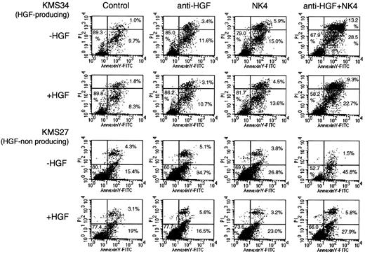
Induction of apoptosis by NK4 was also quantified by flow cytometry using annexin V and propidium iodide staining in KMS27 and KMS34 cells (Figure 3). NK4 treatment increased the annexin V+ fraction in both cell lines. Neutralizing anti-HGF antibody also induced apoptosis in non–HGF-producing KMS27 cells, probably because the antibody reacted with a very small amount of HGF present as a contaminant in FCS (Hyclone Laboratories). Addition of an excess amount of HGF blocked neutralizing antibody-induced apoptosis, whereas addition of exogenous HGF only partially inhibited NK4-induced apoptosis in KMS27 cells. Culture with both NK4 and neutralizing antibody revealed a further increase in the annexin V fraction in KMS27 and KMS34 cells. These results suggested that not only the antagonistic inhibition of HGF but also the additional, unknown effects of NK4 mentioned are important for induction of apoptosis in MM cells.
Flow cytometric analysis for quantification of apoptotic cells. MM cells (2 × 10⁵/mL were cultured in RPMI containing 10% FCS (Hyclone Laboratories) and collected after 4 days of exposure to NK4 (100 nM) or anti–HGF-neutralizing monoclonal antibody (19 μg/mL) in the presence or absence of 100 ng/mL HGF; this concentration of HGF was an excess amount sufficient to counteract NK4 or the neutralizing antibody. Cells were stained with FITC-coupled annexin V and propidium iodide. Induction of apoptosis by NK4 was evaluated by flow cytometry. The percentages of early apoptotic cells (annexin V+/PI−) and late apoptotic cells (annexin V+/PI+) are indicated in the corresponding quadrants.
Flow cytometric analysis for quantification of apoptotic cells. MM cells (2 × 10⁵/mL were cultured in RPMI containing 10% FCS (Hyclone Laboratories) and collected after 4 days of exposure to NK4 (100 nM) or anti–HGF-neutralizing monoclonal antibody (19 μg/mL) in the presence or absence of 100 ng/mL HGF; this concentration of HGF was an excess amount sufficient to counteract NK4 or the neutralizing antibody. Cells were stained with FITC-coupled annexin V and propidium iodide. Induction of apoptosis by NK4 was evaluated by flow cytometry. The percentages of early apoptotic cells (annexin V+/PI−) and late apoptotic cells (annexin V+/PI+) are indicated in the corresponding quadrants.
NK4 regulation of the activation of ERK1/2, STAT3, AKT-1, and c-MET in MM cells
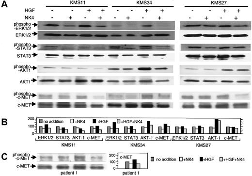
To examine the effects of NK4 on intracellular signaling, the phosphorylation status of ERK1/2, STAT3, and AKT-1 was examined by Western blot analysis, because these 3 signal transducers are key molecules in the growth of MM cells. As shown in Figure 4, baseline as well as HGF-induced phosphorylation of ERK1/2, STAT3, and AKT-1 was inhibited by NK4 treatment in HGF-producing KMS11 and KMS34 cell lines. Immunoprecipitation using anti–c-MET antibody and Western blot analysis using anti–phosphorylated c-MET antibody revealed that phosphorylation of c-MET was also inhibited by NK4 in these cells. The results suggested that in HGF-producing cells ERK1/2, STAT3, and AKT-1 are phosphorylated to different extents through autocrine activation of the c-MET and that NK4 inhibits phosphorylation of these signaling molecules by antagonizing functional association between HGF and c-MET. KMS11 cells showed lower responsiveness to exogenous HGF compared with KMS34 cells probably because KMS11 cells produced much higher amount of HGF by themselves than other cells as shown in Figure 1. Densitometric analysis showed that the phosphorylation of these 3 signal transducers and c-MET was also inhibited weakly by NK4 treatment in non–HGF-producing KMS27 cells. Baseline phosphorylation in the absence of HGF is relatively high in this cell line. Consequently, the phosphorylation level of c-MET was not significantly changed by addition of NK4 or exogenous HGF (Figure 4). Immunoprecipitation and Western blot analysis revealed that NK4 protein inhibited phosphorylation of c-MET in the primary MM cells obtained from patient 1 (Figure 4C).
Western blot analysis of activated ERK1/2, STAT3, and AKT-1, and immunoprecipitation and Western blot analysis of activated c-MET in NK4-treated MM cells. (A) HGF-producing KMS11 and KMS34 cells as well as non–HGF-producing KMS27 cells were treated with 200 nM NK4 protein in RPMI containing 4% dialyzed FCS overnight; the cells were then incubated with or without 50 ng/mL HGF at room temperature for 20 minutes. The cell lysates were subjected to sodium dodecyl sulfate-polyacrylamide gel electrophoresis (SDS-PAGE), followed by immunoblotting with anti–phospho-ERK1/2, anti–phospho-STAT3, and anti–phospho-Ser473 AKT-1 antibodies. Blots were stripped and reprobed with anti-ERK1/2, anti-STAT3, and anti–AKT-1 antibodies. For evaluation of c-MET activation, MM cell lines were also treated with 200 nM NK4 in RPMI containing 4% dialyzed FCS overnight, and then incubated with or without 50 ng/mL HGF at room temperature for 5 minutes. After immunoprecipitation with anti–c-MET antibody, the immunocomplexes were subjected to SDS-PAGE. Immunoblotting was conducted with anti–phospho–c-MET antibody, and then the same filters were stripped and reprobed with anti–c-MET. (B) Densitometric analysis of the activation of c-MET and signal transducers. (C) Evaluation of c-MET activation in primary MM cells obtained from patient 1. Treatment of the MM cells and immunoprecipitation followed by Western blot analysis were performed as described.
Western blot analysis of activated ERK1/2, STAT3, and AKT-1, and immunoprecipitation and Western blot analysis of activated c-MET in NK4-treated MM cells. (A) HGF-producing KMS11 and KMS34 cells as well as non–HGF-producing KMS27 cells were treated with 200 nM NK4 protein in RPMI containing 4% dialyzed FCS overnight; the cells were then incubated with or without 50 ng/mL HGF at room temperature for 20 minutes. The cell lysates were subjected to sodium dodecyl sulfate-polyacrylamide gel electrophoresis (SDS-PAGE), followed by immunoblotting with anti–phospho-ERK1/2, anti–phospho-STAT3, and anti–phospho-Ser473 AKT-1 antibodies. Blots were stripped and reprobed with anti-ERK1/2, anti-STAT3, and anti–AKT-1 antibodies. For evaluation of c-MET activation, MM cell lines were also treated with 200 nM NK4 in RPMI containing 4% dialyzed FCS overnight, and then incubated with or without 50 ng/mL HGF at room temperature for 5 minutes. After immunoprecipitation with anti–c-MET antibody, the immunocomplexes were subjected to SDS-PAGE. Immunoblotting was conducted with anti–phospho–c-MET antibody, and then the same filters were stripped and reprobed with anti–c-MET. (B) Densitometric analysis of the activation of c-MET and signal transducers. (C) Evaluation of c-MET activation in primary MM cells obtained from patient 1. Treatment of the MM cells and immunoprecipitation followed by Western blot analysis were performed as described.
Growth inhibition of MM cells in vivo by adenovirus-mediated gene transfer of NK4
To examine the antimyeloma effects of NK4 in vivo, the NK4 gene was transduced to tumor-bearing lcr/scid mice using the recombinant adenovirus. Because MM is a systemic disease, the systemic delivery of NK4 protein is necessary. Thus, instead of intratumoral injection, we chose to apply intramuscular injection at the femur. To confirm that the adenoviral vector indeed expressed the NK4 protein, an immunoblot analysis was initially performed, and 67 kDa of the NK4 protein was identified in the culture medium of the NK4-infected cells (Figure 5A). The next question to be addressed was whether the injected recombinant adenovirus produced NK4 protein in vivo. In a pharmacokinetic study, the time-course changes in the concentrations of NK4 protein in the plasma and various organs were examined by ELISA, and the results are shown in Figure 5B. After a single intramuscular injection of 5 × 10⁸ infectious particles/mL AdCMV.NK4, the plasma concentration of NK4 protein reached a maximum at day 10 and slowly declined over the following 2 weeks. NK4 protein was also detected in the liver, kidneys, and lungs. The highest concentration of NK4 was observed in the liver as compared to that in the lungs and kidneys. Treatment with NK4 was well tolerated, and no signs of toxicity such as death or weight loss were observed (Figure 5C).
Production of the NK4 gene product by the AdCMV.NK4 vector and intramuscular injection of AdCMV.NK4 in lcr/scid mice. (A) AdCMV.NK4 was used to infect KMS 11 cells at multiplicity of infection (MOI) 30. Supernatant (5 μL) was subjected to Western blot analysis. Rabbit anti-HGF polyclonal antibody enabled the detection of a 67-kDa NK4 gene product. (B) Pharmacokinetic study of the NK4 gene product. Infectious particles (5 × 10⁸/mL) of AdCMV.NK4 were injected into the femoral muscle of 5-week-old male lcr/scid mice on day 0 (arrow). On days 0, 3, 7, 10, 14, and 21, 3 mice were killed. Tissue lysates were obtained by immersing various organs in the lysis buffer overnight. The concentrations of NK4 in the plasma and tissue lysates were examined by ELISA. (C) Change in the weight of AdCMV.NK4-treated mice. A total of 5 × 10⁸ infectious particles/mL AdCMV.NK4 or AdCMV.lacZ were injected into the femoral muscle of 5-week-old male lcr/scid mice. Changes in weight are shown. Each group consists of 3 mice.
Production of the NK4 gene product by the AdCMV.NK4 vector and intramuscular injection of AdCMV.NK4 in lcr/scid mice. (A) AdCMV.NK4 was used to infect KMS 11 cells at multiplicity of infection (MOI) 30. Supernatant (5 μL) was subjected to Western blot analysis. Rabbit anti-HGF polyclonal antibody enabled the detection of a 67-kDa NK4 gene product. (B) Pharmacokinetic study of the NK4 gene product. Infectious particles (5 × 10⁸/mL) of AdCMV.NK4 were injected into the femoral muscle of 5-week-old male lcr/scid mice on day 0 (arrow). On days 0, 3, 7, 10, 14, and 21, 3 mice were killed. Tissue lysates were obtained by immersing various organs in the lysis buffer overnight. The concentrations of NK4 in the plasma and tissue lysates were examined by ELISA. (C) Change in the weight of AdCMV.NK4-treated mice. A total of 5 × 10⁸ infectious particles/mL AdCMV.NK4 or AdCMV.lacZ were injected into the femoral muscle of 5-week-old male lcr/scid mice. Changes in weight are shown. Each group consists of 3 mice.
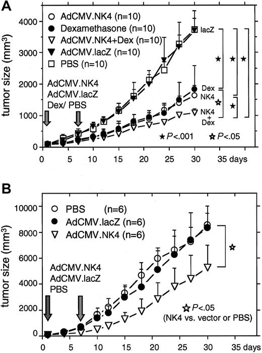
HGF-producing KMS11 and KMS34 cells (1 × 10⁷) were transplanted subcutaneously into 5- to 6-week-old male lcr/scid mice, and a skin plasmacytoma was readily established. In 3 to 4 weeks after the inoculation, that is, when these tumors reached 100 mm3, 5 × 10⁸ infectious particles per milliliter adenovirus was injected into the femoral muscle on days 1 and 7. As shown in Figure 6A, the growth of tumors derived from KMS11 cells was significantly inhibited in AdCMV.NK4-injected mice compared with that in AdCMV.lacZ- or PBS-treated mice (day 30, P < .001). In Dex-sensitive KMS11 cells, coinjection of AdCMV.NK4 with Dex enhanced the antitumor effects of AdCMV.NK4 (day 30, P < .001; Figure 6A). Ad-NK4 also significantly inhibited the growth of Dex-resistant KMS34 cell tumors (day 30, P < .05, compared with the other 2 control groups; Figure 6B).
In vivo growth inhibition of plasmacytoma by intramuscular injection of AdCMV.NK4. A total of 1 × 10⁷ HGF-producing KMS11 (A) and KMS34 cells (B) were inoculated subcutaneously into lcr/scid mice and a plasmacytoma was established. When the tumors reached 100 mm3 (day 0), 5 × 10⁸ infectious particles/mL AdCMV.NK4 or AdCMV.lacZ was injected into the femoral muscle on days 0 and 7. In Dex-sensitive KMS11 tumors, 0.8 μg/g Dex was also injected into the femoral muscle on days 0 and 7. The width and length of the plasmacytoma was measured every 3 days. Sizes on day 30 in each arm were compared and evaluated by the Student t test.
In vivo growth inhibition of plasmacytoma by intramuscular injection of AdCMV.NK4. A total of 1 × 10⁷ HGF-producing KMS11 (A) and KMS34 cells (B) were inoculated subcutaneously into lcr/scid mice and a plasmacytoma was established. When the tumors reached 100 mm3 (day 0), 5 × 10⁸ infectious particles/mL AdCMV.NK4 or AdCMV.lacZ was injected into the femoral muscle on days 0 and 7. In Dex-sensitive KMS11 tumors, 0.8 μg/g Dex was also injected into the femoral muscle on days 0 and 7. The width and length of the plasmacytoma was measured every 3 days. Sizes on day 30 in each arm were compared and evaluated by the Student t test.
Histopathologic examination of MM cells treated with NK4
Subcutaneous tumors derived from KMS11 cells were isolated and microscopically observed. A significant number of MM cells exhibited signs of cell death in AdCMV.NK4-treated mice, as compared with those observed in AdCMV.lacZ- or PBS-treated mice (Figure 7A). Tumor cells treated with Dex alone also underwent extensive cell death due to the direct antimyeloma effects of Dex. TUNEL staining demonstrated that a significant number of MM cells treated with AdCMV.NK4 fell into apoptosis (Figure 7B). The microvessel density in the tumors was also evaluated by immunohistochemical staining of cross-sections with anti-VWF antibody (Figure 7C). In AdCMV.NK4-injected mice, the density of microvessels in the tumors was significantly reduced compared with that in AdCMV.lacZ-treated tumors (P = .029). These results, taken together with our previous observation that NK4 significantly inhibits the proliferation of endothelial cells in vitro, NK4 is considered to possess antiangiogenic activity.15,16 The change of phosphorylation status of c-MET was also examined by immunohistochemical staining with antiphosphorylated c-MET antibody. As shown in Figure 7D, c-MET in AdCMV.NK4-treated tumors was less potently activated compared with that in AdCMV.lacZ-treated tumors.
Histopathologic examination of KMS11-derived tumors treated with AdCMV.NK4. (A) Hematoxylin and eosin staining of AdCMV.lacZ-treated, AdCMV.NK4-treated, Dex-treated, and AdCMV.NK4 plus Dex-treated KMS11 tumors. The arrows indicate areas of tumor cell death. (B) TUNEL assay of KMS11 tumors. The numbers of TUNEL+ cells per one visual field (0.148 mm2) were counted and shown in the form of a bar graph. * P = .001, ** P = .008 compared with AdCMV.lacZ-treated tumors. (C) Immunohistochemical analysis of blood vessels in subcutaneous tumors derived from KMS11 cells. Vascular endothelial cells were stained with anti-VWF antibody. The mean number of blood vessels per one visual field is indicated by the bar graph. *P = .029 compared with that in AdCMV.lacZ-treated tumors. (D) Immunohistochemical staining of activated c-Met in AdCMV.NK4-treated tumors using anti–phosphorylated c-MET. To eliminate background staining, human IgG was used as a control. In panels B and C the mean ± 1 SD (n-7 for B and n-5 for C) is shown.
Histopathologic examination of KMS11-derived tumors treated with AdCMV.NK4. (A) Hematoxylin and eosin staining of AdCMV.lacZ-treated, AdCMV.NK4-treated, Dex-treated, and AdCMV.NK4 plus Dex-treated KMS11 tumors. The arrows indicate areas of tumor cell death. (B) TUNEL assay of KMS11 tumors. The numbers of TUNEL+ cells per one visual field (0.148 mm2) were counted and shown in the form of a bar graph. * P = .001, ** P = .008 compared with AdCMV.lacZ-treated tumors. (C) Immunohistochemical analysis of blood vessels in subcutaneous tumors derived from KMS11 cells. Vascular endothelial cells were stained with anti-VWF antibody. The mean number of blood vessels per one visual field is indicated by the bar graph. *P = .029 compared with that in AdCMV.lacZ-treated tumors. (D) Immunohistochemical staining of activated c-Met in AdCMV.NK4-treated tumors using anti–phosphorylated c-MET. To eliminate background staining, human IgG was used as a control. In panels B and C the mean ± 1 SD (n-7 for B and n-5 for C) is shown.
Discussion
Coexpression of HGF and c-MET has been reported in various types of cancers.13,20 HGF is produced by tumor cells and stromal cells and leads to the growth and metastasis of tumor cells via both autocrine and paracrine mechanisms. Figure 1 shows that in the present study, all of the myeloma cells expressed c-MET protein, and 2 cell lines also produced and secreted HGF, suggesting that the HGF–c-MET axis contributes to the growth of MM cells. Recently, it was reported that a selective c-MET tyrosine kinase inhibitor blocks HGF-induced intracellular signaling and regulates the proliferation and migration of MM cells in vitro.21 In our study, NK4 protein also stabilized the growth of HGF-producing KMS11 and KMS34 cells in vitro as well as in vivo (Figures 2 and 6). NK4 also inhibited the growth of primary tumor cells obtained from patients with MM (Figure 2D). Immunoblot analysis revealed that NK4 inhibited the activation of HGF receptor/c-MET and the downstream signaling such as ERK1/2, STAT3, and AKT-1 in the presence or absence of HGF (Figure 4). Thus, the NK4 molecule is considered to directly regulate the proliferation of MM cells, presumably by interfering with autocrine and paracrine loops.
Growth suppression was also observed in non–HGF-producing cells such as KMS27 cells, as determined by an MTT assay conducted in HGF-free medium even though c-MET activation was weakly decreased by NK4 treatment (Figures 2A and 4). Addition of an excess amount of HGF to KMS27 cells only partially inhibited NK4-induced apoptosis, whereas HGF clearly blocked apoptosis induced by neutralizing antibody (Figure 3). In our previous reports, NK4 was shown to inhibit the proliferation of endothelial cells stimulated not only by HGF, but also by VEGF and FGF-2, even though NK4 does not inhibit the binding of VEGF or FGF-2 to their respective receptors.16 KMS27 and other cells have been shown to produce and secrete VEGF or FGF-2 into culture medium (Y.H., unpublished data, March 2002). Subsequent study has also demonstrated that a deletion mutant of NK4 in the N-terminal hairpin domain lost HGF antagonist activity, but still inhibited the proliferation and migration of endothelial cells induced by VEGF, FGF-2, and HGF.15 It is speculated that the biologic activities of NK4 involve not only the antagonism of HGF/c-Met interactions, but also the inhibition of an HGF/c-Met–independent pathway. Thus, NK4 is likely bifunctional.13 Association of NK4 to a putative binding molecule other than c-Met receptor may participate in the signal transduction for the growth inhibition of endothelial cells by NK4. However, the precise molecular mechanisms of the latter activity remain to be determined. Taken together, we speculate that the similar activity, as seen in case of endothelial inhibition, may be involved in inhibitory effect of NK4 on proliferation of MM cells.
In previous studies, it has been reported that vascular endothelial cells express c-Met and HGF induces the proliferation of endothelial cells, suggesting that HGF contributes to tumor angiogenesis.16 Recently, a positive correlation between serum HGF concentration and bone marrow microvessel density was reported in patients with MM.22 NK4 is structurally similar to angiostatin, a potent angiogenesis inhibitor, and previous reports have demonstrated that NK4 inhibits endothelial cell growth.14,16,18,19,23 In the present study, a significant decrease in vascular endothelial cells in tumors was observed in AdCMV.NK4-treated mice. Thus, antiangiogenesis is expected to be among the reasons for the antimyeloma effects of NK4 in vivo.
Glucocorticoid is a key drug in the treatment of MM and is included in most of the currently applied chemotherapeutic regimens. To establish a more effective antimyeloma therapy, we examined the effects of NK4 on glucocorticoid-resistant MM cells, as well as the effects of a combination treatment using NK4 together with glucocorticoid in glucocorticoid-sensitive cells. The present in vitro and in vivo studies revealed that NK4 is effective at regulating the growth of Dex-resistant KMS34 cells. In future clinical trials, it will be worthwhile to attempt using NK4 to treat patients with MM resistant to conventional chemotherapy. As shown in Figures 2C and 6A, NK4 enhanced the antimyeloma effect of Dex in Dex-sensitive KMS11 cells in vitro as well as in vivo. Thus, NK4 treatment in combination with Dex or with another therapeutic approach may be more efficient at achieving antitumor effects. Along these lines, a previous study has already demonstrated that a combination of NK4 administration with immunotherapy using dendritic cells was effective at countering B-cell malignancy.24
Here, mice were exposed to a challenge with an injection of adenovirus containing NK4 cDNA into a femoral muscle remote from the tumor; the findings demonstrated the systemic delivery of secreted NK4 protein, which in turn led to antimyeloma effects in vivo. In addition, AdCMV.NK4-treated mice did not show any weight loss (Figure 5C) or features of significant tissue damage (eg, venous thrombosis), as based on both macroscopic and microscopic observations (data not shown). These results suggest the feasibility of NK4 gene therapy for patients with MM. Indeed, the tissue and plasma concentrations of NK4 were less than 1 ng/mL. Kushibiki and colleagues also demonstrated the antitumor activities of NK4 using a plasmid vector system in breast cancer-bearing mice, even though the plasma concentration of NK4 was much lower in their model than that observed in our model.25 Even at low concentrations, the long-term exposure of tumor cells to NK4 may delay the growth of tumor cells. In addition, NK4 exhibited antimyeloma effects via multiple mechanisms in vivo; for example, not only direct antitumor effects such as serving as an HGF antagonist, but also antiangiogenic effects and unknown HGF-independent mechanisms were suggested as putative mechanisms, as discussed above in this section. According to the sum effects of these mechanisms, NK4 might have significantly inhibited the proliferation of MM cells inoculated into the SCID mice, even though only low plasma concentrations were achieved.
With the aim of improving future clinical applications, additional studies of the biologic effects of NK4 should be carried out, in particular those involving normal tissues. Alternative safe methods for the delivery of the NK4 gene or gene products should be also explored, including the administration of synthetic NK4 peptide.
Authorship
Contribution: Y.H. designed the study, performed all in vitro and in vivo experiments, analyzed data, wrote the paper, and had all responsibility for this manuscript; W.D. and T.Y. helped with histopathologic examinations; K.M. and T. Nakamura provided the NK4 protein and recombinant adenovirus, and they also helped with the analyzing the data and writing the manuscript; M.S. helped with the FACS study; T.O. provided the myeloma cell lines; T. Niikura contributed to the preparation of the recombinant adenovirus; T. Nukiwa established and provided the recombinant adenovirus; and Y.I. supervised the experiments, data analysis, and provided critical revision of the manuscript.
Conflict-of-interest disclosure: The authors declare no competing financial interests.
Correspondence: Yutaka Hattori, Division of Hematology, Department of Internal Medicine, Keio University School of Medicine, 35 Shinanomachi, Shinjuku-ku, Tokyo 160-8582, Japan; e-mail: yhattori@sc.itc.keio.ac.jp.
The publication costs of this article were defrayed in part by page charge payment. Therefore, and solely to indicate this fact, this article is hereby marked “advertisement” in accordance with 18 USC section 1734.
Acknowledgments
This work was supported by grants from the International Myeloma Foundation Aki Horinouchi Research Grant (Y.H.) and Keio Gijuku Academic Development Funds (Y.H.).


This feature is available to Subscribers Only
Sign In or Create an Account Close Modal