Abstract
We report the results of a randomized study to elucidate whether addition of granulocyte colony-stimulating factor (G-CSF) to immunosuppressive therapy is valuable for the treatment of severe aplastic anemia (SAA) in adults. A total of 101 previously untreated patients (median age, 54 years; range, 19 to 75 years) were randomized to receive antithymocyte globulin (ATG) and cyclosporin A (CyA) (G-CSF− group) or ATG, CyA, and G-CSF (G-CSF+ group). In the G-CSF+ group, the hematologic response rate at 6 months was higher (77% vs 57%; P = .03) than in the G-CSF− group. No differences were observed between the groups in terms of the incidence of infections and febrile episodes. There were no differences between the G-CSF− group and the G-CSF+ group in terms of survival (88% vs 94% at 4 years), and the development of myelodysplastic syndrome (MDS)/acute leukemia (AL) (1 patient vs 2 patients). However, the relapse rate was lower in the G-CSF+ group compared with the G-CSF− group (42% vs 15% at 4 years; P = .01). Further follow-up is required to elucidate the role of G-CSF in immunosuppressive therapy for adult SAA.
Introduction
Acquired aplastic anemia (AA) is a serious hematologic disorder characterized by peripheral blood pancytopenia and hypocellular bone marrow. Bone marrow transplantation (BMT) and immunosuppressive therapy (IST) are standard treatment strategies for severe AA (SAA), and the decision of initial treatment depends largely on an availability of a human leukocyte antigen (HLA)-identical sibling donor and patient age. Antithymocyte globulin (ATG) and cyclosporine A (CyA) are immunosuppressive drugs generally used for AA and had an equivalent efficacy in terms of hematologic response rate and a survival rate.1 It has been also demonstrated that the combination of ATG and CyA is superior to ATG or CyA alone in terms of hematologic response.2,3
Granulocyte colony-stimulating factor (G-CSF) is a hematopoietic growth factor that mainly stimulates the proliferation and differentiation of granulocyte precursors; however, a stimulatory effect of G-CSF on multipotential hematopoietic stem cells has also been demonstrated.4 Clinically, G-CSF can induce a short-term increase in the neutrophil count in most patients with AA.5 In addition, multilineage recovery of hematopoiesis in some patients with AA by G-CSF has been reported.6,7 Therefore, addition of G-CSF to IST may not only decrease the risk for infection but also increase the hematologic response rate. In 1995, a European group showed promising results that ATG, CyA, and G-CSF therapy produced a high response rate (82% at a median follow-up period of 115 days), a high actuarial survival rate (92% with a median follow-up of 428 days), and a relatively low number of early deaths (8%) from infection.8 This encouraging result formed the basis of our prospective randomized trial.
Evolution of AA to myelodysplastic syndrome (MDS) and acute myeloid leukemia (AML) is a major problem in patients undergoing IST.9–11 Because G-CSF can stimulate the growth of leukemic clones, combined use of G-CSF with IST may facilitate the progression of AA to MDS/AML.12,13
To elucidate whether the addition of G-CSF to IST increases the response rate, prevents infections during the treatment, improves the survival or relapse rate, and increases the risk for MDS/AML, we have started the prospective randomized controlled study comparing ATG and CyA therapy with or without G-CSF in adult patients with AA. During the period in which our study has been ongoing, 2 groups have reported the results of similar prospective randomized studies.14,15 However, 1 study focuses on childhood AA,14 and another includes both childhood and adult patients with AA.15 To our knowledge, this is the first prospective randomized study to investigate the role of combined use of G-CSF and IST focusing on adult patients with AA.
Patients and methods
Patients
From June 1996 to June 2000, a total of 101 patients with acquired AA from 43 centers were enrolled. Patients with acquired AA were eligible if they met the following criteria: aged 18 to 75 years, newly diagnosed patients, no specific prior treatment for the disease, and severe AA. The disease was considered severe if at least 2 of the following were fulfilled: a neutrophil count of less than 0.5 × 10⁹/L, a platelet count of less than 20 × 109/L, and a reticulocyte count of less than 20 × 109/L with hypocellular bone marrow.16 Patients were excluded if they had been diagnosed as having Fanconi anemia or dyskeratosis congenita, severe uncontrolled infection, or malignancies. Cytogenetic studies were performed for all patients. We estimated a 30% difference in response rate between the G-CSF− group (ATG + CyA) and the G-CSF+ group (ATG + CyA + G-CSF). To detect a 30% difference, 45 patients per treatment group were required. Compensating for an estimated nonevaluability rate of 10%, it was considered reasonable to enroll at least 100 patients.
Informed written consent was obtained from all patients prior to study entry with Institutional Review Board approval at each of the participating centers and in accordance with the Declaration of Helsinki.
Treatment protocol
Patients were randomized to receive either ATG and CyA or ATG, CyA, and G-CSF. Horse ATG (Lymphoglobuline; Merieux, Lyon, France) was administered at a dose of 15 mg/kg per day for 5 days as a slow intravenous infusion over 12 hours. For the prevention of serum sickness, prednisolone was given orally at a dose of 1 mg/kg per day from day 1 to day 9, 0.5 mg/kg per day from day 10 to day 15, and 0.2 mg/kg per day from day 16 to day 21. CyA, given orally at a dose of 6 mg/kg per day, was started on day 1 and continued for at least 12 weeks. The dose was adjusted to achieve a whole-blood trough level of 150 to 250 ng/mL. In responders, CyA was continued for at least 28 weeks. In patients with a stable hematologic status for at least 4 weeks, gradual tapering of CyA (1mg/kg every 2 weeks) was permitted if hematologic data remained stable during the course of tapering. In patients randomized to receive G-CSF, filgrastim (Gran; Kirin-Sankyo, Tokyo, Japan) or lenograstim (Neutrogin; Chugai, Tokyo, Japan) was given intravenously at a dose of 400 μg/m2 per day and 50 μg/kg per day, respectively, every other day until day 28, and then once or twice a week until day 84. The daily doses of filgrastim and lenograstim were those proved to be effective in clinical studies performed in Japan and approved by the Japanese Ministry for Health, Labor, and Welfare.17,18 The primary end point of the study was the hematologic response at 12 weeks, 3 months, and 1 year after IST, and the secondary end points included the incidence of infections and febrile episodes during the first 12 weeks, survival rate, relapse rate, and incidence of the development of MDS /AL.
Evaluation of response and toxicity
Complete response (CR) was defined as a neutrophil count greater than 1.5 × 10⁹/L, a platelet count greater than 150 × 109/L, and a hemoglobin level of greater than 110 g/L (11.0 g/dL). Partial response (PR) was defined by transfusion independence and no longer meeting criteria for severe disease.19 Relapse was indicated by the requirement for blood transfusion. Toxicity of treatment was evaluated for the first 12 weeks and was graded according to the criteria of the World Health Organization.20
The Fisher Exact test was used to compare categoric variables, and the Mann-Whitney U test or the Student t test was used to compare continuous variables. The probability of survival and relapse was analyzed using the Kaplan-Meier method.21 All statistical analyses were performed using SPSS 15.0 software (SPSS Japan, Tokyo, Japan).
Results
Patient characteristics
A total of 50 patients were randomized to receive ATG and CyA (G-CSF− group), and 51 patients were randomized to receive ATG, CyA, and G-CSF (G-CSF+ group). A total of 6 patients were excluded from analysis because of a diagnosis of lymphoma after randomization (1 patient), or treatment without ATG (5 patients) according to the patient's wishes after enrollment. Patient characteristics of the G-CSF+ and G-CSF− groups are summarized in Table 1. All patients, except 3 with hepatitis-associated AA, had no identifiable cause of AA (idiopathic AA). There were no significant differences between 2 groups in age, sex, hemoglobin level, neutrophil count, platelet count, reticulocyte count, number of patients with a neutrophil count of less than 0.2 × 10⁹/L (ie, very severe AA [vSAA]), and interval between diagnosis and treatment. A total of 8 and 11 patients had a neutrophil count of more than 0.5 × 10⁹/L in the G-CSF− group and in the G-CSF+ group, respectively.
Patient characteristics
| Characteristic . | ATG + CyA . | ATG + CyA + G-CSF . | P . |
|---|---|---|---|
| No. of patients randomized | 50 | 51 | — |
| No. of patients evaluable | 47 | 48 | — |
| Age, median y (range) | 54 (19-75) | 53 (19-74) | .55 |
| Sex, male/female | 21/26 | 23/25 | .75 |
| Cause of AA, no. patients | |||
| Idiopathic | 46 | 46 | .51 |
| Hepatisis | 1 | 2 | — |
| Hemoglobin, median g/L (range) | 60 (35-82) | 60 (31-84) | .67 |
| Neutrophil count, median × 109/L (range) | 0.32 (0.02-1.01) | 0.30 (0.01-1.21) | .45 |
| Platelet count, median × 109/L (range) | 9 (1-38) | 9 (1-31) | .62 |
| Reticulocyte count, median × 109/L (range) | 11 (0-65) | 9 (0-35) | .08 |
| No. of patients with a neutrophil count less than 0.2 × 10⁹/L | 11 | 19 | .07 |
| Interval between diagnosis and treatment, median d (range) | 20 (3-152) | 18 (1-112) | .89 |
— indicates not applicable.
Response
At 12 weeks, CR was observed in 2 (4%) patients, and PR was observed in 22 (47%) patients in the G-CSF− group, for an overall response rate of 51% (24 of 47 patients). In the G-CSF+ group, no patients had a CR and 28 (58%) patients had a PR for an overall response rate of 58% (28 of 48) (Table 2). There were no statistically significant differences in overall response rates at 12 weeks between the 2 groups (P = .31). At 6 months, the overall response rate increased from 51% to 57% in the G-CSF− group, and from 58% to 77% in the G-CSF+ group. The difference in overall response rates at 6 months between the 2 groups was statistically significant (P = .03). At 1 year, the overall response rate increased from 57% to 76% in the G-CSF− group, but did not change (from 77% to 79%) in the G-CSF+ group. There was no statistically significant difference in overall response rate at 1 year between the 2 groups (P = .46). In the G-CSF+ group, there were no differences in overall response rate between the filgrastim-treated group and lenograstim-treated group (data not shown).
Response to treatment at 12 weeks, 3 months, and 1 year after treatment
| Time after treatment . | ATG + CyA, no. (%) . | ATG + CyA + G-CSF, no. (%) . | P . |
|---|---|---|---|
| 12 weeks | |||
| No. of patients evaluable | 47 | 48 | — |
| CR | 2 | 0 | — |
| PR | 22 | 28 | — |
| Total response, CR+PR | 24 (51) | 28 (58) | .31 |
| Death | 0 | 2 | — |
| 6 months | |||
| No. of patients evaluable | 46 | 47 | — |
| CR | 3 | 2 | — |
| PR | 23 | 34 | — |
| Total response, CR+PR | 26 (57) | 36 (77) | .03 |
| Death | 0 | 2 | — |
| 1 year | |||
| No. of patients evaluable | 41 | 47 | — |
| CR | 1 | 3 | — |
| PR | 30 | 34 | — |
| Total response, CR+PR | 31 (76) | 37 (79) | .46 |
| Death | 1 | 2 | — |
— indicates not applicable.
When the overall response rate was analyzed focusing on the vSAA patients (ie, a neutrophil count of less than 0.2 × 10⁹/L), it was 27% (3 of 11 patients) at 12 weeks, 20% (2 of 10 patients) at 6 months, and 63% (5 of 8 patients) at 1 year in the G-CSF− group, and was 42% (8 of 19 patients) at 12 weeks, 63% (12 of 19 patients) at 6 months, and 63% (12 of 19 patients) at 1 year in the G-CSF+ group. Similar to the result obtained in total patients, overall response rate in patients with vSAA at 6 months but not at 12 weeks and 1 year was significantly higher in the G-CSF+ group compared with the G-CSF− group (12 weeks, P = .34; 6 months, P = .03; 1 year; P = .68).
The overall response rate for patients with a neutrophil count of more than 0.5 × 10⁹/L at 12 weeks, 6 months, and 1 year was 50% (4 of 8 patients), 63% (5 of 8 patients), and 86% (6 of 7 patients) in the G-CSF− group, and 64% (7 of 11 patients), 64% (7 of 11 patients), and 91% (10 of 11 patients) in the G-CSF+ group, respectively. There was no significant difference in overall response between the 2 groups (12 weeks, P = .45; 6 months, P = .51; 1 year, P = .64). A total of 7 patients had chromosomal abnormalities at diagnosis, and 4 patients responded to IST.
When patients who responded to immunotherapy but needed continuous administration of CyA to maintain hematologic response were defined as CyA dependent, 8 patients in the G-CSF− group and 6 in the G-CSF+ group were included.
The median neutrophil counts at 4 weeks, 12 weeks, 6 months, and 1 year was 0.36 × 10⁹/L, 1.32 × 10⁹/L, 1.20 × 10⁹/L, and 1.35 × 10⁹/L in the G-CSF− group and 1.03 × 10⁹/L, 1.19 × 10⁹/L, 1.61 × 10⁹/L, and 1.57 × 10⁹/L in the G-CSF+ group, respectively. At 4 weeks but not 12 weeks, 6 months, and 1 year, the median neutrophil count in the G-CSF+ group was significantly higher compared with the G-CSF− group (P = .004).
A total of 3 patients (1 in the G-CSF− group and 2 in the G-CSF+ group) who failed to respond to initial therapy received a second course of IST; however, no patients responded. A total of 2 patients (both patients were in the G-CSF− group) who failed to respond to initial therapy received a BMT. One patient who received a transplant from an HLA-matched sibling is alive at 44 months after transplantation, but another patient who received a transplant from an HLA-matched unrelated donor died of pulmonary bleeding 2 months after transplantation.
Infectious complications
During the first 12 weeks, infections developed in 19 (40%) patients in the G-CSF− group and in 28 (58%) patients in the G-CSF+ group (Table 3). There was no significant difference in the proportion of patients with documented infection between the 2 groups (P = .07). Severe infections (grade 3 or 4) developed in 5 patients in the G-CSF− group and 8 patients in the G-CSF+ group, and the difference in the proportion of patients who contracted severe infections between the 2 groups was not statistically significant (P = .29). There were 31 infectious events in the G-CSF− group, including 6 severe infectious events such as bacteremia (5 events) and pneumonia (1 event), and 39 infectious events in the G-CSF+ group, including 11 severe infections such as bacteremia (7 events), pneumonia (3 events), and fungemia (1 event). There was no difference in the incidence of infectious events between the 2 groups (P = .07). Among 13 infectious events, including bacteremia, pneumonia, and fungal infection, observed in the G-CSF+ group, 7 (54%) occurred during the period of a neutrophil count less than 0.5 × 10⁹/L.
Documented infections and febrile episodes during 12 weeks after initiation of treatment
| . | G-CSF− group . | G-CSF+ group . | P . |
|---|---|---|---|
| n | 47 | 48 | — |
| No. of patients with documented infections*; severe infection, grade 3 or 4 | 19; 5 | 28; 8 | .07; .30 |
| Accumulated number of infectious events† | 31 | 39 | .07 |
| Bacteremia | 5 | 7 | — |
| Pneumonia | 1 | 3 | — |
| Upper respiratory tract infection | 5 | 6 | — |
| Intestinal infection | 2 | 4 | — |
| Urinary tract infection | 1 | 0 | — |
| Genital infection | 0 | 2 | — |
| Cellulitis | 1 | 2 | — |
| Herpes zoster | 1 | 0 | — |
| Herpes simplex | 2 | 4 | — |
| Fungal infection | 0 | 3 | — |
| Gingivitis | 1 | 0 | — |
| Fever of unknown origin | 12 | 8 | — |
| Febrile days over 38°C, median no. (range) | 4 (1-17) | 4 (1-38) | .83 |
| Death due to infection, no. patients | 0 | 2 | .25 |
— indicates not applicable.
Patients who had fever of unknown origin were included.
Events of fever of unknown origin were included
To investigate the correlation between infection and the degree of neutropenia, the morbidity of infection in patients with vSAA and non-vSAA was compared, and was higher in patients with vSAA than in those with non-vSAA (61% in vSAA, 45% in non-vSAA); however, the difference was not statistically significant (P = .07).
The median number of febrile days (38°C or higher) was 4 days in both groups. Deaths due to infection during the first 12 weeks occurred in 2 patients (bacteremia and fungemia) in the G-CSF+ group.
Survival
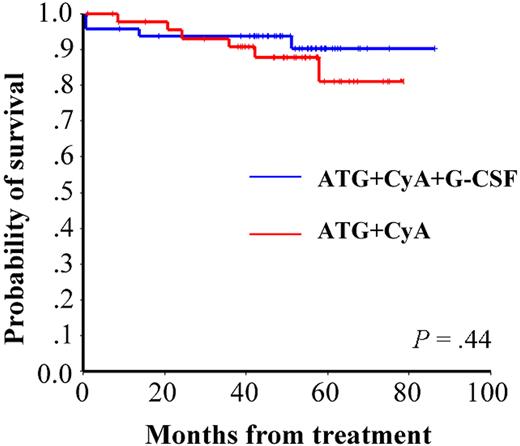
The overall probability of survival at 4 years is 88% for the G-CSF− group and 94% for the G-CSF+ group, with a median follow-up period of 52 months (range, 1 to 78 months) and 54 months (range, 1 to 86 months), respectively (Figure 1). There was no significant difference in the overall probability of survival between the 2 groups (P = .44). In the G-CSF+ group, there were no differences in survival rate between the filgrastim-treated group and lenograstim-treated group (data not shown). There were 6 deaths in the G-CSF− group, and 4 in the G-CSF+ group. Causes of death were bacteremia (1 patient each in the G-CSF− and G-CSF+ groups), pneumonia (1 patient in the G-CSF− group), fungal infection (1 in the G-CSF+ group), intracranial hemorrhage (1 patient each in the G-CSF− and G-CSF+ groups), BMT-related toxicity (1 patient each in the G-CSF− and G-CSF+ groups), renal failure (1 in the G-CSF− group) and metastatic brain tumor (1 in the G-CSF− group).
Actuarial survival of adult patients with SAA in the G-CSF− and G-CSF+ groups.
Cytogenetic analysis and clonal disease
Before therapy, a clonal cytogenetic abnormality was detected in 7 (8%) of 91 evaluable patients (4 patients with −Y, 2 patients with deletion 13, and 1 patient with trisomy 8). Among 7 patients with cytogenetic abnormalities, 1 patient (−Y) randomized to receive G-CSF developed refractory anemia with ringed sideroblasts (RARS) at 14 months after treatment, and 2 patients (1 with −Y, 1 with trisomy 8) not randomized to receive G-CSF experienced the disappearance of clonal abnormalities during the follow-up period.
In the G-CSF− group, chromosomal abnormalities appeared after treatment in 2 (6%) of 33 evaluable patients (1 with −Y and 1 with trisomy 8), with a subsequent disappearance of the chromosomal abnormality in 1 patient with −Y. In the G-CSF+ group, chromosomal abnormalities appeared after treatment in 6 (15%) of 41 evaluable patients (1 with −Y, 2 with monosomy 7, 1 with inv 7, 1 with trisomy 8, and 1 with monosomy 19), with a subsequent disappearance of the chromosomal abnormalities in 3 patients (1 with −Y, 1 with inv 7, and 1 with monosomy 19). All patients who developed chromosomal abnormalities after IST had revealed normal cytogenetics before IST.
There was no significant difference in the incidence of development of chromosomal abnormalities between the 2 groups (P = .21). In 8 patients in whom chromosomal abnormalities appeared after treatment, 2 patients (1 with −Y in the G-CSF− group and 1 with monosomy 7 in the G-CSF+ group) developed refractory anemia (RA).
As for the development of the clonal diseases, including MDS, AML, and paroxysmal nocturnal hemoglobulinemia (PNH), 1 patient developed RA at 38 months and 1 patient developed PNH at 25 months after treatment in the G-CSF− group, 2 patients developed MDS (RA and RARS) at 14 and 40 months, and 1 patient developed PNH at 41 months after treatment in the G-CSF+ group. No patients had definitive PNH before therapy. The presence of PNH was defined by a positive Ham test or loss of expression of CD55 and CD59 on red blood cells by flow cytometry. The overall risk for MDS/AML at 4 years is 3% for the G-CSF− group and 5% for the G-CSF+ group. There were no significant differences in the overall risk for MDS/AML between the 2 groups (P = .63). One patient who developed RA received peripheral blood stem-cell transplantation from an HLA-matched sibling and died of chronic graft-versus-host disease 5 months after transplantation.
Relapse
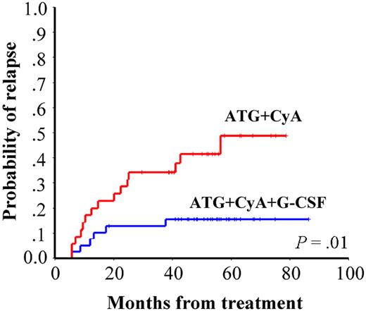
A total of 21 patients (15 in the G-CSF− group and 6 in the G-CSF+ group) relapsed after IST. In relapsed patients, all patients had received CyA at the time of relapse or had a history of CyA administration for at least for 6 months. The risk for relapse at 4 years was 42% in the G-CSF− group, and 15% in the G-CSF+ group (Figure 2). There was a significant difference in relapse rate between the 2 groups (P = .01). A total of 6 patients who relapsed after initial responses received a second course of IST, of whom 4 were in the G-CSF− group and 2 in the G-CSF+ group. Of the 6 patients 2 (33%) responded to the second therapy. Both responders belonged to the G-CSF+ group. A total of 2 patients (both in the G-CSF+ group) who relapsed after initial responses received BMT (1 from a HLA-matched sibling and another from an HLA-matched unrelated donor) and were alive at 41 months and 32 months after transplantation.
Cumulative incidence of relapse in adult patients with SAA in the G-CSF− and G-CSF+ groups.
Cumulative incidence of relapse in adult patients with SAA in the G-CSF− and G-CSF+ groups.
Toxicity
The incidence of toxicity was comparable between G-CSF− and G-CSF+ groups. Acute allergic reaction during ATG therapy was observed in 81% of patients in the G-CSF− group and 92% of patients in G-CSF+ group (P = .11). A total of 2 patients in the G-CSF− group and 4 patients in the G-CSF+ group had serum sickness. Toxicity greater than grade III was noted in 2 patients (anaphylactic reaction to ATG and delirium associated with CyA administration) in the G-CSF− group, and in 1 patient (liver dysfunction related to ATG administration) in the G-CSF+ group.
Discussion
The results of this prospective multicenter study showed that the addition of G-CSF to IST plays some role in the treatment of SAA. The hematologic response rate at 6 months in the G-CSF+ group was significantly higher compared with the G-CSF− group (77% in the G-CSF+ group, 57% in the G-CSF− group), but at 1 year was comparable (79% in the G-CSF+ group, 76% in the G-CSF− group). This indicates that G-CSF can accelerate the recovery of hematopoiesis in patients with AA when used in combination with IST. Our result is inconsistent with a similar study conducted in Japanese children with SAA, which showed no difference between the G-CSF+ and the G-CSF− group in terms of hematologic response.14 A European group also reported the result of a similar study in patients with SAA (age, 1-82 years), which showed that G-CSF only enhanced the recovery of neutrophils.15 The reasons why different results were obtained in these studies are uncertain. However, it is possible that patient's age might influence the results, because the distribution of patient's age is apparently different among the studies.
Our study showed that the accumulated relapse rate was significantly lower in the G-CSF + group (42% in the G-CSF+ group and 15% in the G-CSF− group at 4 years; P = .01). This indicates that G-CSF can reduce the relapse rate in patients who have responded to IST. The reason why G-CSF has an impact on the occurrence of relapse is uncertain. However, as G-CSF has a stimulatory effect on the growth of hematopoietic stem and progenitor cells in AA,22 it is possible that hematopoietic stem and progenitor cells, which can escape from the immune attack, might expand in patients who are successfully treated with IST in combination with G-CSF. If so, it is possible that relapse rate is low in the G-CSF+ group because of the high expansion of immune attack-resistant hematopoietic cells. A Japan childhood AA study group showed that there was a trend of low relapse rate in the G-CSF+ group (the risk for relapse at 4 years was 29% in the G-CSF+ group and 64% in the G-CSF− group), although the difference between the 2 groups was not statistically significant (P = .10), which might be due to a low number of patients enrolled.14 A European group reported that the actuarial risk for relapse after IST was 35% at 10 years, and the relapse occurred at any time from a few months to 10 years after IST without a particular period of higher occurrence.23 Because the median follow-up period of our study was not so long, the possibility that G-CSF only delays the time of relapse cannot be excluded. To elucidate whether G-CSF actually prevents relapse or only delays the time of relapse, further follow-up is required
To date, there was no significant difference between the G-CSF+ and the G-CSF− group in terms of overall survival (94% to 88% at 4 years). This finding is in keeping with that of previous reports.14,15 It has been reported that the overall survival in patients who do not relapse is better than that of patients who relapse.23 Therefore, better survival in the G-CSF+ group will be expected because of the low incidence of relapse in the G-CSF+ group. Further follow-up is necessary to conclude whether a difference in overall survival exists between the G-CSF+ and the G-CSF− group.
There was no difference in the incidence of documented infections and febrile episodes during the first 12 weeks between the G-CSF+ and G-CSF− groups, although the addition of G-CSF to IST resulted in an increase in the neutrophil counts. This result is in agreement with those of previous studies14,15 suggesting that G-CSF has no preventive effect on infections during the IST. In our study, the proportion of patients who contracted infection was relatively higher in the G-CSF+ group compared with the G-CSF− group, although the difference between the 2 groups was not statistically significant (58% vs 40%; P = .07). This might be due to the relatively high proportion of patients with vSAA (neutrophil count, < 0.200 × 10⁹/L [200/μL]) in the G-CSF+ group compared with the G-CSF− group (40% in the G-CSF+ group and 23% in the G-CSF− group) who were more susceptible to infections.
Over the long term, patients with AA who have been treated with IST have an increased risk (10%-47%) of developing MDS or AML.24 However, it is uncertain whether evolution to MDS/AML is a reflection of the natural history of AA or secondary disease related to IST. In addition, it has been reported that administration of G-CSF was associated with the development of MDS/AML,25–27 although conflicting results have been reported.28 In a retrospective study from Japan, it was demonstrated that 4 (22%) of 18 adult patients with AA patients treated with both IST and G-CSF developed MDS, and a high cumulative dose of G-CSF and the use of G-CSF for more than 1 year were the significant risk factors for developing MDS.25 Another study showed that the cumulative incidence of developing MDS/AML was 13.7% in children with AA who received IST and danazol with or without G-CSF, and long-term use of G-CSF and no response to therapy at 6 months were significant risk factors for developing MDS.26 In contrast, the Italian group showed that the risk for developing secondary malignancies at 60 months in patients treated with IST with or without G-CSF treatment was 9% and 7%, respectively (P = .99).28 This study concluded that large doses of G-CSF (36 000 μg/patient) administered over a long period of time (6 months) in conjunction with IST do not increase the actuarial risk of developing MDS/AML. In our study, the risk for developing MDS/AML at 4 years is 3% for the G-CSF− group and 5% for the G-CSF+ group (P = .63). Our finding suggests that use of G-CSF with IST in a relatively short period of time (12 weeks) does not increase the risk for developing MDS/AML, at least during the several years after IST. Longer follow-up (at least 10 years) is necessary to determine the role of G-CSF in the development of MDS/AML. Until then, routine use of G-CSF is not recommended unless part of a clinical trial.
It is well known that chromosomal abnormalities develop in some patients after IST.29 In addition, Japanese studies suggest a close relationship between the use of G-CSF and development of monosomy 7.25,26 In our study, 8 (11%) of 74 evaluable patients who received IST developed chromosomal abnormalities, and there was no significant difference in the incidence of development of chromosomal abnormalities between the G-CSF+ and G-CSF− groups. This result is consistent with data from an Italian group.28 In our study, however, it should be noted that monosomy 7 was only developed in the G-CSF+ group (2 patients), and 1 patient with monosomy 7 developed MDS. It has recently been shown that G-CSF preferentially stimulates the proliferation of monosomy 7 cells expressing the class IV G-CSF receptor, which is defective in signaling cell maturation.29
A transient appearance of chromosomal abnormalities in patients with AA after IST is also a well-documented phenomenon.28.30,31 An Italian group showed 9 patients (4 in the G-CSF+ group, 5 in the G-CSF− group) with transient chromosomal abnormalities.28 In the present study, transient chromosomal abnormalities were observed in 4 patients (3 in the G-CSF+ group, 1 in the G-CSF− group). Therefore, the appearance of abnormal cytogenetic clones after IST does not necessarily mean the subsequent expansion of those clones, and appeared to be unrelated to the combined use of G-CSF.
The present study suggests that combined use of immunosuppressive agents and G-CSF has some benefits in terms of the promotion of hematopoietic recovery and suppression of relapse rate, which result in reducing the need for subsequent treatments such as blood transfusion and second IST. G-CSF support of IST might be feasible for the treatment of adult SAA; however, further follow-up is required to elucidate whether G-CSF increases the risk for MDS/AML. In addition, it is important to discuss whether G-CSF support of IST is appropriate in terms of cost-effectiveness. To address this issue, long-term follow-up is necessary.
The online version of this article contains a data supplement.
The publication costs of this article were defrayed in part by page charge payment. Therefore, and solely to indicate this fact, this article is hereby marked “advertisement” in accordance with 18 USC section 1734.
Acknowledgments
This study was supported, in part, by a grant from Japan Intractable Disease Foundation and by a grant for research on intractable diseases from the Ministry of Health, Labor and Welfare of Japan.
Authorship
Author contributions: M.T., S.N., A.U., M.O., and H.M. designed research; M.T analyzed data and drafted the paper; A.K., S.I, and Y.Y. contributed to the enrollment of patients.
Conflict-of-interest disclosure: The authors declare no competing financial interests.
Correspondence: Masanao Teramura, Department of Hematology, Tokyo Women's Medical University, 8-1, Kawada-cho, Shinjuku-ku, Tokyo 162-8666, Japan; e-mail: teramura@dh.twmu.ac.jp.


This feature is available to Subscribers Only
Sign In or Create an Account Close Modal