Abstract
The mutagenic enzyme activation-induced cytidine deaminase (AID) is required for immunoglobulin class switch recombination (CSR) and somatic hypermutation (SHM) in germinal center (GC) B cells. Deregulated expression of AID is associated with various B-cell malignancies and, currently, it remains unclear how AID activity is extinguished to avoid illegitimate mutations. AID has also been shown to be alternatively spliced in malignant B cells, and there is limited evidence that this also occurs in normal blood B cells. The functional significance of these splice variants remains unknown. Here we show that normal GC human B cells and blood memory B cells similarly express AID splice variants and show for the first time that AID splicing variants are singly expressed in individual normal B cells as well as malignant B cells from chronic lymphocytic leukemia patients. We further demonstrate that the alternative AID splice variants display different activities ranging from inactivation of CSR to inactivation or heightened SHM activity. Our data therefore suggest that CSR and SHM are differentially switched off by varying the expression of splicing products of AID at the individual cell level. Most importantly, our findings suggest a novel tumor suppression mechanism by which unnecessary AID mutagenic activities are promptly contained for GC B cells.
Introduction
To generate highly specific and highly effective antibodies, germinal center (GC) B cells undergo somatic hypermutation (SHM) and class switch recombination (CSR), 2 distinct processes that edit the variable region and the switch region of immunoglobulin (Ig) gene loci, respectively. SHM and CSR involve different types of DNA lesions yet are mediated by the common DNA editing enzyme, activation-induced cytidine deaminase (AID) that converts cytidine to uracil. The resulting uracil-guanidine mismatches are repaired by error-prone DNA repair mechanisms leading to the introduction of mutations.1,2 Given its mutagenic nature, aberrant expression of AID is associated with the development of tumors based on the following observations: (1) AID transgenic mice develop various malignancies3 ; (2) ectopic AID expression in cultured cells leads to genome-wide mutations4 ; (3) aberrant AID expression coincides with accumulation of mutations in many proto-oncogenes of various malignant B cells5 and in the tumor suppressor gene p53 in gastric cancer cells6 ; and (4) in mice, AID is necessary for recapitulating chromosomal translocations involving the IgH locus,7,8 a hallmark feature of many B-cell malignancies,9,10 and plays a direct role in the development of GC-derived B-cell lymphomas.11,12 Considering the mutagenic and oncogenic potential of AID, there is clear need for tight regulation of AID activity.
Besides its aberrant expression, AID is also alternatively spliced into 4 mRNA variants in addition to the full-length (FL) form in B-cell malignancies including a subset of B-cell chronic lymphocytic leukemia (B-CLL)13-15 and various types of B-cell lymphomas.16,17 There is also limited evidence that AID alternative splicing may occur in normal B-lineage cells.15,18 Despite its small size, the human AID protein possesses multiple functional domains. However, study of the functional activity of AID variants has not yet been reported. We hypothesize that alternative splicing would affect one or more of the major functional domains of AID thereby modulating enzymatic activity, which may be important for the physiological functions of AID in CSR and SHM and/or for its potential pathological role in tumorigenesis.
Herein, we show that AID is alternatively spliced in normal human GC B cells and that AID expression in blood B cells is restricted to memory B cells. We also show for the first time that the naturally occurring truncated AID-splicing products possess selective functions in mediating SHM and CSR. Furthermore, we show that each individual B-cell expresses only one mRNA variant, suggesting that different B cells possess different capacities of fostering SHM and CSR at a given time. Individual AID-expressing CLL B cells were also observed to express only a single AID isoform. Finally, CLL B cells, and to a much lesser degree normal memory B cells, exhibited higher levels of variant AID expression than did normal GC B cells. Collectively, these observations have implications for both the regulation of the mutagenic activity of AID as well suggest mechanisms that may underlie or contribute to the malignant transformation of mature B-lineage cells.
Methods
B-CLL patient material
All study subjects provided written consent in accordance with the Declaration of Helsinki and the Mayo Clinic Institutional Review Board. Blood samples from B-CLL patients were processed for peripheral blood mononuclear cells (PBMCs) using density gradient centrifugation.
Human and mouse lymphoid tissue
Human tonsil, spleen, and peripheral blood were obtained from clinical tonsillectomy, splenectomy, and the blood bank, respectively. Wild-type or homozygous AID knockout mice on a C57BL/6 background were used at 2 to 4 months of age as described previously.19 All mouse work was performed in compliance with protocols approved by the Institutional Animal Care and Use Committee.
Isolation and purification of B cells
Human PBMCs from tonsil, spleen, or peripheral blood of healthy donors were isolated by density gradient centrifugation. B cells were enriched to more than 98% purity using a B-cell enrichment kit on the Robosep Separator (StemCell Technologies, Vancouver, BC). In some experiments, blood B cells were fractionated into naive (IgDhi/CD27−) and memory (IgD−/CD27+) B cells subsets using a FACSVantage SE (Becton Dickinson). Post-sort analysis revealed that the purity of the sorted populations was routinely at least 98%. Centroblasts (CB) were purified from pure tonsillar B cells by a magnetic cell separation approach using anti-CD77 as previously reported.20 Mouse B cells were enriched using the EasySep Mouse B-Cell Negative Selection Kit (StemCell Technologies). PBMCs from B-CLL patients were either directly used for RNA extraction or further enriched for CD19+ B cells using a B-cell enrichment kit on the Robosep Separator for limiting dilution PCR.
Total RNA RT-PCR and single-cell PCR
Total RNA from CLL B cells, normal B cells, or normal B-cell subsets was extracted using the Trizol reagent (Invitrogen, Carlsbad, CA) and followed by cDNA synthesis using the first-strand cDNA synthesis kit (Amersham, Piscataway, NJ). First-round polymerase chain reaction (PCR) was performed using a Qiagen HotStart PCR Kit (QIAGEN, Valencia, CA) and the forward primer P1 (5′-AGGCAAGAAGACACTCTGGACACC-3′) and the reverse primer P2 (5′-GTGACATTCCTGGAAGTTGC-3′). For nested PCR, 2 μL the initial PCRs were used for further amplification using nested primers NP1 (5′-GACAGCCTCTTGATGAACCGG-3′) and NP2 (5′-TCAAAGTCCCAAAGTACGAAATGC-3′). Both the first-round and the nested PCRs are capable of amplifying all possible AID variants and were run under the following cycling conditions: 95°C 30 seconds, 55°C 30 seconds, and 72°C 30 seconds for 35 cycles. PCR products were subcloned into the TOPO-TA cloning vector (Invitrogen) for sequencing.
For detection of AID expression in single AID-expressing cells, purified tonsillar or CLL B cells were diluted in PBS at 200 and 1000 cells/μL. The cell suspension was then fractionated into 96-well PCR plates at 1.0 μL per well to achieve 200 and 1000 cells/well, respectively. Cells were lysed with 3.0 μL lysis buffer (0.2% Triton X-100, 1.0 unit/μL RNasin [Promega, Madison, WI], 10 mM Tris-HCl, pH7.4) on ice for 10 minutes followed by RNA denaturation at 70°C for 10 minutes. Reverse transcription was carried out by adding 3.5 μL of the first-strand cDNA synthesis mix and incubating at 37°C for 2 hours. The entire reaction volume was then used in the first-round PCR in a final volume of 50 μL. Nested PCRs were also carried out in a 50-μL volume and included 2.0 μL of the initial PCR as template and PCR cycle conditions described above.
For quantitation of AID-splicing variants in CLL B cells, single-round PCR products were resolved in a 1.5% agarose gel. The gel image was collected with the Bio-Rad (Hercules, CA) gel documentation system XR, and the intensity of each band was quantitated with the Quantity One program (Bio-Rad).
Generation of retroviral constructs
Flag-tagged AID-FL, AID-ΔE4a, AID-ΔE4, and AID-ΔE3E4 cDNAs were cloned into the retroviral vector pMIG upstream of the IRES-YFP cassette.21 To produce recombinant retrovirus, cDNA was transfected into the 293T-derived Plat-E packaging cells22 with lipofectamine (Invitrogen). Viral supernatants were collected 48 hours after transfection and stored at −80°C. For retroviral transduction, 106 target cells were resuspended in 1.0 mL viral supernatants supplemented with 6 μg/mL Polybrene (Sigma-Aldrich, St Louis, MO), centrifuged at 2500g for 2 hours at room temperature, and then cultured as described below.
In vitro assays for CSR and SHM
To measure AID-mediated CSR activity, purified splenic B cells from AID-deficient mice were stimulated with 20 μg/mL LPS (Sigma-Aldrich) and 10 ng/mL recombinant mouse IL4 (R&D Systems, Minneapolis, MN) in RPMI-1640/10% FCS for 24 hours followed by viral infection with AID variant-encoding virus as described above. Cells were cultured 3 more days under the same media conditions before collecting and analyzing for evidence of CSR using anti-IgG1-FITC and B220-APC antibodies (BD Biosciences, Franklin Lakes, NJ). A FACScan (BD Biosciences) was used to detect the appearance of surface IgG1-expressing cells within the gated viable cell population.
To measure AID-mediated SHM activity, murine 70Z/3 pre-B cells stably transfected with green fluorescent protein (GFP) with a premature stop codon at amino acid position 107 were infected with the same viral stocks. Seven days after viral infection, YFP-positive cells were FACS sorted and further cultured for 5 more weeks. FACS analysis was then performed to determine the frequency of GFP-positive cells.
Immunofluorescence staining and microscopy
Immunofluorescence staining and confocal microscopy were performed as described previously.23 Briefly, AID variants were cloned into the pUHD10S expression vector downstream of an in-frame Flag tag sequence. For transient overexpression, HtTA (HeLa tet-off) cells growing on chamber slides were transfected with AID variant constructs using lipofectamine (Invitrogen). Thirty-six hours after transfection, cells were fixed with 4% paraformaldehyde, permeabilized with 0.2% Triton X-100, and stained with anti-Flag antibody (clone M2; Sigma-Aldrich) and Cy3-conjugated secondary antimouse antibody. The slides were then mounted with Vectashield (Vector Laboratories, Burlingame, CA) containing DAPI for DNA counterstaining. Finally, slides were analyzed on a Zeiss LSM 510 confocal microscope, and images were processed using the Adobe Photoshop 7.0 program.
Results
Major functional domains of AID would be affected by alternative splicing
Figure 1 summarizes the alternatively spliced variants previously described in malignant B cells.13-15 Alignment of AID domain organization with exon organization demonstrates that differential splicing has the potential to significantly alter functional activity. For example, the cytidine deaminase domain would be entirely absent in the AID-ΔE3E4 variant, yet the CSR domain would be retained and remain intact. By contrast, the AID-ΔE4 and AID-ivs3 variants both lack the CSR domain yet retain the cytidine deaminase domain. The AID-ivs3 variant can be further distinguished from the AID-ΔE4 variant in that there is retention of intron 3 leading to a C-terminal truncation due to a frame-shift. Thus, the splicing variants would display an assortment of functional defects as compared with the FL counterpart, and because of this, may constitute a novel mechanism of regulation of CSR and SHM. This analysis prompted us to thoroughly examine AID alternative splicing in normal and malignant human B cells.
Schematic comparison of the functional domain organization in AID- splicing variants. The alignment shows that separate AID functional domains are encoded by different exons, some of which are selectively excluded from AID-splicing variants. The numbers are the amino acid positions. Hexagon-shaped stop signs mark the translational stops of the various AID-spliced products. Exon and intron sequences that are not part of the protein coding region are in light gray color.
Schematic comparison of the functional domain organization in AID- splicing variants. The alignment shows that separate AID functional domains are encoded by different exons, some of which are selectively excluded from AID-splicing variants. The numbers are the amino acid positions. Hexagon-shaped stop signs mark the translational stops of the various AID-spliced products. Exon and intron sequences that are not part of the protein coding region are in light gray color.
Alternative splicing of AID occurs in normal B cells
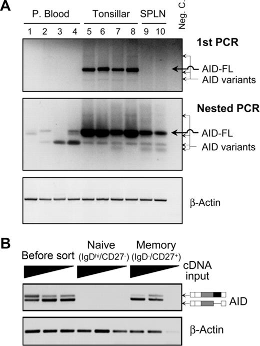
Although normal B cells have been shown to express AID mRNA,15,18,24 previous studies have not examined in detail normal human B-cell expression of the variant AID mRNA species. We examined AID mRNA expression in normal PB, splenic, and tonsillar B cells using single round and nested RT-PCR. Following a single round of RT-PCR, tonsillar B cells were observed to express AID transcripts (Figure 2A top panel). The major product is a 646-bp band that reflects FL AID, and there are some minor bands of various sizes. By contrast, B cells purified from PB or spleen did not yield any product under these conditions. However, when a more sensitive nested PCR method was used, AID transcripts could be detected in PB and splenic B cells, and minor PCR bands became more pronounced in tonsillar B cells (Figure 2A middle panel). Given the current literature suggesting that AID is only expressed in GC B cells,24 it was surprising to find evidence of AID-expressing PB B cells. Moreover, it is interesting that PB B cells predominately expressed AID variants rather than FL AID. However, given the sensitivity of nested PCR, it is possible that this result reflects the presence of a very rare subpopulation of cells. We next subfractionated PB B cells into naive (IgDhi/CD27−) and memory (IgD−/CD27+) populations. Figure 2B demonstrates that AID transcripts were exclusively expressed in memory but not naive B cells, and the shorter transcript dominated over the level FL AID.
Expression of AID-splicing variants in normal B cells. (A) Expression of various transcripts of AID in B cells of peripheral blood, tonsils, and spleens of healthy donors. An agarose gel image of RT-PCR products (top panel), the nested PCR (middle panel), and β-actin PCR loading control (bottom panel) is shown. (B) An agarose gel image of AID-nested RT-PCR on RNAs isolated from FACS sorted pure naive and memory B cells. To ensure the PCR sensitivity, cDNA inputs with 3 different dilutions (1:1, 1:4, and 1:16) were used in PCR amplification.
Expression of AID-splicing variants in normal B cells. (A) Expression of various transcripts of AID in B cells of peripheral blood, tonsils, and spleens of healthy donors. An agarose gel image of RT-PCR products (top panel), the nested PCR (middle panel), and β-actin PCR loading control (bottom panel) is shown. (B) An agarose gel image of AID-nested RT-PCR on RNAs isolated from FACS sorted pure naive and memory B cells. To ensure the PCR sensitivity, cDNA inputs with 3 different dilutions (1:1, 1:4, and 1:16) were used in PCR amplification.
To verify that the various AID PCR products reflected bona fide AID-splicing variants, we subcloned the single round RT-PCR reaction from tonsillar B cells and sequenced 60 clones. Our sequencing data verified that that all of the clones sequenced were exact matches with either FL AID or one of the various AID- splicing isoforms previously identified in malignant B cells.13-15 This experimental approach also permitted an independent assessment of the relative expression levels of each transcript and as summarized in Table 1, although different AID-splicing variants are detectable in tonsillar B cells, the FL AID transcript constituted the majority (77%) of clones sequenced. Our data demonstrate that normal human B cells indeed express multiple bona fide alternatively spliced AID mRNA variants.
Prevalence of AID-splicing variants in normal tonsillar B cells
| Splicing variant . | Accession No. . | No. of clones . | Frequency . |
|---|---|---|---|
| AID-FL | NM_020661 | 46 | 77% |
| AID-ΔE4a | AY536516 | 4 | 7% |
| AID-ΔE4 | AY536517 | 6 | 10% |
| AID-ΔE3E4 | AY534975 | 2 | 3% |
| AID-ivs3 | AY541068 | 2 | 3% |
AID alternative splicing is highly conserved
We then asked if alternative splicing of AID might be a general mechanism utilized by all AID-expressing species. First, we examined the exon organizations of AICDA (AID gene) among various species using available sequences in the database. As depicted in Figure 3A, the number of exons, sizes, and splicing positions of human AICDA are identical to that of chimpanzee (Pan troglodytes), cattle (Bos taurus), mouse (Mus musculus), and chicken (Gallus gallus). Although zebrafish (Danio rerio) AICDA only has 3 exons, all of its splicing sites match perfectly to that of human AICDA exons 3, 4, and 5, which precisely coincide with the locations of the alternative splicing events (Figure 3A). These data suggest that the AICDA gene in all higher vertebrates has the potential to be alternatively spliced in a manner identical to that described in human B cells. To substantiate this hypothesis, we next analyzed AID alternative splicing in murine B cells, and we observed that murine splenic and lymph node B cells expressed FL and AID splice variants, albeit the latter were expressed at very low levels (Figure 3B). Sequence analysis confirmed that the bands appearing in Figure 3B correspond to AID mRNA variants (data not shown).
AID alternative splicing is conserved among vertebrates. (A) Alignment of the AICDA exon organization of human (Homo sapiens), chimpanzee (P troglodytes), cattle (B taurus), mouse (M musculus), chicken (G gallus), and zebrafish (D rerio). (B) Murine B-cell AID alternative splicing. Agarose gel image of AID RT-PCR performed using mouse lymph node (LN) and spleen (SP) B cells.
AID alternative splicing is conserved among vertebrates. (A) Alignment of the AICDA exon organization of human (Homo sapiens), chimpanzee (P troglodytes), cattle (B taurus), mouse (M musculus), chicken (G gallus), and zebrafish (D rerio). (B) Murine B-cell AID alternative splicing. Agarose gel image of AID RT-PCR performed using mouse lymph node (LN) and spleen (SP) B cells.
AID-splicing variants are singly expressed in individual GC B cells
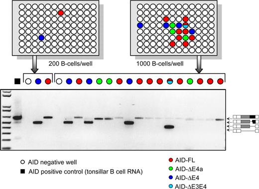
Since alternative splicing has the potential to significantly modify the function of the protein products due to loss or gain of functional domain-encoding sequences, it has been frequently suggested that the alternatively spliced protein products may function as dominant-negative or dominant-positive regulators.25 However, this argument is based on the assumption that splicing variants are physically coexpressed with the FL counterpart within the same cell. We next tested this assumption by asking whether the various AID splice variants are coexpressed with FL AID in GC B cells. Because AID is expressed only in a subpopulation of GC B cells,26 we used limiting dilution as previously described.13 As shown in Figure 4, 17 of 18 PCR positive wells exhibited a single splicing variant, while only 1 well showed evidence of 2 products, for instance, a weak band of FL AID and a strong band of AID-ΔE4. The latter may or may not be due to 2 AID-positive cells co-fractionated in the same well. Consistent with the predominance of the FL AID transcript in tonsillar B cells using the nested PCR approach (Figure 2A), the majority of the wells only yielded a FL AID product. Our surprising results suggest that the majority of B cells in GCs only express a single AID variant at a given time. Therefore, the alternative AID-spliced products are unlikely to play any dominant-negative or dominant-positive role in regulating the normal function of FL AID, but rather function independently in individual B cells.
AID-splicing variants are singly expressed in individual tonsillar B cells. Purified human tonsillar B cells were limiting diluted into 96-well PCR plates at the densities of 200 cells/well and 1000 cells/well (depicted in top panels). The wells filled with various colors indicate where AID was positively detected, and the color codes show the identities of the AID PCR products. (Bottom panel) DNA gel image showing the amplified AID variants from those PCR-positive wells.
AID-splicing variants are singly expressed in individual tonsillar B cells. Purified human tonsillar B cells were limiting diluted into 96-well PCR plates at the densities of 200 cells/well and 1000 cells/well (depicted in top panels). The wells filled with various colors indicate where AID was positively detected, and the color codes show the identities of the AID PCR products. (Bottom panel) DNA gel image showing the amplified AID variants from those PCR-positive wells.
Alternative splicing of AID renders selective modulation of CSR and SHM
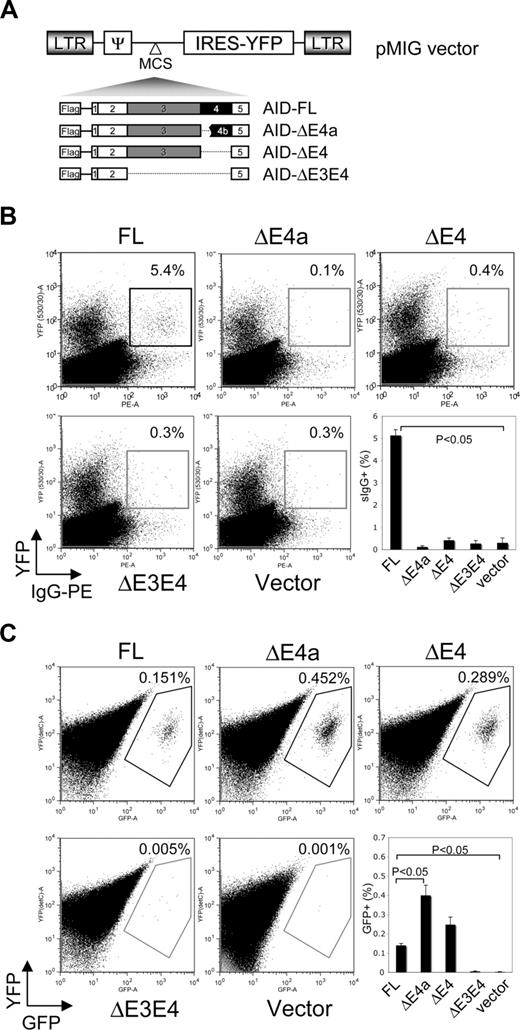
Because individual B cells only express a single type of AID variant, we next tested whether these variants are equally functional in carrying out CSR and SHM functions. Of note, we did not further study the AID-ivs3 mRNA variant because sequence analysis of this AID variant suggested the possibility that AID-ivs3 may be a target of the nonsense-mediated mRNA decay (NMD) surveillance system27 and if so, would be unlikely to yield a protein product due to a premature stop codon (PTC) located 60 nt upstream of the last intron-exon junction (data not shown). To assess CSR activity of the remaining variants, we cloned Flag-tagged AID variants into a retroviral vector upstream of an IRES-YFP cassette (Figure 5A), transduced LPS/IL4 stimulated AID−/− mouse splenic B cells with the viral supernatants and measured CSR as assessed by the expression of surface IgG1. As shown in Figure 5B, human FL AID restored CSR function; however, all 3 AID-splicing variants completely lacked such activity. Our results suggest that alternative splicing is an efficient way to inactivate the CSR activity of AID.
Restoration of SHM and CSR by complementation with expression of AID variants. (A) Cartoon representation of retroviral constructs of AID variants. (B) Flow cytometry assessing CSR in AID-deficient spleen cells stimulated with IL-4 and LPS and infected with retroviruses-expressing FL or alternatively spliced AID variants. CSR was measured by the number of YFP-positive cells acquiring IgG1 cell surface expression in the gated viable population. Numbers indicate percentage of IgG1+GFP+ cells (top right). Dot-plot data are representative of 4 individual experiments that are summarized in the bar-chart panel. The P values were obtained using a 1-tailed t test. (C) Flow cytometry assessing SHM activity by scoring GFP reversion frequencies. Murine 70Z/3 cells stably expressing a GFP transgene with a premature stop codon were infected with retroviruses expressing FL or alternatively spliced AID variants. SHM activity was measured by the number of YFP (Y axis) positive cells acquiring GFP (X axis) expression over 6 weeks of time. Numbers indicate the percentage of YFP+GFP+ cells (top right). Dot-plot data are representative of 4 individual experiments which are summarized in the bar-chart panel. The P values were obtained using a 1-tailed t test.
Restoration of SHM and CSR by complementation with expression of AID variants. (A) Cartoon representation of retroviral constructs of AID variants. (B) Flow cytometry assessing CSR in AID-deficient spleen cells stimulated with IL-4 and LPS and infected with retroviruses-expressing FL or alternatively spliced AID variants. CSR was measured by the number of YFP-positive cells acquiring IgG1 cell surface expression in the gated viable population. Numbers indicate percentage of IgG1+GFP+ cells (top right). Dot-plot data are representative of 4 individual experiments that are summarized in the bar-chart panel. The P values were obtained using a 1-tailed t test. (C) Flow cytometry assessing SHM activity by scoring GFP reversion frequencies. Murine 70Z/3 cells stably expressing a GFP transgene with a premature stop codon were infected with retroviruses expressing FL or alternatively spliced AID variants. SHM activity was measured by the number of YFP (Y axis) positive cells acquiring GFP (X axis) expression over 6 weeks of time. Numbers indicate the percentage of YFP+GFP+ cells (top right). Dot-plot data are representative of 4 individual experiments which are summarized in the bar-chart panel. The P values were obtained using a 1-tailed t test.
Next, we asked whether the splicing variants could mediate SHM using a GFP-based reversion assay to determine the SHM activity of the AID variants in the murine 70Z/3 cell line that does not express endogenous AID.4 The recombinant viruses encoding the FL or splicing isoforms of AID were transduced into 70Z/3 cells that had previously been stably transfected with a GFP transgene with a premature stop codon at amino acid residue 107 position.28 Virally transduced cells were enriched by sorting YFP-positive cells and scored for SHM activity by determining the frequency of GFP-positive cells. As shown in Figure 5C, FL AID is SHM competent, while AID-ΔE3E4 is SHM defective as expected. However, despite complete lack of CSR activity, both AID-ΔE4a and AID-ΔE4 variants exhibited a robust level of SHM activity that exceeded the level of activity supported by FL AID. In an alternative SHM assay, we also sequenced GFP in the transduced cells and observed that the AID-ΔE4a and AID-ΔE4 variants induced more GFP point mutations than that observed in cells transduced with FL AID (data not shown). Our data clearly demonstrate that AID splicing variants differentially express SHM and CSR activity.
It remained formally possible, however, that the varying activities of the AID variants could be alternatively explained by altered nuclear localization as a result of splicing-induced alterations in the nuclear export signal (NES) in its C terminus.29,30 For example, prior studies by McBride et al31 have shown that alteration in subcellular localization by removal of the C-terminal NES also results in defective SHM. To address this possibility, we transiently overexpressed Flag-tagged AID variants into HeLa cells and examined intracellular localization. As shown in Figure 6, AID-FL exhibited predominant cytoplasmic localization, as did the AID-ΔE4a and AID-ΔE3E4 variants. This latter observation was expected since the internal in-frame deletions by splicing do not affect either the NLS or NES domains. However, the C-terminal truncated AID-ΔE4 showed a slightly increased nuclear localization pattern similar to that displayed by a previously described AID C-terminal deletion mutant.30 Collectively, these data suggest that the varying activities of the AID variants are not readily explained by significant changes in intracellular localization properties.
Subcellular localization of overexpressed AID variants. Flag epitope- tagged AID variants (as indicated) were transiently overexpressed in HeLa cells, and cells were then stained with anti-Flag antibody (red) and counterstained with DAPI (blue) to detect nuclear DNA.
Subcellular localization of overexpressed AID variants. Flag epitope- tagged AID variants (as indicated) were transiently overexpressed in HeLa cells, and cells were then stained with anti-Flag antibody (red) and counterstained with DAPI (blue) to detect nuclear DNA.
Alternative splicing of AID in B CLL
Although our studies described above are the first to demonstrate that normal GC B cells express AID splice variants and that memory B cells also exhibit this trait as well albeit at very low levels, several previous reports have suggested that aberrantly expressed and alternatively spliced AID may play important roles in the pathogenesis of CLL and other B-cell lineage malignancies.13-16,32-34 Chiorazzi and colleagues13 originally found that only a small population of B-CLL cells express AID. Here, we further expand their findings by examining AID splicing in individual B-CLL cells. As shown in Figure 7A, individual CLL B cells also appear to only express a single AID variant at a given point in time.
Expression of AID-splicing variants in CLL B cells. (A) Purified CLL B cells from 3 patients were limiting diluted into 96-well PCR plates at the densities of 1000 cells/well (depicted in top panels). The wells filled with various colors indicate where AID was positively detected, and the color codes show the identities of the AID PCR products. (Bottom panels) DNA gel images showing the amplified AID variants from those PCR-positive wells. Data shown are representative of a total of 6 B-CLL patients studied in this manner. (B) Overexpression of AID-splicing variants in CLL B cells. (Top panel) A composite gel image of AID RT-PCR; while the bar chart (bottom panel) is the quantitation of the same image by densitometry. The Ig VH genes in CLL B cells shown in lanes 5, 6, 11, and 13 were mutated and those shown in lanes 7 to 10 and 12 were unmutated.
Expression of AID-splicing variants in CLL B cells. (A) Purified CLL B cells from 3 patients were limiting diluted into 96-well PCR plates at the densities of 1000 cells/well (depicted in top panels). The wells filled with various colors indicate where AID was positively detected, and the color codes show the identities of the AID PCR products. (Bottom panels) DNA gel images showing the amplified AID variants from those PCR-positive wells. Data shown are representative of a total of 6 B-CLL patients studied in this manner. (B) Overexpression of AID-splicing variants in CLL B cells. (Top panel) A composite gel image of AID RT-PCR; while the bar chart (bottom panel) is the quantitation of the same image by densitometry. The Ig VH genes in CLL B cells shown in lanes 5, 6, 11, and 13 were mutated and those shown in lanes 7 to 10 and 12 were unmutated.
Our present study shows that the splicing variants AID-ΔE4a and AID-ΔE4 are defective in CSR but are hyperactive in mediating SHM (Figure 5C). These results imply that enhanced expression of one of these AID variants could result in enhanced SHM. To that end, we quantified expression levels of the AID mRNA variants to determine if the pattern of AID splicing in CLL B cells was different from that exhibited by normal GC B cells. Figure 7B shows that the predominant mRNA species expressed by normal GC B cells was FL AID, accounting for approximately 85–90% of the total AID expressed while the AID-ΔE4 SHM-hyperactive version of AID is only expressed at a minor level (consistent with the subcloning data in Table 1). By contrast, expression of the AID-ΔE4 SHM-hyperactive variant in CLL B cells was much greater, accounting for approximately 35-45% of total AID expression in all patient cells studied. Finally, it is interesting to note that the expression patterns of AID variants in B-CLL cells and in normal memory B cells are strikingly similar. However, expression levels of AID and its variants are markedly lower in normal memory B cells and in limiting dilution and PCR studies not shown, we observed that AID is expressed in memory B cells at a 10-fold lower frequency than in CLL B cells.
Discussion
Through its mutagenic activity, AID mediates physiological CSR and SHM in GC B cells by targeting immunoglobulin gene loci. However, there is accumulating evidence that AID also targets many other endogenous gene loci in normal GC B cells and the frequency of AID-mediated mutagenesis of nonimmunoglobulin genes is greater in malignant B cells.4,5 Recently, AID mistargeting was shown to be counterbalanced by a safety net of DNA repair mechanisms.7,35-37 Indeed, tipping this balance by either increasing AID expression or by decreasing DNA repair activity leads to tumorigenesis as demonstrated in AID transgenic mice3 and in DNA repair knockout mice,38-41 respectively. Additionally, the tumor suppressor p53-dependent DNA damage checkpoint oversees the repair efficiency and eliminates cells with persistent or irreparable DNA damages by inducing apoptosis in non-GC B cells. However, p53 is transcriptionally repressed in GC B cells by Bcl-642 suggesting a more urgent need for the tight regulation and containment of AID activity.43 In this regard, a recent study showed that ∼25% of transcribed genes in murine GC B cells are targeted by AID but are then refurbished by DNA repair systems.36 This raises 2 important questions. First, how is the mutagenic activity of AID promptly inactivated upon completion of CSR and SHM? Second, is there a mechanism that could also heighten AID mutagenic activity possibly predisposing to B-cell malignant transformation and/or promoting accumulation of additional genetic mutations following transformation?
Here we show that alternative splicing of AID selectively generates SHM-inactive (AID-ΔE3E4), or SHM-hyperactive variants (AID-ΔE4a or AID-ΔE4). Similarly, the CSR activity of AID is effectively extinguished by the process of alternative splicing. Furthermore, the splice variants are mutually exclusively expressed in individual B cells, normal or leukemic alike. Therefore, the splicing mechanism provides a novel tailored means to modulate residual AID activity remaining at the end of the GC reaction that is no longer advantageous. On the other hand, alternative splicing also generates the seemingly hazardous SHM-hyperactive variants AID-ΔE4a and AID-ΔE4. It is important to note that additional studies are needed to verify our results at the protein level. We have indeed attempted these studies using a commercially available polyclonal anti-AID antibody that would putatively recognize each of the splicing variants. However, although useful in analysis of AID in an overexpression system, this antibody did not have sufficient specificity to allow conclusions to be made concerning AID variant protein expression in primary B cells (data not shown).
Mutagenesis studies have shown that truncation of the last 10 amino acid residues of the AID C terminus renders CSR deficiency ex vivo44 resembling our AID-ΔE4 data. Furthermore, Honjo and colleagues described a patient carrying a single germ line mutation at the splicing site of the AID intron 4 leading to the production of AID-ΔE4 instead of AID-FL.45 Of interest, the patient exhibited a typical hyper-IgM syndrome suggesting AID-ΔE4 indeed causes CSR deficiency in vivo. These observations together with our present study strongly suggest that AID alternative splicing is an effective mechanism to contain AID CSR activity, presumably to prevent accumulation of DNA double strand breaks (DSB) which normally are detrimental.
We have also made the interesting observation that the ΔE4a and ΔE4 variants express a higher level of SHM activity than FL AID. The ΔE4 variant completely lacks the CSR domain, and its higher SHM activity is consistent with prior work by Nussenzweig and colleagues44 demonstrating the last 10 amino acids (aa) of the AID C terminus may actually attenuate SHM activity. The ΔE4a variant, however, also displays enhanced SHM activity despite retention of the CSR domain, however, it also includes an internal 10-aa in-frame deletion. Recent 3D structural analysis of the AID family member, Apobec3G,46 revealed that an internal 10-aa segment homologous to the spliced out amino acids in the ΔE4a AID variant is critical in forming an α-helical structure. This suggests that the 10-aa in-frame deletion in ΔE4a may similarly disrupt the 3D structure of this AID variant perhaps leading to the structural repositioning of the C-terminal domain that may likewise alleviate the inhibitory influence of an intact CSR domain on SHM. Thus, deletion of the C-terminal segment of AID achieved in the variant AID-ΔE4 or repositioning of the CSR domain away from the deaminase activity center in AID-ΔE4a would have similar effects on SHM.46
Because AID expression correlates well with cytogenetic abnormalities and clinical prognosis in B-CLL,47 and AID can introduce genome-wide mutations,4,36 our observations concerning alternative splicing and restriction of isoform expression at the individual cell level may have relevance to leukemic B cells in B-CLL patients. Moreover, given that primary CLL B cells expressing AID also invariably express relatively higher levels of the AID variants, it is tempting to speculate that heightened expression levels of the SHM-hyperactive variants may be playing some role in CLL pathogenesis and/or disease progression. Paradoxically, initial work by McCarthy et al suggested that AID expression in CLL B cells was a potential surrogate marker for the generally clinically unfavorable unmutated (UM) Ig VH status.14 However, later study of larger cohorts47 as well as our own unpublished data from 70 patients suggests that this correlation is less robust than first described. Nevertheless, expression of AID in UM CLL B cells may instead suggest that AID requires additional cofactors for specific Ig locus targeting as has been previously postulated,15 and if so, UM CLL B cells may lack expression of these factors. Alternatively, AID may be active in UM CLL B cells but cells acquiring SHM in their B-cell receptor may now be at a competitive disadvantage if the specificity of the B-cell receptor is important for disease maintenance and/or progression. This latter model would accommodate the hypothesis that expression of FL- or either of the 2 SHM-competent AID isoforms contributes to acquisition of cytogenetic abnormalities in B-CLL despite the apparent lack of ongoing SHM of Ig loci. Definitive studies in primary CLL B cells designed to rigorously test whether there is a direct causative effect of FL-AID, AID-ΔE4, and/or AID-ΔE4a in introducing common genetic alterations observed in CLL are warranted, however, given the very low number of cells expressing these SHM-competent AID isoforms, current technology does not permit studies of this nature.
Our studies support previous findings by others13 that AID is only expressed in a very small number of CLL B cells. The question now becomes how this apparent small number of AID-expressing cells impacts the overall disease. One of the possible explanations is that although the size of the AID-expressing CLL B-cell pool may be very small in blood, it is conceivable that other microenvironments support a larger population of these cells, given the fact that the frequency of AID-expressing CLL B cells could be increased following in vitro activation.13 Moreover, since AID expression is induced and largely confined to normal GC B cells, CLL B-cell AID expression may simply be one more indication that CLL B cells, particularly UM B-CLL, resemble more closely activated, memory B cells rather than naive B cells.20
In summary, our data suggest a critical role for AID alternative splicing in antibody maturation as well as tumor suppression in B cells. Since there is a precedence for differential signal transduction pathways driving alternative splicing of other genes,48-50 topics of interest for future investigation would include elucidation of the signaling pathway that instructs AID alternative splicing and determination of whether this pathway could be manipulated to influence antibody maturation and lymphomagenesis of B-lineage cells. It would also be interesting to examine SHM and CSR functions as well as lymphomagenesis in mice that are deficient in factors necessary for alternative splicing. Finally, our data demonstrating that AID is expressed in a “one variant per cell” manner raises the question of whether this occurs more frequently in nature, and perhaps has been largely missed because traditional analysis of alternative splicing is performed using total RNA from bulky cell populations or tissue. To our knowledge, the only other reports of alternatively spliced genes expressed in a “one variant per cell” manner are prepro-tachykinin A51 , the calcium channel protein CaV2.1,52 and SCN5A.53 These 3 reports, however, did not discuss the implications of their results from the standpoint of regulation of alternatively spliced genes. Since alternative splicing could generate an enormous amount of functional diversity from limited genomic resources, and is a mechanism that is specific to multicellular organisms,25 this potential functional diversification mechanism in the absence of overtly altering the gene expression spectrum, seems to be ideal for subtly diversifying the same group of cells seen in organogenesis and development.
The publication costs of this article were defrayed in part by page charge payment. Therefore, and solely to indicate this fact, this article is hereby marked “advertisement” in accordance with 18 USC section 1734.
Acknowledgments
We thank Dr Tasuku Honjo for the permission to use the AID-deficient mice and Dr Riccardo Dalla-Favera for providing the mice. We are also grateful to Renee Tschumper, Bonnie Arendt, Cheryl Jankiewicz, and Dr Neil Kay for their valuable assistance and constructive discussions.
This work was supported in part by the National Institutes of Health, Bethesda, MD (CA062242, D.F.J.).
National Institutes of Health
Authorship
Contribution: X.W. designed and conducted most of the experiments; J.R.D. and S.K.C. helped in strategizing the project and isolating tonsillar B-cell subsets; G.N. helped with interpreting and processing the data; and D.F.J. oversaw the entire project. X.W. and D.F.J. wrote the manuscript with comments from all co-authors.
Conflict-of-interest disclosure: The authors declare no competing financial interests.
Correspondence: Diane F. Jelinek, Department of Immunology, Mayo Clinic, 200 1st Street SW, Rochester, MN 55905; e-mail: jelinek.diane@mayo.edu.


This feature is available to Subscribers Only
Sign In or Create an Account Close Modal