Abstract
Phosphoinositide 3-kinase (PI3K) negatively regulates Toll-like receptor (TLR)–mediated interleukin-12 (IL-12) expression in dendritic cells (DCs). We show here that 2 signaling pathways downstream of PI3K, mammalian target of rapamycin (mTOR) and glycogen synthase kinase 3 (GSK3), differentially regulate the expression of IL-12 in lipopolysaccharide (LPS)–stimulated DCs. Rapamycin, an inhibitor of mTOR, enhanced IL-12 production in LPS-stimulated DCs, whereas the activation of mTOR by lentivirus-mediated transduction of a constitutively active form of Rheb suppressed the production of IL-12. The inhibition of protein secretion or deletion of IL-10 cancelled the effect of rapamycin, indicating that mTOR regulates IL-12 expression through an autocrine action of IL-10. In contrast, GSK3 positively regulates IL-12 production through an IL-10–independent pathway. Rapamycin-treated DCs enhanced Th1 induction in vitro compared with untreated DCs. LiCl, an inhibitor of GSK3, suppressed a Th1 response on Leishmania major infection in vivo. These results suggest that mTOR and GSK3 pathways regulate the Th1/Th2 balance though the regulation of IL-12 expression in DCs. The signaling pathway downstream of PI3K would be a good target to modulate the Th1/Th2 balance in immune responses in vivo.
Introduction
Dendritic cells (DCs) recognize pathogens via pattern-recognition receptors, such as Toll-like receptors (TLRs), nucleotide-binding oligomerization domain (NOD)-like receptors, and retinoic acid inducible gene-1 (RIG-I)-like receptors, produce various cytokines, including interleukin-12 (IL-12), and thus activate the innate immune system, which in turn leads to the induction of adaptive immunity.1-5 Bioactive IL-12 is composed of p40 and p35 subunits and functions as a crucial inducer of Th1 responses. IL-12 is typically produced by antigen-presenting cells such as DCs and monocytes-macrophages and plays an important role in infection and tumor immunity.1-5 Because the overproduction of IL-12 gives rise to strong cell-mediated immunity and organ-specific autoimmune diseases via exaggerated Th1 cell differentiation, it is critical that IL-12 levels be tightly controlled.5
Phosphoinositide 3-kinases (PI3Ks) are lipid kinases playing important roles in various signal transduction pathways.6 PI3K family members are classified into 4 subgroups according to their structure and substrate specificity. Among them, class IA heterodimeric PI3Ks are involved in receptor-mediated signaling pathways in the immune system.6,7 Phosphatidylinositol-(3,4)bisphosphate and phosphatidylinositol(3,4,5)trisphosphate produced by class IA PI3Ks recruit specific signaling proteins containing a pleckstrin homology domain to the plasma membrane. These proteins include Akt and phosphoinositide-dependent kinase 1 and are involved in a wide range of cellular responses, such as cell growth, survival, and cytokine production.6,7 PI3K signaling pathways are counteracted by phosphatase and tensin homologue deleted on chromosome 10 (PTEN), a 3-phosphoinositide–specific lipid phosphatase.6
We have previously demonstrated that PI3K negatively regulates IL-12 production in DCs stimulated with TLR ligands.8,9 An enhanced Th1 response was observed on Leishmania major infection,8 and an impaired Th2 response was observed on Strongyloides venezuelensis infection10 in mice deficient for p85α, a major regulatory subunit of class IA PI3K, indicating that PI3K plays a key role in the regulation of Th1/ Th2 balance in vivo. Although several reports confirmed the negative feedback regulation of IL-12 production by PI3K on TLR stimulation,11-13 the molecular mechanism(s) remains controversial.
One downstream substrate of PI3K pathways is a Ser/Thr protein kinase named “mammalian target of rapamycin” (mTOR), which regulates cell growth and protein synthesis by activating p70 S6 kinase (p70S6K) and by inhibiting eukaryotic initiation factor 4E-binding protein 1 (4E-BP1).14 There are 2 functionally distinct mTOR complexes, mTORC1 and mTORC2, and only mTORC1 acts downstream of the PI3K-Akt-tuberous sclerosis complex 2 (TSC2)–Rheb signaling pathway to phosphorylate p70S6K and 4E-BP1 in a rapamycin-sensitive fashion14 (Figure 1A). Although the mTOR pathway is activated in response to not only growth factors but also environmental stresses such as hypoxia,14 the TLR-triggered mTOR function is poorly understood. In the present study, we show that mTOR negatively regulates IL-12 production through the production of IL-10 in DCs. We further demonstrate that glycogen synthase kinase 3 (GSK3), another downstream target of PI3K pathways, is also involved in the PI3K-mediated regulation of IL-12 production in a manner distinct from that of mTOR. We also provide evidence that the inhibition of mTOR and GSK3 pathways in DCs results in the increase and decrease of Th1 responses, respectively.
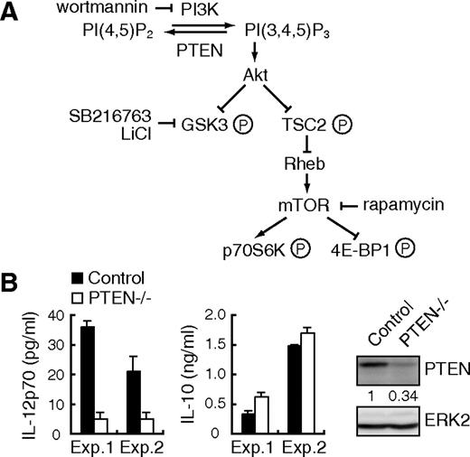
LPS-induced IL-12 production is suppressed in PTEN−/− BMDCs. (A) The overview of PI3K signaling pathway. PI(4,5)P2 indicates phos-phatidylinositol(4,5)bisphosphate; PI(3,4,5)P3, phosphatidylinositol-(3,4,5)tris-phosphate. (B) BMDCs from PTEN−/− mice (n = 2) or their littermate controls (n = 2) were stimulated with 1 μg/mL LPS for 24 hours and assayed for the production of IL-12p70 and IL-10 by ELISA. Data are indicated as median plus or minus SD. Essentially, the same results were obtained with 2 independent experiments (experiments 1 and 2). The expression levels of PTEN and ERK2 in BMDCs were determined by Western blotting (right panel).
LPS-induced IL-12 production is suppressed in PTEN−/− BMDCs. (A) The overview of PI3K signaling pathway. PI(4,5)P2 indicates phos-phatidylinositol(4,5)bisphosphate; PI(3,4,5)P3, phosphatidylinositol-(3,4,5)tris-phosphate. (B) BMDCs from PTEN−/− mice (n = 2) or their littermate controls (n = 2) were stimulated with 1 μg/mL LPS for 24 hours and assayed for the production of IL-12p70 and IL-10 by ELISA. Data are indicated as median plus or minus SD. Essentially, the same results were obtained with 2 independent experiments (experiments 1 and 2). The expression levels of PTEN and ERK2 in BMDCs were determined by Western blotting (right panel).
Methods
Reagents
Lipopolysaccharide (LPS) from Escherichia coli 055:B5 was purchased from Sigma-Aldrich (St Louis, MO). Recombinant human (rh) GM-CSF, rhIL-4, rhIL-10, and recombinant mouse (rm) GM-CSF were purchased from PeproTech (Rocky Hill, NJ). Wortmannin, rapamycin, SB216763, cycloheximide, and brefeldin A (BFA) were purchased from Calbiochem (San Diego, CA). Antibodies to ERK2, IκBα, p70S6K, Rheb, GSK3α/β, and STAT3 were purchased from Santa Cruz Biotechnology (Santa Cruz, CA). Antibody to Akt and phosphorylation-specific antibodies to Akt (Ser473), GSK3α/β (Ser21/Ser9), TSC2 (Thr1462), p38 (Thr180/Tyr182), ERK (Thr202/Tyr204), JNK (Thr183/Tyr185), and STAT3 (Tyr705) were purchased from Cell Signaling Technology (Danvers, MA).
Mice
C57BL/6 and BALB/c mice were purchased from Nihon SLC. IL-10−/− mice on a C57BL/6 background were purchased from The Jackson Laboratory (Bar Harbor, ME). Mice deficient for p85α were reported previously.15,16 Mice had been backcrossed to the BALB/c background for 12 generations. STAT3 mutant (LysM-Cre x STAT3flox/flox) mice17 on a mixed (129 × C57BL/6) background were kindly provided by Dr K. Takeda (Osaka University, Osaka, Japan). STAT3flox/flox or LysM-Cre x STAT3flox/+ mice were used as controls. PTEN mutant (LysM-Cre x PTENflox/flox) mice18,19 on a C57BL/6 background were kindly provided by Dr A. Suzuki (Akita University, Akita, Japan). PTENflox/flox mice were used as controls. LysM-Cre x STAT3flox/flox and LysM-Cre x PTENflox/flox mice were referred to here as STAT3−/− and PTEN−/− mice, respectively. Mice were maintained at Taconic Farms (Germantown, NY) or in our animal facility under specific pathogen-free conditions. All experiments were performed in accordance with our institutional guidelines.
Preparation of dendritic cells
To generate bone marrow (BM)–derived DCs (BMDCs), mouse BM cells were cultured at 106/mL in complete medium (RPMI 1640; Sigma-Aldrich, 10% fetal calf serum, 55 μM 2-mercaptoethanol, 100 U/mL penicillin, 100 μg/mL streptomycin) supplemented with 10 ng/mL rmGM-CSF for 6 days. The culture medium was changed every 2 days. On day 6, BMDCs were isolated using antimouse CD11c magnetic beads (Miltenyi Biotec, Auburn, CA) with an AutoMACS (Miltenyi Biotec). Human peripheral blood mononuclear cells from normal healthy volunteers were isolated by centrifugation on a Ficoll-Metrizoate density gradient (Lymphoprep; Nycomed, Oslo, Norway). The protocol was approved by the local ethics committee at Keio University School of Medicine, and informed consent was obtained from donors in accordance with the Declaration of Helsinki. Monocytes were then isolated using antihuman CD14 magnetic beads (Miltenyi Biotec) with an AutoMACS, followed by incubation at 106 cells/mL in complete medium supplemented with 100 ng/mL rhGM-CSF and 100 ng/mL rhIL-4 for 6 days, to obtain monocyte-derived DCs (MDDCs).
Western blotting
Western blotting was carried out as described.8 To detect phospho-STAT3 and phospho-TSC2, immunoreaction enhancer (Can Get Signal, Toyobo, Japan) was added to the reaction according to the manufacturer's instructions. ERK2 was used as a loading control. A LAS-3000 imaging system (Fuji) was used to produce digital images. Signal intensities (for phospho-STAT3) and signal profiles (for p70S6K) were quantified with Image Gauge software version 4.1 (Fuji).
Enzyme-linked immunosorbent assay
Cytokine concentrations in the culture supernatants were quantified by enzyme-linked immunosorbent assay (ELISA; Quantikine; R&D Systems, Minneapolis, MN).
Quantitative real-time polymerase chain reaction
Total RNA was prepared using NucleoSpin RNA II (Macherey-Nagel, Düreu, Germany), and cDNA was synthesized with Ready-To-Go T-Primed First-Strand kit (GE Healthcare, Chalfont St Giles, United Kingdom). Quantitative real-time polymerase chain reaction (PCR) was performed by applying the real-time SYBR Green PCR technology using SYBR premix Ex Taq (Takara, Otsu, Japan) with specific primers on an iCycler IQ (Bio-Rad, Hercules, CA). PCR cycling was as follows: 95°C for 10 seconds for 1 cycle, 95°C for 5 seconds, 58°C for 20 seconds, 72°C for 15 seconds for 40 cycles, and 70°C for 5 minutes. Amplification of cyclophilin A mRNA was done for each sample as an endogenous control. Primer pairs specific for IL-12p40 (forward, CAGAAGCTAACCATCTCCTGGTTTG; reverse, CCGGAGTAATTTGGTGCTCCACAC), IL-12p35 (forward, TCACATCTCATCTCCCCAAA; reverse, TCTGCTAACACATTGAGGGG), IL-10 (forward, GGTTGCC-AAGCCTTATCGGA; reverse, ACCTGCTCCACTGCCTTGCT), interferon-β (IFN-β) (forward, CCATCCAAGAGATGCTCCAG; reverse, GTGGAGAGCAGTTGAGGACA), suppressor of cytokine signaling 3 (SOCS3) (forward, GGGGGAGGCAGGAGGTGATGGA; reverse, GGGCGGGCTGGAGGTGGATT) and cyclophilin A (forward, ATGGCACTGGCGGCAGGTCC; reverse, TTGCCATTCCTGGACCCAAA) were used.
Preparation of lentiviral vectors
The following constructs were kindly provided by Dr H. Miyoshi (RIKEN, Tsukuba, Japan): CSII-EF-MCS-IRES2-Venus, a self-inactivating lentiviral construct; pCAG-HIVgp and pCMV-VSVG-RSV-Rev, packaging constructs.20 This lentiviral system is designed to express a desired gene under the direction of the elongation factor-1 promoter along with internal ribosomal entry site (IRES)-driven Venus, a derivative of YFP,21 as a marker for monitoring the infection efficiency. Mouse Rheb was amplified by PCR using cDNA from the brain of C57BL/6 mice as a template (5′ primer, ATGCCTCAGTCCAAGTCCCG; 3′ primer, TCACATCACCGAGCACGAAG) and cloned into the pGEM-T Easy vector (Promega, Madison, WI). After sequence verification, the construct was subjected to PCR mutagenesis to obtain Rheb Q64L, a constitutively active form of Rheb, Glu-64 replaced by Leu. The product was verified by DNA sequencing and subcloned into CSII-EF-MCS-IRES2-Venus. For the generation of lentiviral vectors, 293T cells were transfected with CSII-EF-MCS-IRES2-Venus with or without Rheb Q64L insert, pCAG-HIVgp, and pCMV-VSUG-RSV-Rev using Lipofectamine 2000 (Invitrogen, Carlsbad, CA). After 2 days, culture supernatants were passed through a 0.45-μm filter, condensed to 0.5% volume, and used for gene transduction.
Generation of gene-transduced BMDCs
Mouse BM cells were incubated with phycoerythrin-conjugated antibodies against CD3ϵ, CD4, CD8α, CD11b, Gr-1, B220, and TER119 (BD Biosciences, San Jose, CA) along with anti-phycoerythrin microbeads (Miltenyi Biotec), followed by negative selection with an AutoMACS. The remaining cells (0.5 × 10⁵ cells/0.5 mL) were cultured with 10 ng/mL rmGM-CSF in a 24-well plate for 2 days, followed by spin infection (1800 rpm, 2 hours) with 40 μL of each viral vector along with 5 μg/mL polybrene. After infection, each well was split into 2 wells (2 mL/well) and cultured with 10 ng/mL rmGM-CSF for another 4 days. The culture medium was changed every 2 days. The cells were then harvested, washed, and incubated with allophycocyanin-conjugated antimouse CD11c monoclonal antibody (mAb), and Venus-as well as CD11c-positive cells were sorted as gene-transduced BMDCs using a FACSAria (BD Biosciences). The purity was estimated to be more than 85%.
CD4+ T-cell priming
Human MDDCs (1 × 10⁶ cells) were incubated in the presence or absence of 10 μg/mL tuberculin purified protein derivative (PPD) for 1 hour, and subsequently stimulated with or without 1 μg/mL of LPS along with or without 100 nM of rapamycin. After 2 hours of stimulation, the cells were harvested, washed, and cultured (3 × 10⁵ cells) for a week with CD4+ T cells from the same donor (3 × 10⁶ cells), which were isolated from peripheral blood mononuclear cells using antihuman CD4 magnetic beads (Miltenyi Biotec) with an AutoMACS. CD4+ T cells were then restimulated with antihuman CD3ϵ (10 μg/mL) and antihuman CD28 (1 μg/mL) mAbs for 48 hours.
Leishmania major infection
Results
Products of PI3K are involved in the regulation of IL-12 expression
To prove that the lipid kinase activity of PI3K regulates IL-12 production, we examined the role of PTEN, which catalyzes a reaction opposite to PI3K (Figure 1A). BMDCs lacking PTEN produced lower amounts of IL-12 than control BMDCs on LPS stimulation (Figure 1B), indicating that the products of PI3Ks, phosphatidylinositol(3,4,5)trisphosphate in particular, are indeed critical for the regulation of IL-12 expression.
Activation of mTOR and GSK3 pathways by LPS stimulation of DCs
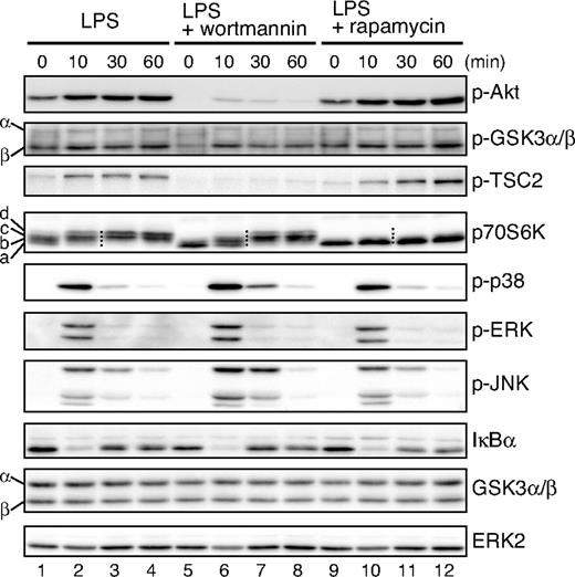
We examined the signaling components of PI3K-Akt pathway in LPS-stimulated BMDCs (Figure 1A). As shown in Figure 2, LPS stimulation induced the phosphorylation of TSC2 and GSK3, known targets of Akt. The activation of mTOR was evaluated by the phosphorylation of p70S6K. Consistent with the fact that mTOR is activated by serum and nutrients, p70S6K was partially phosphorylated even without LPS (Figure 2 lane 1). The hyperphosphorylation of p70S6K examined by electrophoretic mobility shifts was induced within 10 minutes (Figure 2 lane 2) and became clearer up to 60 minutes after LPS stimulation (Figure 2 and Figure S1 lane 4, available on the Blood website; see the Supplemental Materials link at the top of the online article; note that the electrophoretic mobility of p70S6K shifts from a and b up to c and d bands). Rapamycin completely blocked the phosphorylation of p70S6K (Figures 2, S1, compare lanes 1-4 and 9-12). In contrast, rapamycin had no effect on the LPS-induced phosphorylation of Akt and TSC2 (Figure 2 lanes 9-12), which is consistent with the fact that Akt and TSC2 act upstream of mTOR (Figure 1A). Wortmannin, an inhibitor of PI3K, only partially blocked the LPS-induced phosphorylation of p70S6K (Figures 2, S1, lanes 5-8), whereas the phosphorylation of Akt and TSC2 was completely inhibited (Figure 2 lanes 5-8). These data suggest that LPS-induced mTOR activation is mediated by both PI3K-dependent and-independent pathways, the latter of which could involve serum and/or nutrients.
The PI3K signaling pathway is activated by LPS in BMDCs. BMDCs were cultured overnight and then pretreated with either 100 nM wortmannin or 10 ng/mL rapamycin for 20 minutes or left untreated before being stimulated with 1 μg/mL LPS for the indicated times. Cell lysates were analyzed for phospho-Akt, phospho-GSK3α/β, phospho-TSC2, p70S6K, phospho-p38, phospho-ERK, phospho-JNK, IκBα, GSK3α/β, and ERK2 by Western blotting. Four dots added between the 10-minute and 30-minute lanes of p70S6K samples indicate the migration positions of hyperphosphorylated p70S6K caused by multiple phosphorylation events, which are represented as a through d on the left side (Figure S1).
The PI3K signaling pathway is activated by LPS in BMDCs. BMDCs were cultured overnight and then pretreated with either 100 nM wortmannin or 10 ng/mL rapamycin for 20 minutes or left untreated before being stimulated with 1 μg/mL LPS for the indicated times. Cell lysates were analyzed for phospho-Akt, phospho-GSK3α/β, phospho-TSC2, p70S6K, phospho-p38, phospho-ERK, phospho-JNK, IκBα, GSK3α/β, and ERK2 by Western blotting. Four dots added between the 10-minute and 30-minute lanes of p70S6K samples indicate the migration positions of hyperphosphorylated p70S6K caused by multiple phosphorylation events, which are represented as a through d on the left side (Figure S1).
GSK3 activity is negatively regulated by Akt-mediated phosphorylation.23 We found that GSK3β was predominantly phosphorylated on LPS stimulation in BMDCs, whereas both GSK3α and GSK3β were expressed (Figure 2 lanes 1-4). The effect of wortmannin on GSK3β phosphorylation was partial (Figure 2 lanes 5-8), indicating that the LPS-induced phosphorylation of GSK3β was also mediated by both PI3K-dependent and -independent pathways. As expected, rapamycin had no effect on the LPS-induced phosphorylation of GSK3β (Figure 2 lanes 9-12).
We also examined the effects of rapamycin and wortmannin on LPS-activated MAPK and NF-κB pathways. Rapamycin had little effect on the phosphorylation status of MAPK family members or the degradation of IκBα, a measure of NF-κB activation (Figure 2, compare lanes 1-4 and lanes 9-12). In contrast, consistent with previous reports,8,24,25 wortmannin slightly but reproducibly enhanced the LPS-induced phosphorylation of p38 as well as JNK (Figure 2, compare lanes 3 and 7), indicating that a PI3K-dependent but mTOR-independent pathway(s) negatively regulates the activation of p38 and JNK. Wortmannin had little effect on IκBα degradation, indicating that the NF-κB pathway is probably not the target of PI3K pathway in BMDCs.
Rapamycin augments LPS-induced IL-12 production but suppresses IL-10 production in an mTOR-dependent manner
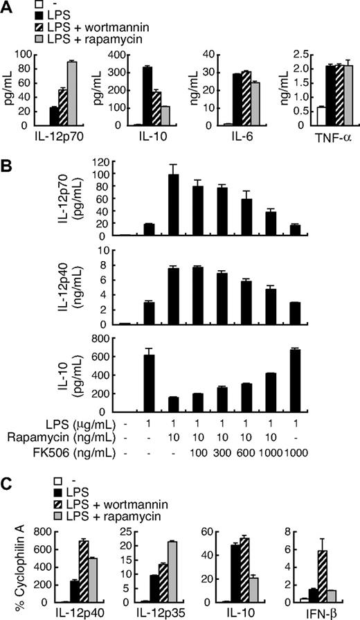
We next examined the role of mTOR in the regulation of IL-12 expression. As shown in Figure 3A, the treatment of BMDCs with rapamycin as well as wortmannin enhanced LPS-induced IL-12p70 production. In contrast, LPS-induced IL-10 production was suppressed by rapamycin or wortmannin, and the effect of rapamycin was more potent than wortmannin (Figure 3A). Rapamycin had the same effect on human MDDCs (Figure S2). On the other hand, rapamycin and wortmannin had little effect on the production of IL-6 and tumor necrosis factor-α (TNF-α; Figure 3A). PTEN−/− BMDCs produced slightly more IL-10 compared with control BMDCs (Figure 1B), but the effect of PTEN deficiency is more pronounced on IL-12 compared with IL-10 production.
The effect of rapamycin on LPS-induced cytokine expression in BMDCs. (A) BMDCs were stimulated with 1 μg/mL LPS in the presence or absence of either 100 nM wortmannin or 10 ng/mL rapamycin for 24 hours and assayed for the production of IL-12p70, IL-10, IL-6, and TNF-α by ELISA. (B) BMDCs were stimulated with 1 μg/mL LPS with or without 10 ng/mL rapamycin along with the indicated concentrations of FK506 for 24 hours and assayed for the production of IL-12p70, IL-12p40, and IL-10 by ELISA. (C) BMDCs were pretreated with or without either 100 nM wortmannin or 10 ng/mL rapamycin for 20 minutes before stimulation with 1 μg/mL LPS. After 4 hours, total RNA was isolated, and IL-12p40, IL-12p35, IL-10, and IFN-β mRNA levels were assessed by real-time PCR using cyclophilin A mRNA as a reference. All data are indicated as median plus or minus SD of duplicate samples. Similar results were obtained with 2 to 4 independent experiments.
The effect of rapamycin on LPS-induced cytokine expression in BMDCs. (A) BMDCs were stimulated with 1 μg/mL LPS in the presence or absence of either 100 nM wortmannin or 10 ng/mL rapamycin for 24 hours and assayed for the production of IL-12p70, IL-10, IL-6, and TNF-α by ELISA. (B) BMDCs were stimulated with 1 μg/mL LPS with or without 10 ng/mL rapamycin along with the indicated concentrations of FK506 for 24 hours and assayed for the production of IL-12p70, IL-12p40, and IL-10 by ELISA. (C) BMDCs were pretreated with or without either 100 nM wortmannin or 10 ng/mL rapamycin for 20 minutes before stimulation with 1 μg/mL LPS. After 4 hours, total RNA was isolated, and IL-12p40, IL-12p35, IL-10, and IFN-β mRNA levels were assessed by real-time PCR using cyclophilin A mRNA as a reference. All data are indicated as median plus or minus SD of duplicate samples. Similar results were obtained with 2 to 4 independent experiments.
A complex of rapamycin and FK506-binding protein 12 (FKBP12) binds to and inhibits mTOR. Because FK506 competes with rapamycin for binding to FKBP12, the excess amounts of FK506 cancel the biologic actions of the rapamycin-FKBP12 complex.26 Indeed, FK506 prevented the rapamycin-induced augmentation of IL-12p70 and IL-12p40 production in a dose-dependent manner (Figure 3B). The suppression of IL-10 production by rapamycin was also canceled with excess FK506 (Figures 3B, S3). Excess amounts of FK506 partially restored the hyperphosphorylation of p70S6K (Figure S3). These results confirm that the effect of rapamycin on cytokine production is mediated by the inhibition of mTOR function.
We further examined the effects of wortmannin and rapamycin on LPS-induced cytokine mRNA expression, including IL-12p40, IL-12p35, IL-10, and IFN-β by real-time RT-PCR (Figure 3C). Consistent with these results, the expression of both IL-12p40 and IL-12p35 mRNAs was enhanced by wortmannin and rapamycin. In contrast, IL-10 mRNA expression was suppressed only by rapamycin. Interestingly, wortmannin but not rapamycin enhanced LPS-induced IFN-β mRNA expression. These results collectively suggest that the PI3K regulates the expression of distinct sets of cytokine genes expression in mTOR-dependent and -independent pathways.
Constitutively active Rheb affects the regulation of cytokine production
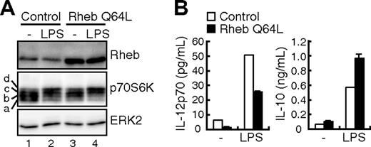
To further confirm the role of mTOR in cytokine gene regulation, we used a lentiviral vector-mediated gene delivery system20 to activate mTOR by expressing a constitutively active form of Rheb (Rheb Q64L).27 The phosphorylation-induced electrophoretic mobility shift of p70S6K in untreated and LPS-stimulated DCs was augmented in BMDCs expressing Rheb Q64L compared with control BMDCs (Figure 4A and Figure S4 lanes 1 and 3, lanes 2 and 4), confirming the activity of Rheb Q64L. As shown in Figure 4B, LPS-induced IL-12p70 production was decreased in Rheb Q64L-expressing BMDCs compared with mock-infected BMDCs. In contrast, LPS-induced IL-10 production was increased in Rheb Q64L-expressing BMDCs. No difference in IL-6 production was observed (data not shown). These results indicate that mTOR is indeed involved in the regulation of IL-12 and IL-10 production in LPS-stimulated BMDCs.
The effect of Rheb Q64L on LPS-induced cytokine production. BMDCs were infected with a lentivirus vector expressing a constitutively active form of Rheb (Rheb Q64L) or vector alone (control). Gene-transduced BMDCs were isolated (“Methods”) and stimulated with or without 1 μg/mL LPS for 24 hours. (A) The cell lysates were analyzed for Rheb, p70S6K, and ERK2 by Western blotting. Note that the mobility shifts of p70S6K caused by multiple phosphorylation are represented as a through d (Figure S4). (B) The production of IL-12p70 and IL-10 in culture supernatants was assayed by ELISA.
The effect of Rheb Q64L on LPS-induced cytokine production. BMDCs were infected with a lentivirus vector expressing a constitutively active form of Rheb (Rheb Q64L) or vector alone (control). Gene-transduced BMDCs were isolated (“Methods”) and stimulated with or without 1 μg/mL LPS for 24 hours. (A) The cell lysates were analyzed for Rheb, p70S6K, and ERK2 by Western blotting. Note that the mobility shifts of p70S6K caused by multiple phosphorylation are represented as a through d (Figure S4). (B) The production of IL-12p70 and IL-10 in culture supernatants was assayed by ELISA.
The effect of rapamycin on IL-12 expression involves IL-10 in an autocrine manner
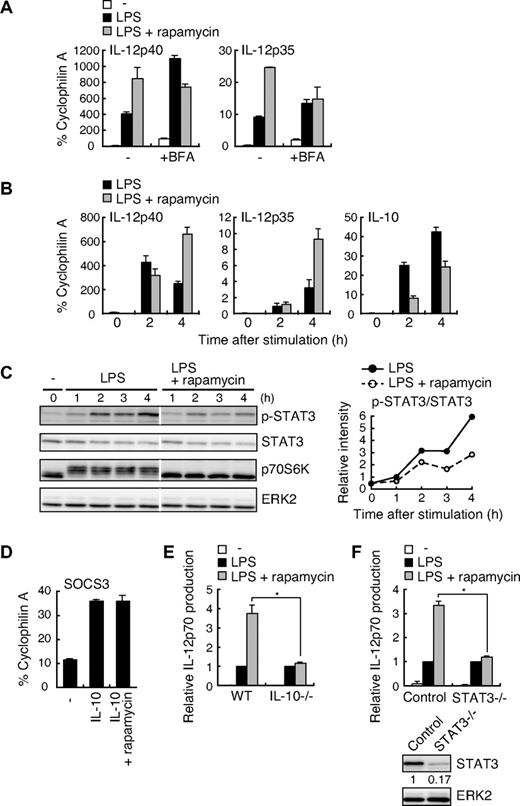
Because mTOR is involved in diverse biologic processes, including protein synthesis and gene expression,14 it is possible that mTOR regulates IL-12 gene expression through new protein synthesis. When BMDCs were treated with cycloheximide to inhibit de novo protein synthesis, the effect of rapamycin was reduced (data not shown). BFA, which blocks protein secretion, abrogated the effect of rapamycin on LPS-induced IL-12p40 and IL-12p35 mRNA expression (Figure 5A). These results strongly suggest that rapamycin controls LPS-induced IL-12 expression through a newly synthesized autocrine mediator(s).
The effect of rapamycin on LPS-induced IL-12 production depends on the IL-10–STAT3 signaling pathway. (A) BMDCs were pretreated with or without 10 ng/mL rapamycin along with or without 5 μg/mL BFA for 20 minutes before stimulation with 1 μg/mL LPS. After 4 hours, total RNA was isolated, and IL-12p40 and IL-12p35 mRNA levels were assessed by real-time PCR using cyclophilin A mRNA as a reference. (B) BMDCs were pretreated with or without 10 ng/mL rapamycin for 20 minutes and then stimulated with 1 μg/mL LPS for the indicated times. Total RNA was isolated, and IL-12p40, IL-12p35, and IL-10 mRNA levels were assessed by real-time PCR using cyclophilin A mRNA as a reference. (C) BMDCs were pretreated with or without 100 ng/mL rapamycin for 20 minutes and then stimulated with 1 μg/mL LPS for the indicated times. The cell lysates were analyzed for phospho-STAT3, STAT3, p70S6K, and ERK2 by Western blotting. The white lines indicate that intervening lanes have been removed. The right panel indicates relative intensities of tyrosine-phosphorylated STAT3 normalized by STAT3 signals. (D) BMDCs were pretreated with or without 100 ng/mL rapamycin for 20 minutes before stimulation with 10 ng/mL IL-10. After 1 hour, total RNA was isolated, and SOCS3 mRNA levels were assessed by real-time PCR using cyclophilin A mRNA as a reference. In panels A, B, and D, data are indicated as mean plus or minus SD of duplicate samples. Data are representative of 2 (B,C) or 3 (A,D) independent experiments with similar results. (E) BMDCs from WT or IL-10−/− mice were stimulated with 1 μg/mL LPS in the presence or absence of 10 ng/mL rapamycin for 24 hours and assayed for the production of IL-12p70 by ELISA. Absolute IL-12p70 levels in the stimulation of LPS alone: WT, 24.1 plus or minus 3.6 pg/mL; IL-10−/−, 1120 plus or minus 230 pg/mL. Data are indicated as median plus or minus SD of 3 independent experiments. *P < .05 by Mann-Whitney U test comparing WT with IL-10−/− groups. (F) BMDCs from STAT3−/− mice or littermate controls were stimulated with 0.1 μg/mL LPS in the presence or absence of 100 ng/mL rapamycin for 24 hours. Cytokine production was evaluated as in panel E. Absolute IL-12p70 levels in the stimulation of LPS alone: control, 11.8 plus or minus 3.7 pg/mL; STAT3−/−, 299 plus or minus 67 pg/mL. *P < .05 by Mann-Whitney U test comparing control with STAT3−/− groups. Indicated below are the expression levels of STAT3 and ERK2 in BMDCs determined by Western blotting.
The effect of rapamycin on LPS-induced IL-12 production depends on the IL-10–STAT3 signaling pathway. (A) BMDCs were pretreated with or without 10 ng/mL rapamycin along with or without 5 μg/mL BFA for 20 minutes before stimulation with 1 μg/mL LPS. After 4 hours, total RNA was isolated, and IL-12p40 and IL-12p35 mRNA levels were assessed by real-time PCR using cyclophilin A mRNA as a reference. (B) BMDCs were pretreated with or without 10 ng/mL rapamycin for 20 minutes and then stimulated with 1 μg/mL LPS for the indicated times. Total RNA was isolated, and IL-12p40, IL-12p35, and IL-10 mRNA levels were assessed by real-time PCR using cyclophilin A mRNA as a reference. (C) BMDCs were pretreated with or without 100 ng/mL rapamycin for 20 minutes and then stimulated with 1 μg/mL LPS for the indicated times. The cell lysates were analyzed for phospho-STAT3, STAT3, p70S6K, and ERK2 by Western blotting. The white lines indicate that intervening lanes have been removed. The right panel indicates relative intensities of tyrosine-phosphorylated STAT3 normalized by STAT3 signals. (D) BMDCs were pretreated with or without 100 ng/mL rapamycin for 20 minutes before stimulation with 10 ng/mL IL-10. After 1 hour, total RNA was isolated, and SOCS3 mRNA levels were assessed by real-time PCR using cyclophilin A mRNA as a reference. In panels A, B, and D, data are indicated as mean plus or minus SD of duplicate samples. Data are representative of 2 (B,C) or 3 (A,D) independent experiments with similar results. (E) BMDCs from WT or IL-10−/− mice were stimulated with 1 μg/mL LPS in the presence or absence of 10 ng/mL rapamycin for 24 hours and assayed for the production of IL-12p70 by ELISA. Absolute IL-12p70 levels in the stimulation of LPS alone: WT, 24.1 plus or minus 3.6 pg/mL; IL-10−/−, 1120 plus or minus 230 pg/mL. Data are indicated as median plus or minus SD of 3 independent experiments. *P < .05 by Mann-Whitney U test comparing WT with IL-10−/− groups. (F) BMDCs from STAT3−/− mice or littermate controls were stimulated with 0.1 μg/mL LPS in the presence or absence of 100 ng/mL rapamycin for 24 hours. Cytokine production was evaluated as in panel E. Absolute IL-12p70 levels in the stimulation of LPS alone: control, 11.8 plus or minus 3.7 pg/mL; STAT3−/−, 299 plus or minus 67 pg/mL. *P < .05 by Mann-Whitney U test comparing control with STAT3−/− groups. Indicated below are the expression levels of STAT3 and ERK2 in BMDCs determined by Western blotting.
IL-10 is an anti-inflammatory cytokine capable of inhibiting the LPS-induced production of proinflammatory cytokines, including IL-12 in DCs.28 When we examined the kinetics of LPS-induced IL-12 and IL-10 expression, rapamycin augmented IL-12p40 and IL-12p35 mRNA expression at 4 hours but not 2 hours after LPS stimulation (Figure 5B). On the other hand, the rapamycin-induced suppression of IL-10 mRNA expression was already observed at 2 hours after LPS stimulation (Figure 5B). Consistent with these results, LPS-induced IL-10 production evaluated by ELISA was significantly reduced by rapamycin at 4 hours after LPS stimulation, whereas IL-12p40 production was little affected (data not shown). These results suggest that the suppression of IL-10 expression by rapamycin subsequently augments IL-12 expression.
To test this hypothesis further, we examined whether rapamycin attenuates the IL-10 signaling pathway in LPS-stimulated DCs. For this purpose, we analyzed the phosphorylation status of STAT3 at Tyr705 as a measure of IL-10 signaling. We found that the tyrosine phosphorylation of STAT3 was markedly induced at 2 hours after LPS stimulation, which was inhibited by rapamycin (Figure 5C). Similar results were obtained in human MDDCs (Figure S5). To rule out the possibility that rapamycin directly inhibits the STAT3 signaling pathway, we examined the effect of rapamycin on the IL-10–induced expression of SOCS3, a well-known target of the STAT3 signaling pathway.29 As shown in Figure 5D, SOCS3 mRNA expression induced by IL-10 stimulation was unaffected by rapamycin. Collectively, these results indicate that rapamycin directly suppresses LPS-induced IL-10 expression but not the STAT3 signaling pathway.
To confirm that rapamycin works through the IL-10-STAT3 pathway to down-regulate IL-12 expression, we used IL-10−/− and STAT3−/− DCs. As shown in Figure 5E, the effect of rapamycin was virtually absent in IL-10−/− BMDCs (1.2 ± 0.03-fold increase) compared with WT BMDCs (3.8 ± 0.1-fold increase). Essentially the same result was obtained with STAT3−/− BMDCs (Figure 5F, 3.3 ± 0.2-fold increase in control BMDCs vs 1.2 ± 0.1-fold increase in STAT3−/− BMDCs). The production of IL-12p70 from IL-10−/− DCs was strongly suppressed by the addition of exogenous IL-10, and such inhibition was rapamycin-independent (Figure S6). These results clearly indicate that rapamycin enhances IL-12 production through the inhibition of autocrine IL-10 action in LPS-stimulated BMDCs.
mTOR and GSK3 cooperatively regulate LPS-induced IL-12 production
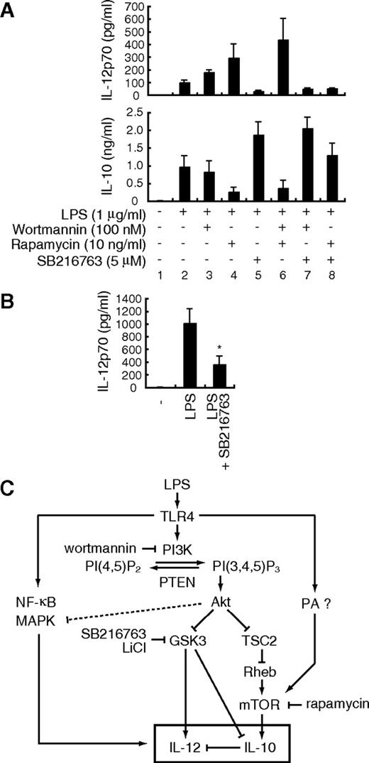
As wortmannin had only a marginal effect on LPS-induced phosphorylation of p70S6K (Figure 2) as well as IL-10 expression (Figure 3A,C), we examined pathways other than mTOR that lie downstream of PI3K. Indeed, GSK3 has been reported to regulate the TLR-mediated production of cytokines, such as IL-12p40 and IL-10 in human monocytes and DCs.30,31 We therefore examined whether GSK3 regulates LPS-induced cytokine production in mouse BMDCs using SB216763, a specific GSK3 inhibitor. SB216763 attenuated IL-12p70 production but enhanced IL-10 production by LPS-stimulated DCs (Figure 6A, lane 5). We obtained similar results with another GSK3 inhibitor LiCl (data not shown). These data indicate that PI3K regulates IL-12 production through both mTOR and GSK3 pathways and that GSK3 positively regulates LPS-induced IL-12p70 and negatively regulates IL-10 production in BMDCs.
mTOR and GSK3β regulate LPS-induced IL-12 production through distinct mechanisms. (A) BMDCs were stimulated with 1 μg/mL LPS together with the indicated inhibitors for 24 hours and assayed for the production of IL-12p70 and IL-10 by ELISA. Data are indicated as median plus or minus SD of 3 independent experiments. (B) BMDCs from IL-10−/− mice were stimulated with 1 μg/mL LPS in the presence or absence of 5 μM SB216763 for 24 hours and assayed for the production of IL-12p70 by ELISA. Data are indicated as median plus or minus SD of 5 independent experiments. *P < .05 by Wilcoxon t test compared with LPS alone. (C) The schematic diagram of the PI3K-mediated regulation of IL-12 production. PA indicates phosphatidic acid; PI(4,5)P2, phosphatidylinositol(4,5)bisphosphate; PI(3,4,5)P3, phosphatidylinositol(3,4,5)trisphosphate.
mTOR and GSK3β regulate LPS-induced IL-12 production through distinct mechanisms. (A) BMDCs were stimulated with 1 μg/mL LPS together with the indicated inhibitors for 24 hours and assayed for the production of IL-12p70 and IL-10 by ELISA. Data are indicated as median plus or minus SD of 3 independent experiments. (B) BMDCs from IL-10−/− mice were stimulated with 1 μg/mL LPS in the presence or absence of 5 μM SB216763 for 24 hours and assayed for the production of IL-12p70 by ELISA. Data are indicated as median plus or minus SD of 5 independent experiments. *P < .05 by Wilcoxon t test compared with LPS alone. (C) The schematic diagram of the PI3K-mediated regulation of IL-12 production. PA indicates phosphatidic acid; PI(4,5)P2, phosphatidylinositol(4,5)bisphosphate; PI(3,4,5)P3, phosphatidylinositol(3,4,5)trisphosphate.
Given that the PI3K-Akt pathway negatively regulates GSK3 (Figure 1A), the treatment of cells with wortmannin should activate GSK3. Interestingly, wortmannin augmented the effect of rapamycin on LPS-induced IL-12p70 production (Figure 6A lanes 2-4 and 6). On the other hand, consistent with the marginal effect of wortmannin alone (Figure 6A lane 3), wortmannin failed to augment the suppressive effect of rapamycin on LPS-induced IL-10 production (Figure 6A lanes 2-4 and 6), suggesting that mTOR and GSK3 differentially regulate IL-12 production. Wortmannin had little effect on LPS-induced IL-12p70 and IL-10 production in the presence of SB216763 (Figure 6A lanes 5 and 7). It is probable that the contribution of GSK3 is similar to or greater than that of mTOR for the regulation of IL-12 production in DCs. Consistent with those observations, LPS-induced IL-12p70 production was decreased in the presence of SB216763 in IL-10−/− BMDCs (Figure 6B). These results collectively indicate that GSK3 directly regulates LPS-induced IL-12 production independent of IL-10 (Figure 6C).
Attenuation of mTOR and GSK3 affects Th1/Th2 balance
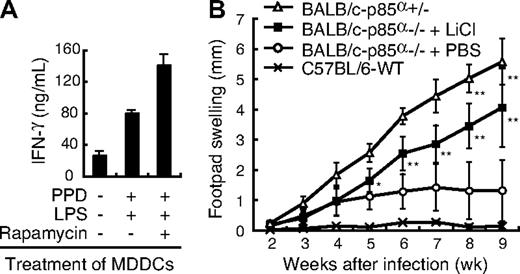
Because IL-12 is critical for triggering Th1 responses, our results raise an interesting possibility that blocking mTOR and GSK3 may enhance and diminish Th1 responses, respectively. Human peripheral CD4+ T cells stimulated with MDDCs pretreated with LPS plus PPD in the presence of rapamycin produced more IFN-γ on restimulation with anti-CD3 plus anti-CD28 antibodies than CD4+ T cells stimulated with DCs pretreated in the absence of rapamycin (Figure 7A). It is thus probable that treatment of DCs with rapamycin results in the augmentation of a Th1 response presumably through enhanced IL-12 production and reduced IL-10 production. We next examined the effect of GSK3 inhibition in an in vivo infection model with L major, in which adequate Th1 development is required for disease control.22 We have previously shown that Th2 prone BALB/c mice can elicit a reasonable Th1 response on L major infection in the absence of p85α (Fukao et al8 and Figure 7B, compare open triangles and open circles). Because GSK3 is expected to have an increased activity in the absence of p85α, we administered a GSK3 inhibitor, LiCl, into footpad of p85α−/− mice on a BALB/c background when infected with L major. As shown in Figure 7B, whereas p85α−/− BALB/c mice were resistant to infection, the administration of LiCl to p85α−/− BALB/c mice resulted in increased footpad swelling and animals were no longer able to control the infection. These results collectively indicate that the attenuation of mTOR and GSK3 by inhibitors affects the balance between Th1 and Th2 responses.
The attenuation of mTOR and GSK3 affects Th1/Th2 balance. (A) Human MDDCs were pretreated with PPD and LPS with or without rapamycin. CD4+ T cells from the same donor were cultured with those MDDCs in the absence of rapamycin. After 1 week of incubation, CD4+ T cells were stimulated with anti-CD3ϵ and anti-CD28 mAbs for 48 hours and assayed for the production of IFN-γ by ELISA. Data are indicated as median plus or minus SD of duplicate samples. (B) The footpad swelling of L major–infected BALB/c-p85α−/− mice treated with LiCl (n = 8) or with PBS (n = 6) was monitored on a weekly basis. BALB/c-p85α+/− mice (n = 5) or C57BL/6-WT mice (n = 2) were used as positive and negative controls for L major infection, respectively. Data are indicated as median plus or minus SD. *P < .05, **P < .01 by Mann-Whitney U test compared with PBS-treated BALB/c-p85α−/− mice. There was no significant difference in footpad swelling between LiCl-treated BALB/c-p85α−/− mice and untreated BALB/c-p85α+/− mice.
The attenuation of mTOR and GSK3 affects Th1/Th2 balance. (A) Human MDDCs were pretreated with PPD and LPS with or without rapamycin. CD4+ T cells from the same donor were cultured with those MDDCs in the absence of rapamycin. After 1 week of incubation, CD4+ T cells were stimulated with anti-CD3ϵ and anti-CD28 mAbs for 48 hours and assayed for the production of IFN-γ by ELISA. Data are indicated as median plus or minus SD of duplicate samples. (B) The footpad swelling of L major–infected BALB/c-p85α−/− mice treated with LiCl (n = 8) or with PBS (n = 6) was monitored on a weekly basis. BALB/c-p85α+/− mice (n = 5) or C57BL/6-WT mice (n = 2) were used as positive and negative controls for L major infection, respectively. Data are indicated as median plus or minus SD. *P < .05, **P < .01 by Mann-Whitney U test compared with PBS-treated BALB/c-p85α−/− mice. There was no significant difference in footpad swelling between LiCl-treated BALB/c-p85α−/− mice and untreated BALB/c-p85α+/− mice.
Discussion
We have previously shown that PI3K negatively regulates TLR-induced pro-inflammatory cytokine production by DCs and gut epithelial cells.8,9,25 Because LPS-induced IL-12 production was decreased in PTEN−/− BMDCs (Figure 1B), the PI3K-Akt pathway triggered by the lipid product of PI3K is indeed important for the suppression of LPS-induced IL-12 production.
Furthermore, our results show that the PI3K-Akt pathway positively regulates IL-10 production. IL-10 is produced by various cell types and plays anti-inflammatory roles in many immune responses.32 In particular, it has been shown that DC-derived IL-10 is involved in a variety of responses, such as infectious diseases,33 the induction of tolerance,34 and cytotoxic T-lymphocyte–mediated antitumor activity.32 Considering that IL-10 plays a pivotal role in immune regulation, the elucidation of the molecular mechanism underlying PI3K-mediated IL-10 regulation would shed new light on therapeutic approaches toward cancer as well as autoimmune diseases.
Indeed, signaling molecules involved in the PI3K path-way (ie, mTOR and GSK3) seem reasonable targets for appropriately modulating the Th1/Th2 balance. As shown here, the treatment of DCs with rapamycin augments a Th1 response (Figure 7A). Because rapamycin does not alter antigen uptake and presentation,35 or the expression level of costimulatory molecules such as CD80 and CD86 in DCs,36 the enhancement of a Th1 response by rapamycin is probably the result of the augmentation of IL-12 production. In addition, the inhibition of GSK3 by LiCl suppressed a Th1-mediated immune response against L major in vivo (Figure 7B). These results raise the possibility that the attenuation of these signaling pathways may provide new therapeutic approaches for human diseases.
Based on our present studies as well as other reports, we propose that signal transduction pathways downstream of Akt regulating IL-12 production are composed of at least 3 components: mTOR, GSK3, and MAPK (Figure 6C). First, the mTOR pathway negatively regulates IL-12 production through the induction of IL-10 gene expression. Wortmannin did not completely inhibit the LPS-induced phosphorylation of p70S6K (Figure 2), and a combination of wortmannin and rapamycin further enhanced LPS-induced IL-12 production (Figure 6A), suggesting that the LPS-induced mTOR activation depends only partially on the PI3K pathway. It should be noted that phosphatidic acid produced by LPS stimulation in macrophages activates mTOR in a PI3K-independent manner37 (Figure 6C).
Second, the GSK3 pathway positively regulates IL-12 production in a more direct manner. Using human monocytes, Martin et al30 showed that GSK3 positively regulates LPS-induced IL-12p40 production. GSK3 augments the binding of NF-κB p65 to a coactivator “cAMP response element-binding protein” (CREB)-binding protein by competitively inhibiting the binding of CREB to CREB-binding protein.30 Rodionova et al31 have also shown that GSK3 enhances IL-12p70 and IL-12p40 production by human Escherichia coli–activated MDDCs. Considering that the GSK3-mediated regulation of IL-12 production was independent of IL-10 (Figure 6B), it is probable that GSK3 controls LPS-induced IL-12 production by a mechanism distinct from mTOR.
Third and finally, we and others have reported that the treatment of monocytes or DCs with SB203580, a specific inhibitor of p38, suppressed LPS-induced IL-12 production.8,11,38 In addition, consistent with the fact that Akt negatively regulates p38,39 the inhibition of PI3K during LPS activation enhanced p38 phosphorylation and activation8,24,25 (Figure 2). Conversely, p38 phosphorylation on LPS stimulation was decreased in macrophages derived from PTEN−/− mice.40 These observations suggest that the PI3K-mediated suppression of p38 results in the attenuation of IL-12 production as well. It has also been reported that the PI3K-Akt pathway negatively regulates JNK activity.41 Indeed, the treatment of BMDCs with wortmannin slightly enhanced LPS-induced JNK phosphorylation (Figure 2). Studies regarding the function of JNK in LPS-induced IL-12 production using human monocyte cell lines are controversial,42,43 and the role of JNK in LPS-induced IL-12 production should be evaluated in primary cells, such as BMDCs derived from JNK-deficient mice. Given that rapamycin (Figure 2) and SB216763 (data not shown) had no effect on LPS-induced phosphorylation of p38 and JNK, it seems improbable that mTOR or GSK3 is involved in the PI3K-Akt pathway-mediated negative regulation of p38 and JNK activity. These results taken together indicate that mTOR, GSK3, and MAPK cooperatively regulate TLR-induced IL-12 production in DCs (Figure 6C).
In contrast to IL-10, IFN-β enhances IL-12 production in an autocrine manner.44 Because wortmannin augments the expression of IFN-β in response to LPS (Figure 3C), it is possible that wortmannin enhances IL-12 production through IFN-β. However, whereas wortmannin enhances the expression of both IL-12p40 and IL-12p35, IFN-β enhances the expression of IL-12p35 but not IL-12p40,44 suggesting that the effect of wortmannin is probably not mediated by IFN-β. In contrast to wortmannin, rapamycin affected only IL-10 but not IFN-β gene expression (Figure 3C).
Because IL-10 inhibits the LPS-induced production of a diverse array of cytokines, such as IL-6 and TNF-α in addition to IL-12,28 rapamycin may influence the production of these cytokines. However, we found that rapamycin had no effect on LPS-induced IL-6 and TNF-α production (Figure 3A), consistent with a previous observation that the LPS-induced mRNA expression of IL-12p40, but not TNF-α, is decreased in BMDCs expressing a constitutively active STAT3.45 Although not shown, rapamycin also augmented IL-12p40 production and suppressed IL-10 production in mouse BMDCs in response to other TLR ligands, such as zymosan (for TLR2/6) and CpG-ODN (for TLR9), whereas it had no effect on IL-6 production. It is possible that the expression of those cytokines has different sensitivities to the negative regulation by IL-10.
What is the molecular mechanism underlying the PI3K-mediated regulation of LPS-induced IL-10 production? p70S6K, one of the targets regulated by mTOR, is able to increase the translation of a subset of mRNAs that contain a 5′ tract of oligopyrimidine.46 However, the 5′ tract of oligopyrimidine was not detected in mouse or human IL-10 mRNA. Another target molecule, 4E-BP1, regulates eIF-4E, which stimulates translational initiation but whose function is thought to be general and not restricted to a subset of genes. mTOR is also involved in gene transcription through the regulation of transcriptional factors without a translational event.14,47,48 This mechanism is more probable because our observations indicate that rapamycin suppresses LPS-induced IL-10 production at a transcriptional level (Figure 3C), rather than through translational regulators. However, the DNA binding of transcriptional factor Sp1 to the IL10 promoter in LPS-stimulated mice BMDCs was unaffected in the presence of rapamycin (data not shown). The regulation by transcription factors downstream of mTOR remains to be elucidated in future studies. In addition to mTOR, GSK3 is involved in a regulatory pathway for IL-10 production. Indeed, GSK3 negatively regulated LPS-induced IL-10 production (Figure 6A), presumably by inactivation of CREB,30 which is involved in the transcriptional activation of the IL10 gene.
It should be noted that many previous studies on the regulation of IL-10 or IL-12 gene expression were performed using macrophage or DC cell lines. We have been aware that cell lines and primary cells often showed different results. One obvious problem is the fact that many cell lines have some defect or alteration in PI3K-PTEN regulation, such that many immortalized cell lines lack PTEN expression and show enhanced Akt activity. These cells are obviously not suitable for studying PI3K pathways. Detailed analysis with primary cells is important in future studies.
The online version of this article contains a data supplement.
The publication costs of this article were defrayed in part by page charge payment. Therefore, and solely to indicate this fact, this article is hereby marked “advertisement” in accordance with 18 USC section 1734.
Acknowledgments
The authors thank Drs A. Suzuki, H. Miyoshi, and K. Takeda for valuable materials, Dr T. Luft of German Cancer Research Center for valuable discussion and sharing unpublished data, and Dr Linda K. Clayton of Harvard Medical School for critically reading the manuscript.
This work was supported by the Uehara Memorial Foundation, a Keio Gijuku Academic Development Fund (S. Matsuda), a Grant-in-Aid for Scientific Research (C 19590499; S. Matsuda) from the Japan Society for the Promotion of Science, a Grant-in-Aid for Scientific Research on Priority Areas (14021110 and 18073015), a National Grant-in-Aid for the Establishment of a High-Tech Research Center in a private university, and a Scientific Frontier Research Grant from the Ministry of Education, Culture, Sports, Science and Technology, Japan.
Authorship
Contribution: M.O. designed the research, performed experiments, and wrote the paper; S.N., S. Kondo, S. Mizuno, K.N., and M.T. performed experiments; T.T. and S. Matsuda designed the research; and S. Koyasu designed the research and wrote the paper.
Conflict-of-interest disclosure: The authors declare no competing financial interests.
Correspondence: Shigeo Koyasu, Department of Microbiology and Immunology, Keio University School of Medicine, 35 Shinanomachi, Shinjuku-ku, Tokyo 160-8582, Japan; e-mail: koyasu@sc.itc.keio.ac.jp.

This feature is available to Subscribers Only
Sign In or Create an Account Close Modal