Abstract
Abstract 4371
The classical immunophenotype of Chronic Lymphocytic Leukaemia (CLL) involves weak or moderate expression of surface immunoglobulin (sIg) M and D. Expression of sIgG has generally been considered to be rare (published estimates 4-28%). The B Cell Receptor is thought to be central to the pathophysiology of CLL.
We present data from an analysis of 748 patients with CLL who have had flow cytometry performed at Barts and the London NHS trust. This includes patients from several other clinical centres serving a population of 3.7 million. A cohort of cases (n=26) displaying differing patterns of sIgG and sIgM expression was investigated further by: 4-colour flow cytometry; detection of constant (C) gene mRNA transcripts including Ca, Cm, Cg, or Cd to confirm isotype status; variable heavy chain somatic mutation rate (VH status); and functionality of the sIg using calcium flux and cell proliferation assays ([H3]- thymidine incorporation) after BCR crosslinking using isotype-specific F(ab')2 anti-human Ig. We also analysed whether differing patterns of sIgG and sIgM expression conveyed prognostic impact.
399 of the 748 patients with CLL had complete sIgG and sIgM expression data. We found that 33% of all CLL cases express sIgG (19% with co-expression of sIgG and sIgM). 19% appeared to have neither sIgG nor sIgM expression. When tested for, sIgD appeared to be present in most of this IgG-IgM- subset. The expression pattern appeared to be stable over time in the majority of cases.
To explore the signalling capacity of the sIg, a cohort of 26 patients with readily available cells was chosen that displayed a variety of expression patterns of sIgG and sIgM (6 IgG-IgM+, 3 IgG+IgM-, 15 IgG+IgM+, 2 IgG-IgM- ). This cohort underwent further testing:
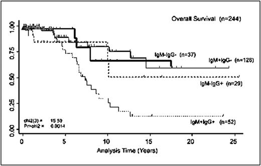
Four-colour flow cytometry confirmed the presence of both sIgG and sIgM on single CLL cells in cases of IgG+IgM+ identified in the diagnostic laboratory, as well as the sIgG and sIgM expression of other cases. In the IgG-IgM+ cases 3 of 6 were VH mutated, one had Cg mRNA transcripts detectable, with all expressing Cm. The IgG+IgM- cases were VH mutated and contained only mRNA for Cg gene transcripts. Of the IgG+IgM+ cases 10/15 were VH mutated. All had detectable Cg and Cm transcripts, with several expressing Ca and Cd. One IgG-IgM- case had no detectable C-gene transcripts but detectable VDJ-genes, the other had Cm and Cd transcripts detectable in the absence of sIgG and sIgM. Both IgG-IgM- cases were VH mutated. Crosslinking of the sIg with appropriate anti sIgG or sIgM F(ab')2 resulted in calcium flux and cell proliferation. These data confirm that the expressed sIgG and sIgM is produced by the CLL cell, and are functionally competent to signal. Prognostic impact: Survival and treatment data were available on 382 patients in our database. Of these, 244 had complete sIgG and sIgM expression determination. Analyses of the sIgG and sIgM expression subgroups suggested that while there is no difference in time to first treatment (median 4.5 years), the IgG+IgM+ group (n=52, median OS 7 years) had a significantly shorter overall survival (OS) compared to the IgG-IgM+ (n=126, median survival not reached), IgG+IgM- (n=29, median OS 11 years), or IgG-IgM- (n=37, median survival not reached) groups.
Overall survival of 244 patients with CLL, as defined by expression of IgG and IgM.
Overall survival of 244 patients with CLL, as defined by expression of IgG and IgM.
In a large cohort of patients with CLL, we show that expression of sIgG is common, with a large number of cases co-expressing sIgG and sIgM simultaneously. When stimulated, both isotypes are capable of signal transduction resulting in cellular activation. These malignant cells appear to have deranged mechanisms of Class Switch Recombination. The IgG+IgM+ group of patients appears to have a more treatment resistant disease, conferring shortened survival. Many different groups have suggested the B Cell Receptor to be an important component of CLL pathophysiology. Our data support these models and provide further insight into the underlying biology of CLL, as well as offering another prognostic marker and possible target for treatment.
No relevant conflicts of interest to declare.
Author notes
Asterisk with author names denotes non-ASH members.


This feature is available to Subscribers Only
Sign In or Create an Account Close Modal